ISSN 2234-3806 • eISSN 2234-3814
https://doi.org/10.3343/alm.2018.38.1.54
Application of Multigene Panel Sequencing in Patients with Prolonged Rate-corrected QT Interval and No
Pathogenic Variants Detected in KCNQ1, KCNH2, and SCN5A
Soo Hyun Seo, M.D.1, So Yeon Kim, M.D.2, Sung Im Cho, M.T.3, Hyunwoong Park, M.D.4, Seungjun Lee, M.D.5, Jong-Moon Choi, M.D.6,7, Man Jin Kim, M.D.3, Jee-Soo Lee, M.D.8, Kyung Jin Ahn, M.D.9, Mi Kyoung Song, M.D.9, Eun-Jung Bae, M.D.9, Sung Sup Park, M.D.3, and Moon-Woo Seong, M.D.3
Department of Laboratory Medicine1, Seoul National University Bundang Hospital, Seongnam; Department of Laboratory Medicine2, National Medical Center, Seoul; Department of Laboratory Medicine3, Seoul National University Hospital, Seoul National University College of Medicine, Seoul; Department of Laboratory Medicine4, Gyeongsang National University Changwon Hospital, Changwon; Department of Laboratory Medicine5, Kangbuk Samsung Hospital, Sungkyunkwan University School of Medicine, Seoul; Green Cross Genome6, Yongin; Green Cross Laboratories7, Yongin; Department of Laboratory Medicine8, Hallym University Sacred Heart Hospital, Anyang; Department of Pediatrics9, Seoul National University Children’s Hospital, Seoul National University College of Medicine, Seoul, Korea
Long QT syndrome (LQTS) is an inherited cardiac disease characterized by a prolonged heart rate-corrected QT (QTc) interval. We investigated the genetic causes in patients with prolonged QTc intervals who were negative for pathogenic variants in three major LQTS- related genes (KCNQ1, KCNH2, and SCN5A). Molecular genetic testing was performed using a panel including 13 LQTS-related genes and 67 additional genes implicated in other cardiac diseases. Overall, putative genetic causes of prolonged QTc interval were identified in three of the 30 patients (10%). Among the LQTS-related genes, we detected a previously reported pathogenic variant, CACNA1C c.1552C>T, responsible for cardiac- only Timothy syndrome. Among the genes related to other cardiac diseases, a likely patho- genic variant, RYR2 c.11995A>G, was identified in a patient with catecholaminergic poly- morphic ventricular tachycardia. Another patient who developed dilated cardiomyopathy with prolonged QTc interval was found to carry a likely pathogenic variant, TAZ c.718G>A, associated with infantile dilated cardiomyopathy. Comprehensive screening of genetic vari- ants using multigene panel sequencing enables detection of genetic variants with a possi- ble involvement in QTc interval prolongation, thus uncovering unknown molecular mecha- nisms underlying LQTS.
Key Words: Multigene panel sequencing, Prolonged heart rate-corrected QT interval, Long QT syndrome
Received: March 23, 2017 Revision received: June 29, 2017 Accepted: September 14, 2017 Corresponding author: Moon-Woo Seong Department of Laboratory Medicine, Seoul National University Hospital, Seoul National University College of Medicine, 101 Daehak-ro, Jongno-gu, Seoul 03080, Korea Tel: +82-2-2072-4180
Fax: +82-2-747-0359 E-mail: [email protected]
© Korean Society for Laboratory Medicine This is an Open Access article distributed under the terms of the Creative Commons Attribution Non-Commercial License (http://creativecom- mons.org/licenses/by-nc/4.0) which permits unrestricted non-commercial use, distribution, and reproduction in any medium, provided the original work is properly cited.
Long QT syndrome (LQTS) is an inherited cardiac disease char- acterized by a prolonged heart rate-corrected QT (QTc) interval on the electrocardiogram (ECG). Besides genetic factors, sev- eral other conditions can lengthen the QTc interval such as elec- trolyte imbalance, use of QTc-prolonging drugs, and structural heart diseases.
To date, 15 genes (KCNQ1, KCNH2, SCN5A, ANK2, KCNE1, KCNE2, KCNJ2, CACNA1C, CAV3, SCN4B, AKAP9, SNTA1, KCNJ5, CALM1, and CALM2) have been implicated in LQTS.
KCNQ1, KCNH2, and SCN5A are known to be responsible for 60–75% of genotype-positive LQTS cases [1, 2], while the other 12 genes linked to LQTS susceptibility collectively account for
2017-03-16 https://crossmark-cdn.crossref.org/widget/v2.0/logos/CROSSMARK_Color_square.svg
<5%. Because genetic predisposition often factors in cases of acquired LQTS, genetic profiling is important, even for patients with mild QTc interval prolongation [3].
Here, we applied targeted sequencing based on a multigene panel containing known LQTS-associated genes to investigate the genetic background of patients with prolonged QTc interval, but negative for mutations in KCNQ1, KCNH2, and SCN5A by Sanger sequencing. The multigene panel also included 67 genes related to other cardiac diseases, which were used to target pa- tients without pathogenic variants of the LQTS-associated genes.
For this study, 30 participants were selected among patients with prolonged QTc interval who underwent genetic testing at the Seoul National University Hospital, Korea, between 2005 and 2011. DNA samples had been archived after the routing screening of KCNQ1, KCNH2, and SCN5A variants for the pur- pose of this research. Most participants exhibited prolonged QTc interval on the resting ECG (>450 ms) and/or positive results on the provocative tests; a few patients with borderline QTc intervals,
but suspected LQTS, were also included. The study was approv ed by the institutional review board of Seoul National University Hos- pital.
The multigene test panel included 13 known LQTS-associated genes (AKAP9, ANK2, CACNA1C, CAV3, KCNE1, KCNE2, KCNH2, KCNJ2, KCNJ5, KCNQ1, SCN4B, SCN5A, and SNTA1) and 67 genes related to other cardiac diseases (see Supplemental Table S1). DNA samples were enriched using the TruSeq Custom En- richment Kit and sequenced with MiSeq (Illumina, Inc., San Di- ego, CA, USA); sequencing data were analyzed using the Next- GENe® Software (Softgenetics, State College, PA, USA). Patho- genic/likely pathogenic variants or variants of uncertain signifi- cance were further confirmed by Sanger sequencing. Copy num- ber variation was not analyzed. Exonic variants with non-synon- ymous changes and intronic variants in 10-bp exon-flanking re- gions were analyzed. Each variant was assessed by considering allele frequencies in normal controls from the 1,000 Genomes database, Exome Aggregation Consortium (ExAC), and Korean
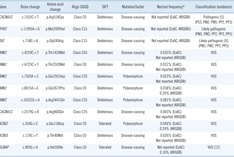
Table 1. Pathogenic, likely pathogenic variants and variants of uncertain significance detected in this study Gene Base change Amino acid
change Align GVGD SIFT MutationTaster Normal frequency* Classification (evidence) CACNA1C c.1552C>T p.Arg518Cys Class C0 Deleterious Disease-causing Not reported (ExAC, KRGDB) Pathogenic [5]
(PS3, PM2, PM5, PP2, PP3) RYR2 c.11995A>G p.Met3999Val Class C15 Deleterious Disease-causing Not reported (ExAC, KRGDB)) Likely pathogenic
(PM1, PM2, PP2, PP3, PP5) TAZ c.718G>A p.Gly240Arg Class C15 Deleterious Disease-causing Not reported (ExAC, KRGDB) Likely pathogenic [6]
(PM1, PM2, PP3, PP5) ANK2 c.4259C>T p.Thr1420Met Class C65 Deleterious Disease-causing 0.035% (ExAC)
Not reported (KRGDB) VUS
ANK2 c.6725C>T p.Thr2242Met Class C0 Deleterious Disease causing 0.031% (ExAC) Not reported (KRGDB)
VUS ANK2 c.7503A>C p.Glu2501Asp Class C35 Deleterious Polymorphism 0.023% (ExAC)
Not reported (KRGDB) VUS
ANK2 c.8015A>C p.Gln2672Pro Class C0 Deleterious Polymorphism 0.058% (ExAC) 0.24% (KRGDB)
VUS ANK2 c.10322G>A p.Arg3441Gln Class C35 Deleterious Polymorphism 0.081% (ExAC)
Not reported (KRGDB) VUS
CACNA1C c.2579G>A p.Arg860Gln Class C35 Deleterious Disease-causing 0.003% (ExAC) Not reported (KRGDB)
VUS KCNJ2 c.354G>C p.Glu118Asp Class C0 Tolerated Polymorphism 0.046% (ExAC)
0.24% (KRGDB) VUS
KCNJ5 c.119C>T p.Thr40Met Class C0 Deleterious Disease-causing 0.026% (ExAC) Not reported (KRGDB)
VUS SLMAP c.805G>A p.Val269Ile Class C0 Tolerated Disease-causing Not reported (ExAC)
0.16% (KRGDB) VUS [12]
*Highest minor allele frequency among the different populations (ExAC).
Abbreviation: VUS, variant of uncertain significance.
Reference Genome DB (KRGDB) and in silico prediction results (Align GVGD, SIFT, and MutationTaster). The highest minor al- lele frequency (MAF) reported in the databases for any popula-
tion was taken into account, and variants with MAF >0.1% were filtered out. Each retained variant was classified according to the American College of Medical Genetics (ACMG) guidelines [4].
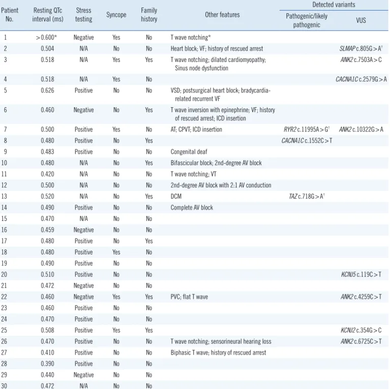
Table 2. Clinical characteristics of the patients included in this study Patient
No. Resting QTc
interval (ms) Stress
testing Syncope Family
history Other features
Detected variants Pathogenic/likely
pathogenic VUS
1 >0.600* Negative Yes No T wave notching*
2 0.504 N/A No No Heart block; VF; history of rescued arrest SLMAP c.805G>A†
3 0.518 N/A Yes Yes T wave notching; dilated cardiomyopathy;
Sinus node dysfunction ANK2 c.7503A>C
4 0.518 N/A Yes No CACNA1C c.2579G>A
5 0.626 Positive No No VSD; postsurgical heart block; bradycardia- related recurrent VF
6 0.460 Negative No Yes T wave inversion with epinephrine; VF; history of rescued arrest; ICD insertion
7 0.500 Positive Yes No AT; CPVT; ICD insertion RYR2 c.11995A>G† ANK2 c.10322G>A
8 0.480 Positive No Yes CACNA1C c.1552C>T
9 0.483 Positive No No Congenital deaf
10 0.480 N/A No Yes Bifascicular block; 2nd-degree AV block
11 0.420 N/A No No T wave notching; VT
12 0.500 N/A No No 2nd-degree AV block with 2:1 AV conduction
13 0.520 N/A No Yes DCM TAZ c.718G>A†
14 0.490 Positive No No Complete AV block
15 0.470 N/A No No
16 0.459 Negative No No
17 0.480 Positive No Yes
18 0.480 Positive Yes No
19 0.490 Positive No No
20 0.510 Positive No No KCNJ5 c.119C>T
21 0.472 Negative No No
22 0.460 Negative Yes Yes PVC; flat T wave ANK2 c.4259C>T
23 0.460 Positive No No
24 0.470 Positive No No
25 0.508 Positive Yes Yes KCNJ2 c.354G>C
26 0.470 Positive No No T wave notching; sensorineural hearing loss ANK2 c.6725C>T
27 0.410 Positive No No Biphasic T wave; history of rescued arrest
28 0.390 Positive No No
29 0.440 Negative No No
30 0.472 N/A No No
*Prolonged QTc interval > 0.600 ms with T wave notching was described in a medical record transferred from another healthcare center; †Variants detected in the expanded panel of other cardiac disease-related genes.
Abbreviations: AT, atrial tachycardia; AV, atrioventricular; CPVT, catecholaminergic polymorphic ventricular tachycardia; DCM, dilated cardiomyopathy; ICD, implantable cardioverter defibrillator; PVC, premature ventricular contraction; VF, ventricular fibrillation; VSD, ventricular septal defect; VT, ventricular tachy- cardia; and VUS, variant of uncertain significance.
For previously reported variants, segregation and functional test results were reviewed. When no pathogenic or likely pathogenic variants were found among the LQTS-linked genes, the genes related to other cardiac diseases were sequentially analyzed.
The average coverage depth in target regions of the multigene panel was 235×, representing 1,831 exons in total; 99.77% and 99.95 % of the bases had ≥30× and ≥5× coverage, respec- tively, the latter was the minimal level of acceptable coverage considered in this study. Regions with coverage <5× were not subjected to Sanger sequencing. The median patient age at ex- amination was 10 years (1 month–30 years), and the average QTc interval was 485 ms (468–502 ms; 95% confidence inter- val). Of the study participants, only one (patient No. 8) was con- firmed to have a pathogenic variant of an LQTS-related gene, CACNA1C c.1552C >T (p.Arg518Cys), which was reported to be associated with Timothy syndrome (Table 1) [5]. This patient showed a phenotype similar to that of an originally reported pro- band: he had a ventricular septal defect (VSD) detected at birth and QTc interval prolongation of 480 ms accompanied by notched T-wave detected on his ECG at age five. The patient did not have any other extracardiac symptoms frequently found in Timothy syndrome, which could have been caused by the pathogenic variant in the CACNA1C gene. He had a family history of sud- den cardiac death, but none of the other family members were tested (Table 2).
Next, genes related to other cardiac diseases were examined in the rest of the patients. One patient (No. 7), who had baseline QTc interval prolongation of 500 ms, exhibited polymorphic ven- tricular tachyarrhythmia, suggesting a diagnosis of catecholamin- ergic polymorphic ventricular tachycardia (CPVT). He was found to carry the ANK2 c.10322G>A (p.Arg3441Gln) variant of un- certain significance; however, an examination of the CPVT-re- lated genes revealed that he also harbored a likely pathogenic variant, RYR2 c.11995A>G, (p.Met3999Val), which could have caused CPVT.
Patient No. 13, who developed dilated cardiomyopathy with QTc interval prolongation of 520 ms, carried a likely pathogenic variant, TAZ c.718G>A (p.Gly240Arg), previously reported as a causative mutation in infantile dilated cardiomyopathy [6]. The patient’s deceased brother had been similarly diagnosed.
Prior to this study, we tested 57 patients with prolonged QTc interval for pathogenic point mutations and large deletions/du- plications in KCNQ1, KCNH2, and SCN5A. Twenty-six patients (45.6%) were identified with pathogenic variants associated with QTc interval prolongation: 14 carried pathogenic mutations in KCNQ1 (24.6%), six in KCNH2 (10.5%), and six in KCNH2
(10.5%) (unpublished data). The detected proportion of patho- genic variants (45.6%) was relatively low compared with that in a previous study demonstrating that pathogenic variants in KCNQ1, KCNH2, and SCN5A accounted for nearly 75% of clinically de- fined LQTS cases and up to 80%, if copy number variant or ge- nomic rearrangement data were included [7]. Another study tested a panel of 12 LQTS-related genes and reported a molec- ular diagnostic level of 30%, which was probably due to the in- clusion of patients with borderline QTc intervals without other clinical symptoms [8]. More stringent criteria considering longer QTc intervals would increase the detection rate; however, in clini- cal laboratory settings, physicians may request diagnostic test- ing for patients with prolonged QTc interval and an intermediate probability of LQTS. Therefore, mutation analysis of genes related to a broader spectrum of cardiac diseases that can cause QTc interval prolongation should improve the diagnostic rate.
In this study, we detected pathogenic or likely pathogenic vari- ants relevant to QTc interval prolongation in three out of 30 pa- tients (10%) negative for KCNQ1, KCNH2, and SCN5A patho- genic variants. Only one patient was confirmed to have a patho- genic variant in an LQTS-related gene. When we expanded the targets to other cardiac disease-related genes, we detected likely pathogenic variants in two patients who showed cardiac mani- festations other than prolonged QTc interval. Similar to the pa- tient No. 7, who carried RYR2 c.11995A>G, several other cases with RYR2 mutations suspected of LQTS were diagnosed as CPVT in a previous study [9]. It should be noted that RYR2 has been proposed as a candidate gene involved in LQTS pathogen- esis as it exhibits interactions with several genes with an estab- lished role in LQTS pathogenesis [10, 11].
In addition, the SLMAP c.805G>A variant, originally reported in a Japanese patient with Brugada syndrome [12], was detected in one of our participants who exhibited an ECG with a complete AV block, but did not exhibit Brugada syndrome features. A pre- vious functional analysis study showed that SLMAP c.805G>A affects the membrane expression of cardiac sodium channel hNav1.5 and reduces hNav1.5-dependent current [12]. How- ever, we found that this variant had a MAF of 0.16% by KRGDB, which was higher than expected for the disorder; therefore, it was recategorized as a variant of uncertain significance.
Because multigene panel sequencing has become widely available, mutation screening of cardiac disease-related genes that may cause QTc interval prolongation can be conducted in parallel, providing more comprehensive molecular analysis and improving the diagnosis rate in patients negative for mutations in LQTS-related genes. Incidental findings of well-described genes
included in the expanded gene panel should be taken into con- sideration, especially if they are present in the minimum list rec- ommended by the ACMG [13].
In conclusion, the multigene panel sequencing performed in this study enabled comprehensive screening of genetic variants with possible involvement in prolonged QTc interval and helped identify additional patients with genotypes that may lead to QTc interval prolongation. Thus, this approach expands the spectrum of genetic causes underlying QTc interval prolongation and can help prevent adverse outcomes, such as sudden cardiac death, in patients and their relatives [14].
Acknowledgements
This study was supported by grant no. 03-2013-0600 from the SNUH Research Fund.
Authors’ Disclosures of Potential Conflicts of Interest
The authors declare no conflict of interest.
REFERENCES
1. Alders M and Christiaans I. Long QT Syndrome. In: Pagon RA, Adam MP, et al., eds. GeneReviews(R). Seattle (WA), University of Washing- ton, 1993, 1993-2017.
2. Giudicessi JR and Ackerman MJ. Genotype- and phenotype-guided management of congenital long QT syndrome. Curr Probl Cardiol 2013;
38:417-55.
3. Itoh H, Crotti L, Aiba T, Spazzolini C, Denjoy I, Fressart V, et al. The ge- netics underlying acquired long QT syndrome: impact for genetic screen- ing. Eur Heart J 2016;37:1456-64.
4. Richards S, Aziz N, Bale S, Bick D, Das S, Gastier-Foster J, et al. Stan- dards and guidelines for the interpretation of sequence variants: a joint
consensus recommendation of the American College of Medical Genet- ics and Genomics and the Association for Molecular Pathology. Genet Med 2015;17:405-24.
5. Boczek NJ, Ye D, Jin F, Tester DJ, Huseby A, Bos JM, et al. Identifica- tion and Functional Characterization of a Novel CACNA1C-Mediated Cardiac Disorder Characterized by Prolonged QT Intervals With Hyper- trophic Cardiomyopathy, Congenital Heart Defects, and Sudden Cardiac Death. Circ Arrhythm Electrophysiol 2015;8:1122-32.
6. D’Adamo P, Fassone L, Gedeon A, Janssen EA, Bione S, Bolhuis PA, et al. The X-linked gene G4.5 is responsible for different infantile dilated cardiomyopathies. Am J Hum Genet 1997;61:862-7.
7. Ackerman MJ, Priori SG, Willems S, Berul C, Brugada R, Calkins H, et al. HRS/EHRA expert consensus statement on the state of genetic test- ing for the channelopathies and cardiomyopathies: this document was developed as a partnership between the Heart Rhythm Society (HRS) and the European Heart Rhythm Association (EHRA). Europace 2011;
13:1077-109.
8. Lieve KV, Williams L, Daly A, Richard G, Bale S, Macaya D, et al. Re- sults of genetic testing in 855 consecutive unrelated patients referred for long QT syndrome in a clinical laboratory. Genet Test Mol Biomark- ers 2013;17:553-61.
9. Stattin EL, Bostrom IM, Winbo A, Cederquist K, Jonasson J, Jonsson BA, et al. Founder mutations characterise the mutation panorama in 200 Swedish index cases referred for Long QT syndrome genetic test- ing. BMC Cardiovasc Disord 2012;12:95.
10. Kauferstein S, Kiehne N, Erkapic D, Schmidt J, Hamm CW, Bratzke H, et al. A novel mutation in the cardiac ryanodine receptor gene (RyR2) in a patient with an unequivocal LQTS. Int J Cardiol 2011;146:249-50.
11. Shigemizu D, Aiba T, Nakagawa H, Ozaki K, Miya F, Satake W, et al.
Exome Analyses of Long QT Syndrome Reveal Candidate Pathogenic Mutations in Calmodulin-Interacting Genes. PLoS One 2015;10:e0130329.
12. Ishikawa T, Sato A, Marcou CA, Tester DJ, Ackerman MJ, Crotti L, et al.
A novel disease gene for Brugada syndrome: sarcolemmal membrane- associated protein gene mutations impair intracellular trafficking of hNav- 1.5. Circ Arrhythm Electrophysiol 2012;5:1098-107.
13. Kalia SS, Adelman K, Bale SJ, Chung WK, Eng C, Evans JP, et al. Rec- ommendations for reporting of secondary findings in clinical exome and genome sequencing, 2016 update (ACMG SF v2.0): a policy statement of the American College of Medical Genetics and Genomics. Genet Med 2017;19:249-55.
14. Nakano Y and Shimizu W. Genetics of long-QT syndrome. J Hum Genet 2016;61:51-5.
Supplementary Table S1. Genes included in the multigene panel
Panel Gene Reference_NM Disease
LQTS genes AKAP9 NM_005751.4 LQTS
ANK2 NM_001148.4 CPVT, LQTS
CACNA1C NM_000719.6 BS, LQTS
CAV3 NM_033337.2 DCM, HCM, LQTS
KCNE1 NM_000219.4 LQTS
KCNE2 NM_172201.1 LQTS
KCNH2 NM_000238.3 LQTS
KCNJ2 NM_000891.2 CPVT, LQTS
KCNJ5 NM_000890.3 LQTS
KCNQ1 NM_000218.2 LQTS
SCN4B NM_174934.3 LQTS
SCN5A NM_198056.2 BS, DCM, LQTS
SNTA1 NM_003098.2 LQTS
Other cardiac disease-related genes ABCC9 NM_005691.2 DCM
ACTC1 NM_005159.4 DCM, HCM
ACTN2 NM_001103.3 DCM, HCM
ANKRD1 NM_014391.2 DCM, HCM
BAG3 NM_004281.3 DCM, HCM
CACNB2 NM_201590.2 BS
CASQ2 NM_001232.3 CPVT
CRYAB NM_001885.1 DCM, HCM
CSRP3 NM_003476.4 DCM, HCM
CTF1 NM_001330.3 DCM
DES NM_001927.3 ARVC, DCM
DMD NM_004006.2 DCM
DSC2 NM_024422.3 ARVC
DSG2 NM_001943.3 ARVC, DCM
DSP NM_004415.2 ARVC, DCM
EMD NM_000117.2 DCM
EYA4 NM_004100.4 DCM
FHL2 NM_201555.1 DCM
FKTN NM_001079802.1 DCM
GATAD1 NM_021167.4 DCM
GLA NM_000169.2 HCM
GPD1L NM_015141.3 BS
HCN4 NM_005477.2 BS
JPH2 NM_020433.4 HCM
JUP NM_002230.2 ARVC, DCM
KCNE3 NM_005472.4 BS
LAMA4 NM_001105206.2 DCM
LAMP2 NM_002294.2 DCM, HCM
(continued to the next page)
Panel Gene Reference_NM Disease
LDB3 NM_001080116.1 DCM, HCM
LMNA NM_005572.3 DCM
MYBPC3 NM_000256.3 DCM, HCM
MYH6 NM_002471.3 DCM, HCM
MYH7 NM_000257.2 DCM, HCM
MYL2 NM_000432.3 HCM
MYL3 NM_000258.2 HCM
MYLK2 NM_033118.3 HCM
MYOZ2 NM_016599.4 HCM
MYPN NM_032578.3 DCM
NEBL NM_006393.2 DCM
NEXN NM_144573.3 DCM, HCM
OBSCN NM_052843.3 HCM
PKP2 NM_004572.3 ARVC
PLB1 NM_153021.4 DCM
PLN NM_002667.3 DCM, HCM
PRKAG2 NM_016203.3 HCM
PSEN1 NM_000021.3 DCM
PSEN2 NM_000447.2 DCM
RBM20 NM_001134363.1 DCM
RYR2 NM_001035.2 ARVC, CPVT, HCM
SCN1B NM_001037.4 BS
SCN3B NM_018400.3 BS
SDHA NM_004168.2 DCM
SGCD NM_000337.5 DCM
SLMAP NM_007159.2 BS
TAZ NM_000116.3 DCM
TCAP NM_003673.3 DCM, HCM
TGFB3 NM_003239.2 ARVC
TMEM43 NM_024334.2 ARVC
TMPO NM_003276.2 DCM
TNNC1 NM_003280.2 DCM, HCM
TNNC2 NM_003279.2 DCM, HCM
TNNI3 NM_000363.4 DCM, HCM
TNNT2 NM_001001430.2 DCM, HCM
TPM1 NM_001018005.1 DCM, HCM
TTN NM_001267550.1 DCM, HCM
TTR NM_000371.3 DCM, HCM
VCL NM_014000.2 DCM, HCM
Abbreviations: ARVC, arrhythmogenic right ventricular cardiomyopathy; BS, Brugada syndrome; CPVT, catecholaminergic polymorphic ventricular tachycar- dia; DCM, dilated cardiomyopathy; HCM, hypertrophic cardiomyopathy; and LQTS, long QT syndrome.
Supplementary Table S1. Continued