Cavo-caval intervention stent insertion after deceased-donor liver transplantation using side-to-side piggyback technique:
report of a case
In-Gyu Kim1, Byung Seup Kim1, Jang Yong Jeon1, Jae Woo Kwon1, Joo Seop Kim1, Doo Jin Kim1, Jae Pil Jung1, Seong Eun Chon1, Han Joon Kim1, Eui Yong Jeon2,
Min-Jeong Kim2, and Kwanseop Lee2
Departments of 1Surgery and 2Radiology, Hallym University Medical Center, Hallym University College of Medicine, Anyang, Korea
Liver transplantation with preservation of the recipient vena cava (piggyback technique) has been performed as an alternative to the conventional method. Outflow disturbance or obstruction of the vena cava in the early period after liver transplantation is associated with high morbidity and mortality. We used side-to-side cavo-caval anastomosis (modified piggyback technique) in a deceased-donor liver transplantation (DDLT) for venous outflow reconstruction.
On postoperative day 9, the patient developed abdominal discomfort, and abnormal liver function showing serum total bilirubin of 6.2 mg/dl and serum AST/ALT of 297/597 IU/L. Doppler ultrasound showed mono-phasic wave forms of the hepatic vein. Computed tomography showed focal narrowing of 9.5 mm×12 mm in diameter at the cavo-caval anastomosis site. Liver biopsy was showed that there was no evidence of acute allograft rejection. Direct venogram showed stenosis of the cavo-caval anastomosis with a pressure gradient of 12 mmHg. An interventional stent was inserted in the stenotic site of the inferior vena cava, and the pressure gradient decreased to 2 mmHg. He was dis- charged from hospital on postoperative day 23 without any other complications. Herein we report a case of de- ceased-donor liver transplantation using the modified piggyback technique, who received an inferior vena cava stent due to stricture of the reconstructed orifice of the vena cava. (Korean J Hepatobiliary Pancreat Surg 2011;15:184-188) Key Words: Liver transplantation; Outflow obstruction; Anastomotic stenosis; Stent
Received: July 30, 2011; Revised: August 4, 2011; Accepted: August 14, 2011 Corresponding author: Jang Yong Jeon
Department of Surgery, Hallym University Medical Center, Hallym University College of Medicine, 896, Pyeonchon-dong, Dongan-gu, Anyang 431-070, Korea
Tel: +82-31-380-3772, Fax: +82-31-385-0157, E-mail: jjy1030@hallym.or.kr
Copyright Ⓒ 2011 by The Korean Association of Hepato-Biliary-Pancreatic Surgery Korean Journal of Hepato-Biliary-Pancreatic Surgery ∙ pISSN: 1738-6349
INTRODUCTION
Liver transplantation with preservation of the recipient vena cava (piggyback technique) has been performed as an alternative to conventional method. As another piggy- back technique, side-to-side cavo-caval anastomosis has been proposed. Outflow disturbance or obstruction of the vena cava in the early period after liver transplantation is associated with high morbidity and mortality. These complications occur more frequently when the piggyback technique is used.1,2
In deceased-donor liver transplantation (DDLT) with a classic piggyback technique, it was reported venous out- flow obstruction with an incidence of 4.6%, and 40% of these patients required re-transplantation.2 In DDLT with
a modified piggyback technique, it was also reported an incidence of obstruction of 1.4%.1 Another report pre- sented that the incidence of outflow obstructions in the classic piggyback technique, the end-to-side technique, and the modified piggyback technique were 2.2%, 6.6%, and 0.7%, respectively.3 Reported incidences of hepatic venous outflow occlusion following living-donor liver transplantation (LDLT) range from 3.9% to 16.6%.4 There are various treatment methods for outflow obstruction, in- cluding endoluminal anastomotic dilation.3
We have used a side-to-side piggyback technique (modified piggyback technique) for venous outflow reconstruction. We had one case of DDLT using a modi- fied piggyback technique recently, but he had to undergo inferior vena cava stenting after operation. We did not
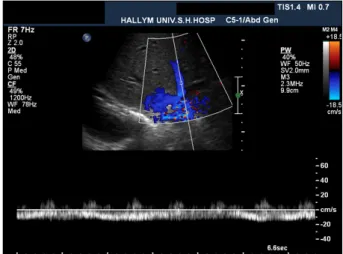
Fig. 1. Doppler sonogram obtained on the fourth post- operative day. The spectral Doppler waveform of the middle hepatic vein shows a mono-phasic flow pattern suggesting hepatic outflow stenosis.
Fig. 2. CT scan images of portal phase at postoperative day 6. (A) There is focal luminal narrowing (arrow) at the side-to-side anastomotic site of the intrahepatic inferior vena cava. (B) Mild focal luminal narrowing (arrow) is also seen at the end-to-end anastomotic site of the main portal vein.
find any reports of inferior vena cava stents for the modi- fied piggyback technique in literature in English.
Herein we report a case of DDLT using the modified piggyback technique, who received inferior vena cava stent due to outflow disturbances of the reconstructed or- ifice of the vena cava.
CASE
A 50-year-old male was admitted to the hospital due to jaundice for 7 days. He had been diagnosed with liver cirrhosis from chronic viral hepatitis B for 8 years. He
had been a heavy smoker (20 pack-years), but had quit 20 years before. The patient had no history of diabetes mellitus, essential hypertension, hyperlipidemia, heart dis- ease, trauma, vasculitis, and connective tissue disease. His blood pressure was 120/80 mmHg, and pulse rate was 80 /min. His body weight was 78.2 kg, and his height was 160 cm. All his close family members were infected with viral hepatitis B. Physical examination showed icteric sclera, but no anemic conjunctiva. He had no tenderness or rebound tenderness of the abdomen. Routine laboratory tests showed total bilirubin of 32 mg/dl, BUN/Cr of 11.4/0.5 mg/dl, and prothrombin time of INR 6.39.
Preoperative abdominal computed tomography (CT) showed liver cirrhosis. Model for end-stage liver disease (MELD) score was 40, and his mental status was hepatic encephalopathy of stage III to IV.
Regarding the operation, both ends of the graft inferior vena cava were closed with staplers. After tangential clamping of the inferior vena cava, the recipient cava was incised approximately 25 mm. Venous outflow reconstruc- tion was performed using a side-to-side cavo-caval anasto- mosis (modified piggyback) method. The total operation time was 8 hour 30 minutes, cold ischemic time was 6 hour 35 minutes, anhepatic time was 72 minutes, and warm ischemic time was 38 minutes. His condition was stable enough to be moved to the general ward on post- operative day 5. But, he showed psychotic activity on postoperative day 7. He kept refusing to take the medi- cine, and spat out his immunosuppressive agents.
Fig. 4. Follow-up CT images at 1 day after stent insertion. (A) The CT image shows a patent stent without narrowing in the cavo-caval anastomosis. (B) The CT image reveals resolution of the stenosis at the portal vein anastomotic site after stenting.
Fig. 3. Interventional stent place- ment at the cavo-caval anasto- mosis site on postoperative day 11. (A) Right hepatic venog- raphy shows contrast pooling in the donor inferior vena cava (IVC) and faint filling of the re- cipient IVC and right atrium (thick arrows) due to tight steno- sis (thin arrows). (B) Hepatic ve- nography after stent insertion across the stenosis (left anterior oblique 44o view). Early contrast flow into the right atrium (arrows) is visible. The pressure gradient between the right hep- atic vein and the recipient IVC decreased from 12 mmHg to 2 mmHg after stent insertion.
On postoperative day 9, he developed symptoms of ab- dominal discomfort, and abnormal liver functions: serum total bilirubin of 6.2 mg/dl and AST/ALT of 297/597 IU/L. Doppler ultrasound examination on postoperative day 4 showed mono-phasic waveforms of the hepatic vein (Fig. 1). Dynamic CT was performed on postoperative day 6 and showed focal narrowing at the cavo-caval anasto- mosis site and mild luminal narrowing at the portal vein anastomosis site (Fig. 2).
Liver biopsy was performed on postoperative day 10, and there was no evidence of acute allograft rejection. On postoperative day 11, he underwent hepatic venogram to check the pressure gradient under the suspicion of hepatic outflow obstruction. The venogram showed stenosis of the cavo-caval anastomosis with a pressure gradient of 12 mmHg. A SMART nitinol stentⓒ (12 mm in diameter and
4 cm in length, self-expandable metallic stent, Cordis, J
& J Medical Systems, Miami Lakes, FL, USA) was in- serted in the stenotic site of the inferior vena cava. After the procedure, the pressure gradient between the right hepatic vein and the recipient inferior vena cava decreased from 12 mmhg to 2 mmHg (Fig. 3). Another stent was inserted into the portal vein because of mild anastomotic stenosis. A follow-up CT obtained 1 day after stent in- sertion demonstrated resolution of the stenosis in the cavo-caval anastomotic site and the portal vein anasto- motic site (Fig. 4). He was discharged from hospital on postoperative day 23 with serum total bilirubin of 2.3 mg/dl and AST/ALT of 69/239 IU/L, and without any other symptoms. A liver function test 6 months after liver transplantation showed total bilirubin of 0.8 mg/dl and AST/ALT of 19/15 IU/L.
DISCUSSION
Multiple factors are known to play important roles in the outcome after liver transplantation. Among them, some are partly or completely related to the trans- plantation technique. The operative time, including the warm ischemic time and anhepatic time, hemorrhage, and hemodynamic instability are the most important ones.
Additionally, it has been reported that the outflow is as important a factor as the inflow.5,6
With the clinical establishment of the piggyback techni- que by Tzakis et al.,7 some complications of conventional liver transplantation could be overcome. Their classic pig- gyback technique has several advantages over conven- tional liver transplantation, such as avoiding the use of ex- tra-corporeal veno-venous bypass and inferior vena cava anastomosis. This leads to shortening of the total oper- ation time and warm ischemic time, and a reduction in complications associated with dissection of the retro- hepatic inferior vena cava, such as phrenic nerve injury and hemorrhage. In addition, the classic piggyback techni- que allows an improvement in intraoperative hemody- namic stability by maintenance of venous return, a reduc- tion of intraoperative blood and fluid administration, im- proved postoperative renal function, better maintenance of oxygen delivery to tissues throughout the surgery, and a reduction in hospital stay length.1 Moreover, the classic piggyback technique does not compress the inferior vena cava and does not cause kinking of the anastomosis be- cause the anastomosis is located in the right and anterior portion of the recipient inferior vena cava.8
There have been criticisms of the classic piggyback technique. Bismuth et al.9 reported that his method had an advantage over the classic piggyback technique be- cause the classic piggyback technique could cause a low flow area followed by thrombosis and a pendulous graft with downward displacement. The classic piggyback tech- nique has been associated with some disadvantages and complications, including hepatic venous outflow ob- struction and thrombosis in up to 10%, because of the in- appropriate size of the hepatic vein outlet, which results in venous congestion of the liver allograft and post-trans- plant ascites, and even Budd-Chiari syndrome. These problems led to technical modifications of the classic pig- gyback technique, in which a side-to-side cavo-caval
anastomosis was proposed.10 This reduces the risk of up- per caval outflow obstruction and Budd-Chiari-like syn- drome, permits the avoidance of caval occlusion, and al- most eliminates the indications of veno-venous bypass for patients who do not tolerate the inferior vena cava cross-clamping test.1,11 One of the other advantages of the modified piggyback is the ease of retransplantation. If re- transplantation is necessary, the non-functional liver can be conveniently removed with preservation of the anasto- mosis and a cuff of the donor’s inferior vena cava. The caval anastomosis can be performed between the cuff and the second donor’s inferior vena cava in a side-to-side fashion.1,12
Outflow obstruction has been variably attributed to the creation of too small outflow tract, twisting of the anasto- mosis, compression of the liver on the inferior vena cava, kinking of the hepatic veins due to long graft suprahepatic inferior vena cava, other technical errors, abnormalities of the inferior vena cava, and adhesions between the liver and the retrohepatic inferior vena cava.1,13 But most of all, a voluminous donor graft or an enlarged caudate lobe be- comes the anatomic limitation that is important in sit- uations that limit the use of caval preserving procedures.
In the modified piggyback technique, the incision on the inferior vena cava should have enough distance from the hepatic veins and the closed cranial part of the donor in- ferior vena cava (approximately 10 mm), to avoid graft outflow obstruction and a functional Budd-Chiari-like syndrome.1
There are various treatment modalities for outflow ob- struction, such as simple rotation of the graft, caval anas- tomosis reconstruction, omentoplasty, diaphragmatic pla- cement of the graft, a second caval anastomosis, endolu- minal anastomotic dilation, conversion to standard ortho- topic liver transplantation, and retransplantation.3 In tradi- tional conventional anastomosis which does not use the piggyback technique, stent insertion has been reported for the treatment of outflow obstruction.
They were concerned about the risk of disruption of a relatively fresh anastomosis during balloon angioplasty because a hepatic venous outflow abnormality was diag- nosed in the early post-transplant period (less than 4 weeks). Balloon angioplasty frequently needed stent placement or surgical reposition because of recurrences of outflow obstruction in the late period.4 Navarro et al.3 re-
ported that of 1,361 liver transplantation cases, 21 had outflow obstruction after the operation. They underwent multiple treatments including endoluminal anastomotic di- latation, but did not mention stent insertion. Thus we think they did not perform interventional stent insertion for those patients.
We also inserted a portal vein stent in present case. The portal vein stent was not the focus in this study, but there have been a few cases of portal vein stents after liver transplantation, and they were performed not infre- quently.14
Despite the widespread acceptance of piggyback techni- que, many surgeons have performed numerous diverse in- cision length of inferior vena cava, and they have diffi- culties in deciding what incision length of inferior vena cava of the donated liver. One underlying reason for this may be the rare studies of the proper incision length of the inferior vena cava. Mehrabi et al.1 insisted that it was performed as 60 mm approximately, but they did not identify the specific reason to do that. In the clinical sit- uation, it is not always so easy to make 60 mm incision length of inferior vena cava. Although Belghiti introduced the modified piggyback technique for the first time, re- cently he stopped doing his modified piggyback technique due to the problem of inferior vena cava exposure.15 The normal diameter of the inferior vena cava is re- ported to be 30 mm × 20 mm.16 Some surgeons insist the incision length of the inferior vena cava the same as its diameter. In present case, the incision length of the in- ferior vena cava was 25 mm. Some experiments suggested that appreciable changes in pressure and flow do not oc- cur until the cross-sectional area of a vessel has been re- duced by more than 75%. The reduction in cross-sectional area is symmetric, and corresponds to at least a 50% re- duction in vessel diameter.17,18 We could not find a reli- able incision length of the inferior vena cava, searching for articles on liver transplantation. We carefully suggest that we have to decide on the incision length of the in- ferior vena cava based on that golden rule of the ex- perimental vascular work above-mentioned.
REFERENCES
1. Mehrabi A, Mood ZA, Fonouni H, et al. A single-center experi- ence of 500 liver transplants using the modified piggyback tech- nique by Belghiti. Liver Transpl 2009;15:466-474.
2. Cescon M, Grazi GL, Varotti G, et al. Venous outflow re- constructions with the piggyback technique in liver trans- plantation: a single-center experience of 431 cases. Transpl Int 2005;18:318-325.
3. Navarro F, Le Moine MC, Fabre JM, et al. Specific vascular complications of orthotopic liver transplantation with preserva- tion of the retrohepatic vena cava: review of 1361 cases.
Transplantation 1999;68:646-650.
4. Ko GY, Sung KB, Yoon HK, et al. Early posttransplant hepatic venous outflow obstruction: Long-term efficacy of primary stent placement. Liver Transpl 2008;14:1505-1511.
5. Lee SG, Park KM, Hwang S, et al. Modified right liver graft from a living donor to prevent congestion. Transplantation 2002;74:54-59.
6. Lee S, Park K, Hwang S, et al. Anterior segment congestion of a right liver lobe graft in living-donor liver transplantation and strategy to prevent congestion. J Hepatobiliary Pancreat Surg 2003;10:16-25.
7. Tzakis A, Todo S, Starzl TE. Orthotopic liver transplantation with preservation of the inferior vena cava. Ann Surg 1989;210:
649-652.
8. Cherqui D, Lauzet JY, Rotman N, et al. Orthotopic liver trans- plantation with preservation of the caval and portal flows.
Technique and results in 62 cases. Transplantation 1994;58:
793-796.
9. Bismuth H, Castaing D, Sherlock DJ. Liver transplantation by
"face-à-face" venacavaplasty. Surgery 1992;111:151-155.
10. Belghiti J, Panis Y, Sauvanet A, Gayet B, Fékété F. A new tech- nique of side to side caval anastomosis during orthotopic hepatic transplantation without inferior vena caval occlusion. Surg Gynecol Obstet 1992;175:270-272.
11. Kim JU, Wang HJ, Lee WH, et al. Clinical experience of side to side caval anastomosis during orthotopic liver transplantation without inferior vena caval occlusion. Korean J Hepatobiliary Pancreat Surg 2001;5:9-14.
12. Mosimann F, Gillet M. Retransplantation of the liver after side-to-side caval anastomosis. Transpl Int 1995;8:157-158.
13. Aucejo F, Winans C, Henderson JM, et al. Isolated right hepatic vein obstruction after piggyback liver transplantation. Liver Transpl 2006;12:808-812.
14. Kim YJ, Ko GY, Yoon HK, Shin JH, Ko HK, Sung KB.
Intraoperative stent placement in the portal vein during or after liver transplantation. Liver Transpl 2007;13:1145-1152.
15. Belghiti J, Ettorre GM, Durand F, et al. Feasibility and limits of caval-flow preservation during liver transplantation. Liver Transpl 2001;7:983-987.
16. Akilli B, Bayir A, Kara F, Ak A, Cander B. Inferior vena cava diameter as a marker of early hemorrhagic shock: a comparative study. Ulus Travma Acil Cerrahi Derg 2010;16:113-118.
17. May AG, Van de berg L, Deweese JA, Rob CG. Critical arterial stenosis. Surgery 1963;54:250-259.
18. Moore WS, Malone JM. Effect of flow rate and vessel calibre on critical arterial stenosis. J Surg Res 1979;26:1-9.