388
©The Korean Society of Food Science and Technology
감마선 조사로 유도된 세포 손상 마우스에서 루틴 투여 후의
방사선 방호 효과
강정애
1,2·윤선혜
1,3·노종국
1,4·최대성
1·장범수
1,3·박상현
1,3,*
1한국원자력연구원 첨단방사선연구소, 2충남대학교 식품영양학과,
3한국과학기술연합대학원대학교 방사선동위원소 응용 및 생명공학, 4광명당제약 부설연구소
Radioprotective Effect of Post-treatment with Rutin on
γ-Irradiation-induced Cellular Damage in Mice
Jung Ae Kang1,2, Seon Hye Yoon1,3, Jong Kook Rho1,4,Dae Seong Choi1, Beom-Su Jang1,3, and Sang Hyun Park1,3,*
1Advanced Radiation Technology Institute, Korea Atomic Energy Research Institute 2Department of Food and Nutrition, Chungnam National University
3Department of Radiation Biotechnology and Applied Radioisotope Science, Korea University of Science and Technology R&D Center 4Kwangmyungdang Pharmaceutical Co. LTD.
Abstract This study aimed to investigate the therapeutic effect of rutin against whole-body γ-irradiation in BALB/c mice. BALB/c mice were randomly divided into four groups and exposed to 6 Gy γ-irradiation. One hour later, mice were orally
administered rutin (50 and 100 mg/kg) for seven consecutive days. γ-Irradiation (6 Gy) resulted in cellular damage as
manifested by elevated levels of plasma hepatic marker enzymes and lipid peroxidation in liver tissue, accompanied with decreased spleen and thymus indices, and white blood cell count. In addition, γ-irradiation significantly decreased the levels of antioxidant enzymes such as superoxide dismutase, glutathione peroxidase and catalase. Rutin treatment significantly
protected against γ-irradiation-induced cellular damage, which was evident by the improvement in the status of most of
the investigated parameters. Therefore, rutin has beneficial effects against radiation-induced damage. Keywords: rutin, γ-irradiation, cellular damage
서
론
우주방사선 및 자연방사선 등 방사선은 우리 주변에 항상 존 재하며, 현대의학의 발전으로 암 환자들의 방사선 치료 증가와 최근 발생한 일본 후쿠시마 원전 사고로 인체의 방사선 노출 위 험도가 증가하고 있다. 이에 따라 방사선에 대한 인체장애나 인 체방어에 대한 관심이 집중되고 있다(1-3). 방사선에 의한 세포 손상은 방사선이 세포에 직접 이온화시키는 직접작용과 방사선 이 세포내의 원자 또는 분자와의 상호작용을 통해 생성된 활성 산소(reactive oxygen species, ROS)를 통해 세포를 손상시키는 간 접작용으로 나눠진다. 방사선에 의한 손상의 대부분은 간접작용 에 의해 이루어지며, 생체는 ROS의 유해한 작용에 대하여 생체 내에 존재하는 항산화 물질이나 항산화 효소와 같은 방어체계를 통해서 제거하거나 생성을 억제한다. 이때 항산화 방어체계가 고 갈되고, ROS생성이 지나치게 많아지면 DNA, 단백질 및 세포막 등에 손상을 입혀 세포 사멸 및 암을 유도한다(4-6). 따라서 방사선에 의한 부작용 경감과 생체 손상을 보호하기 위 해 방사선 보호제에 대한 연구가 진행되고 있다. 현재까지 많은 연구를 통해 WR2721 (amifostine) 등의 SH기를 갖고 있는 합성 물질, 사이토카인(cytokines), 그리고 면역조절제(immunomodulator) 가 방사선 보호제로 개발되어 있다. 그러나 합성물질 같은 경우 인체에 독성이 강하며, 면역조절제는 가격이 고가여서 사용에 한 계가 있어 인체에 독성이 없고 부작용이 적은 천연소재의 방사 선 방호제 개발이 필요하다(7). 최근 천연물질 중에서 항산화 기 능을 갖고 있는 플라보노이드(flavonoid)가 방사선 조사로 유도된 활성산소를 제거하여 방사선에 의해 유도된 세포의 산화적 스트 레스를 방지하는 것으로 보고되었다(8,9). 플라보노이드는 야채와 과일 등의 식물에 널리 존재하는 폴리 페놀(polyphenol) 화합물로써 치환기의 구조와 위치에 따라 안토 시아니딘(anthocyanidin), 플라본(flavone), 플라보놀(flavonol), 플라 바논(flavanol), 플라바노올(flavanolol), 차르콘(chalcone), 카테친 (catechin) 그리고 이소플라본(isoflavone)으로 나누어지며, 지금까 지 알려진 종류만 6,500가지가 넘는다(10). 메밀과 감귤류에 풍부 한 루틴(quercetin-3-rhamnosyl glucoside, rutin, Fig 1)은 비타민 P 라고도 불리며 플라보놀 배당체(glycoside)의 한 종류로서 3-수산 기 그룹 대신 이당류 루티노오스(rutinose)와 결합되어 있다(11,12). 루틴은 항고혈압, 항염증, 혈관 투과성 강화, 혈소판 안정화 등의 기능을 가지고 있다(13,14). 또한, 강력한 항산화 작용 및 라디칼 소거기능, 세포 보호 효과가 있으며, 자외선 조사로 증가된 멜라 닌과 NO (nitric oxide)를 억제하는 것으로 알려져 있다(15-17). 이 *Corresponding author: Sang Hyun Park, Advanced RadiationTech-nology Institute, Atomic Energy Research Institute, Jeongeup, Jeol-labukdo 580-185 Korea
Tel: +82-63-570-3370 Fax: +82-63-570-3371 E-mail: parksh@kaeri.re.kr
Received March 2, 2015; revised April 2, 2015; accepted April 9, 2015
를 바탕으로 방사선에 의한 산화적 세포 손상에 대한 루틴의 방 사선 보호효과를 살펴보고자 한다. 본 연구에서는 BALB/c 마우 스에서 감마선 조사로 유도된 간세포와 조혈계 손상 회복에 대 한 루틴 투여 후의 효과를 평가하였다.
재료 및 방법
시약 및 재료루틴, carboxyl methyl cellulose (CMC), 2,4-dinitro phenylhy-drazine (DNPH), bovine serum albumin (BSA), sodium dode-cyl sulfate (SDS), trichloroacetic acid (TCA), thiobarbituric acid (TBA), aspartate, alanine은 Sigma-Aldrich (St. Louis, MO, USA) 로부터 구입하였다. 실험동물 실험동물은 6주령(18.3±1 g)의 BALB/c 마우스 암컷을 오리엔 트바이오(주)(Seoul, Korea)에서 구입하여 한국원자력연구원 첨단 방사선연구소 동물 사육실에서 식이 및 일정한 조건(23±2oC의 온 도, 55±5%의 습도, 명암순환은 12시간)으로 1주일간 적응시킨 후 실험에 사용하였다. 적응기간 동안 고형사료와 물은 자유로이 공 급하였으며 식수는 멸균하여 사용하였다. 본 연구를 위한 동물실 험은 한국원자력연구원 첨단방사선연구소의 동물실험 윤리위원 회의 승인(KAERI-IACUC-2014-006) 하에 수행되었다. 감마선 조사 감마선 조사는 한국원자력연구원 첨단방사선연구소 내 Gammacell 40 exactor (MDS Nordion, Ottawa, Canada)을 사용하여 1.1 Gy/ min의 선량율이 되게 조사하였다. 정상대조군을 제외한 BALB/c 마우스는 아크릴 상자에 넣고 137Cs선의 흡수선량이 6 Gy가 되 도록 마우스 몸 전체에 조사하였다. 동물실험 수행 감마선 조사 후 루틴의 투여가 감마선 조사로 유도된 손상에 대한 회복 효과를 보기 위하여 BALB/c 마우스를 7마리씩 4개 그룹으로 나누어 실험을 진행하였다. 실험군은 7일 동안 CMC를 제공한 정상대조군, 6 Gy의 감마선을 조사한 후 CMC를 제공한 감마선 조사군, 6 Gy의 감마선을 조사한 후 7일 동안 루틴 50, 100 mg/kg을 경구투여 한 군으로 나누었다. 시료는 감마선 조사 1시간 뒤 투여하여 1일 1회씩 7일 동안 존대(gastric tube)를 이용 하여 식도 하단부까지 삽입하여 경구투여 하였으며, 마지막 시료 투여 후 12시간 절식시킨 뒤 희생하여 분석시료를 채취하였다. 시료채취 감마선 조사 후 루틴의 투여가 감마선 조사로 유도된 손상에 대한 회복 효과를 보기 위해 감마선 조사 후 7일이 지난 다음 12시간 절식시킨 후 BALB/c 마우스를 흡입마취제(2% isoflurane) 로 전신 마취시켰다. 그 후 개복하여 하대정맥에서 혈액을 채취 하고, 간, 비장, 흉선을 적출하여 냉식염수로 세척한 후 건조시켜 무게를 측정하였다. 비장 및 흉선 지수는 (비장 및 흉선 무게/체 중)×100에 의해 계산되었다. 생화학 분석 채취한 혈액을 혈액응고 방지제인 헤파린(heparin)이 처리된 튜 브에 채취하여 3,000×g으로 20분 동안 원심분리하고 혈장을 분 리하여 4oC에서 저장하였다. 혈장 중 aspartate aminotransferase
(AST)와 alanine aminotransferase (ALT)의 측정은 Bergmeye 등 (18)의 방법에 따라 측정하였다. AST와 ALT 기질액 1.0 mL를 각 각 시험관에 가하여 37oC에서 5분간 방치한 다음 혈장 0.2 mL를
넣고 잘 혼합한 후 37oC에서 반응시킨 뒤 DNPH 1 mL를 첨가하
여 잘 혼합하여 실온에서 20분간 방치하여 반응을 종료시켰다. 마지막으로 0.4 M NaOH 용액을 가하여 잘 혼합한 다음 실온에 서 약 10분간 방치하였다가 Multiskan FC microplate photometer (Thermo, Vantaa, Finland)를 이용하여 540 nm에서 흡광도의 변화 를 측정하였다.
혈구 분석
채취한 혈액을 혈액응고 방지제인 ethylendiamin tetraacetic acid (EDTA)가 처리된 튜브에 채취하여 전혈 내 백혈구 수를 혈액학 적 분석기(HEMAVET HV950, Drew Scientific, Inc., Dallas, TX, USA)를 이용하여 측정하였다.
간 조직의 생화학 분석
간 조직에 인산염 완충액(0.1 M, pH 7.4)을 넣은 후, Precellys 24-dual (Bertin, Villeurbanne, France)을 이용하여 10% 균질화 용 액으로 만들었으며, 4oC에서 10,000×g로 10분 동안 원심분리시켜 상층액을 생화학적 분석에 사용하였다. 방사선 조사에 의한 생체 조직의 지질과산화를 알아보기 위해 Ohakawa 등(19)의 방법에 준하여 malondialdehyde (MDA)를 측정하였다. 상층액 100 µL을 취하여 8.1% SDS lysis 용액 100 µL, 20% TCA 용액 150 µL, 0.8% TBA 용액 150 µL을 넣고 100에서 1시간 동안 반응 한 후, 상온에서 식혀 4oC에서 3,000×g로 10분 동안 원심분리 하였으며, 상층액 300 µL과 n-butanol 300 µL을 섞어 10,000×g로 5분 동안 원심분리 한 후, 상층액 200 µL을 취하여 Multiskan FC micro-plate photometer (Thermo, Vantaa, Finland)를 이용하여 540 nm에 서 흡광도의 변화를 측정하였다. superoxide dismutase (SOD), catalase, glutathione peroxidase (GPx)는 측정용 키트 시약(Biovi-sion, Milpitas, CA, USA)으로 측정하였다. 간 조직의 단백질 함 량 측정은 Bradford 방법(20)에 따라 BSA을 표준품으로 사용하 여 측정하였다.
통계분석
실험에서 얻어진 결과는 SPSS (ver. 18.0, IBM, Chicago, IL, USA)를 이용하였다. 방사선에 의해 유도된 손상에 대한 루틴의 동물실험 결과는 일원배치 분산분석(one-way ANOVA)을 이용하 였고, 내부 군 비교는 Tukey 다중범위분석(Tukey’s multiple range test)를 이용하여 수행되었다. 수치는 각 군의 7마리에 대하여 평 균±표준편차로 표현하였고, p<0.05인 경우 유의한 것으로 판단하였다. Fig. 1. Chemical structure of rutin
결과 및 고찰
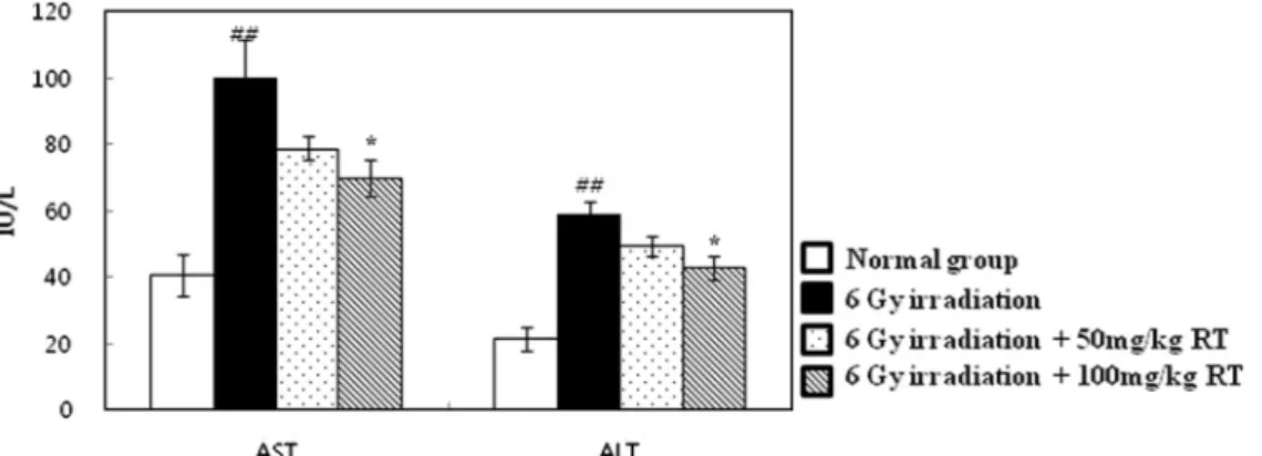
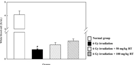
조혈 조직 지수 변화 중선량의 방사선 조사 후 생체 내에서는 방사선에 민감한 조 혈 면역계 기관인 비장 및 흉선이 방사선 장해를 받게 되고, 림 프구 괴사가 일어나 비장 및 흉선의 부피 및 중량이 감소하는 것 으로 알려져 있다(21). 본 연구에서는 정상대조군의 비장 및 흉 선 지수는 각각 0.436과 0.356으로 나타났으며, 감마선 조사군의 비장 및 흉선 지수는 각각 0.086과 0.037로 정상대조군보다 유의 적으로 감소하였다(p<0.05). 본 연구에서는 감마선을 조사 한 후 루틴 100 mg/kg을 투여 한 군의 비장지수는 0.127로 감마선 조사 군에 비해 유의적으로 비장지수가 증가하였다(p<0.05). 또한, 루 틴 100 mg/kg을 투여 한 군의 흉선지수는 0.093으로 감마선 조사 군에 비해 유의적으로 흉선지수가 증가하였다(p<0.05). Pradeep 등(22)은 플라바논 배당체인 헤스페리딘이 방사선 조사로 손상된 비장 지수 감소를 회복하는 것으로 보고하였으며, 본 연구에서도 플라보놀 배당체인 루틴이 방사선 조사 후 방사선 조사에 의한 조혈 면역계 기관 손상에 대해 회복시키는 것을 확인하였다(Table 1). 간세포 손상의 혈장 표식 효소 변화 AST와 ALT는 간세포가 손상을 받았을 경우 활성이 증가되는 아미노기 전이효소로서 간독성 검사에 주로 사용되며, 특히 ALT 는 대부분 간 조직에 분포하여 간 손상과 직접적으로 연결되는 것으로 알려져 있다(23). 또한, 랫트에 감마선을 조사할 경우 혈 중 AST와 ALT의 효소 활성이 급격히 증가됨을 보였다(22). 본 실험에서는 감마선 조사군의 혈장 중 AST와 ALT는 정상대조군 과 비교하였을 때 유의적으로 증가하여 간독성이 나타남을 보여 주었다(p<0.05). 감마선 조사 후 루틴 100 mg/kg을 투여 한 군에 서는 감마선 조사군에 비해 ALT와 AST가 유의적으로 감소하였 다(p<0.05, Fig. 2). Nafees 등(24)은 루틴이 cyclophosphamide로 유도된 간세포 손상에 대해 유의적으로 보호하는 것으로 보고하 였다. 감마선 조사 후 silymarin을 경구 또는 정맥 주사 한 결과 에서 AST와 ALT가 감소하였다는 보고는 본 연구의 실험 결과 와 유사함을 알 수 있었다(25). 따라서 방사선 조사 후 루틴의 투 여가 방사선 조사에 의한 혈장 내 간 손상 지표 효소의 활성 상 승을 유의적으로 억제하여 세포 손상을 억제하고 세포를 보호하 는 것을 알 수 있다. 혈구 성분 변화 방사선 조사 후에는 골수의 혈구모세포 손상으로 혈구생성이 정지되므로 조사 후 일정시간이 지나면 말초혈액내 혈구의 수적 변화가 나타나며, 이러한 세포수의 회복 및 성장은 이들의 회복 에 있어 중요한 요인이 된다(26). 감마선 조사군은 정상대조군에 비해 백혈구(white blood cell, WBC)가 유의적으로 감소하였다 (p<0.05). Lu 등(27)이 보고한 감마선 조사 전 ICR 마우스에서 betalains을 30일간 투여한 실험결과에서 혈구의 수가 증가한 것 을 확인할 수 있었다. 본 실험에서는 루틴을 투여한 군의 백혈구 수치가 감마선 조사군과 비교했을 때 유의성은 나타나지 않았지 만 증가하는 경향을 보였다(Fig. 3). 간 조직의 항산화 효소 변화 방사선에 의해 생성된 자유 라디칼은 생체 내에서 존재하는 항 산화 물질이나 항산화 효소, 또는 섭취되는 항산화제에 의하여 제거된다(28). ROS는 SOD에 의해 과산화수소(H2O2)로 전환되며 과산화수소는 catalase와 GPx에 의해서 물로 전환되어 해독화 된 다(29). 동물에 감마선을 조사할 경우 위와 같은 항산화 효소 활 성의 감소가 나타나는 것으로 알려져 있다(22,30). 감마선 조사 7 일 후 감마선 조사군의 SOD, catalase, GPx는 정상대조군에 비해 유의적으로 감소하였다(p<0.05). 감마선 조사 후 루틴 100 mg/kg 을 투여한 군에서 SOD, catalase, GPx가 감마선 조사군에 비해 유의적으로 증가함을 보였다(p<0.05, Fig. 4). Wang 등(31)은 루 틴이 β-amyloid로 유도된 항산화 효소 활성 감소를 증가시키는 것으로 보고하였다. 홍삼성분이 감마선 조사된 생쥐 간에서 항산Fig. 2. Effect of post-treatment with rutin on the status of AST and ALT of control and experimental animals at day 7 after 6 Gy of γ-irradiation. Values are expressed as mean±SD for seven mice in each group. ##p<0.01 represents significant differences compared with normal group. *p<0.05 represents significant differences compared with 6 Gy of irradiation group.
Table 1. Effect of post-treatment with rutin on the status of spleen index and thymus index of control and experimental animals at day 7 after 6 Gy of γ-irradiation
Groups Spleen index Thymus index Normal group 0.436±0.016 0.356±0.01 6 Gy irradiation 0.086±0.002## 0.037±0.001## 6 Gy irradiation+50 mg/kg RT 00.12±0.008 0.076±0.008 6 Gy irradiation+100 mg/kg RT 0.127±0.003* 0.093±0.009** Values are expressed as mean±SD for seven mice in each group. ##p<0.01 represents significant differences compared with normal group. *p<0.05 and **p<0.01 represent significant differences compared with 6 Gy of irradiation group.
화 효소의 활성도를 증가시켜 방사선 방호 효과가 나타났다는 보 고(32)는 본 연구의 실험 결과와 유사함을 알 수 있었다. 따라서, 루틴이 방사선 조사에 따른 간 조직 내 항산화 효소의 감소를 회 복시킨 것을 확인 할 수 있었다. 간 조직의 지질과산화 변화 세포에 이온화 방사선이 조사되면 생체막을 구성하고 있는 인 지질의 불포화지방산이 활성산소에 의해서 쉽게 손상을 입어 지 질과산화를 유도하여 MDA를 만들어 내며, 이는 산화적 스트레 스의 지표가 된다(33). 본 실험에서는 감마선 조사 7일 후 감마 선 조사군의 MDA는 정상대조군과 비교하였을 때 유의적으로 증 가하였다(p<0.05). 감마선 조사 후 루틴 100 mg/kg을 투여한 군에 서 감마선 조사군에 비해 MDA가 유의적으로 감소하였다(p<0.05, Fig. 5). Kim 등(34)은 루틴이 생쥐 간 마이크로솜의 지질과산화 반응을 억제하고, 수산화 라디칼(hydroxyl radical) 소거능이 있는 항산화제임을 보고하여 본 실험결과와 유사함을 알 수가 있었다. 따라서, 루틴이 방사선 조사에 따른 간 조직 내 지질과산화 상승 을 억제한 것을 확인할 수 있었다.
Fig. 3. Effect of post-treatment with rutin on the status of WBC of control and experimental animals at day 7 after 6 Gy of γ-irradiation. Values are expressed as mean±SD for seven mice in each group. #p<0.05 represents significant differences compared with normal group
Fig. 4. Effect of post-treatment with rutin on the status of SOD (A), catalase (B) and GPx (C) in the liver of control and experimental animals at day 7 after 6 Gy of γ-irradiation. Values are expressed as mean±SD for seven mice in each group. #p<0.05 represent significant differences compared with normal group. *p<0.05 represents significant differences compared with 6 Gy of irradiation
요
약
본 연구는 BALB/c 마우스에서 감마선 조사 후 루틴을 7일 동 안 경구투여하여 감마선 조사로 인한 조혈계 및 간세포 손상에 대한 방사선 회복 효과를 검토하였다. 루틴의 투여는 감마선 조 사군에 비해 조혈 면역계 손상을 억제하여 보호하였으며, 증가된 간독성 지표 효소도 유의적으로 감소하여 간세포 손상에 대한 보 호효과를 확인하였다. 방사선 조사에 의해 체내에서 생성된 자유 라디칼은 생체물질과 결합하여 지질과산화를 일으키고 산화적 스 트레스를 유도하여 조직을 손상시킨다. 루틴을 투여한 군은 감마 선 조사군에 비해 지질과산화가 유의적으로 낮게 나타나 방사선 에 의한 장해를 감소시킨 것으로 사료된다. 루틴을 투여한 군의 항산화 효소의 활성은 감마선 조사군에 비해 유의적으로 증가하 여 생체 내에 항산화 활성을 회복시켰다. 이 상의 결과를 통해 방사선 조사 후에 루틴의 투여는 방사선에 의한 조혈계 및 간세 포 손상에 대해 회복 효과가 있어 방사선 보호제로 유용하게 사 용될 수 있다.감사의 글
이 연구는 미래창조과학부에서 시행하는 방사선기술개발사업 (Grant No. 2012M2A2A601135)의 지원으로 수행되었습니다.References
1. Srinivasa E, Rangaswamy DR, sannappa J. Study on natural gamma radiation hazards in and around Hassan district, Karna-taka state, India. Int. J. Adv. Res. Sci. Technol. 4: 237-240 (2015)
2. Shimura T, Yamaguchi I, Terada H, Okuda K, Svendsen ER, Kunugita N. Radiation occupational health interventions offered to radiation workers in response to the complex catastrophic disaster at the Fukushima Daiichi nuclear power plant. J. Radiat. Res. 56: 413-421 (2015)
3. Huda W. CT radiation exposure: an overview. Curr. Radiol. Rep. 3: 1-16 (2014)
4. Khan A, Manna KC, Das DK, Sinha M, Kesh SB. Das U, Dey RS, Banerji A, Dey S. Seabuckthron (Hippophae rhamnoides L.) leaf extract ameliorates the gamma radiation mediated DNA dam-age and hepatic alterations. Indian J. Exp. Biol. 52: 952-964
(2014)
5. Havaki S, Kotsinas A, Chronopoulos E, Kletsas D, Georgakilas A, Gorgoulis VG. The role of oxidative DNA damage in radia-tion induced bystander effect. Cancer Lett. 356: 43-51 (2015) 6. Riklis E. Radioprotection of DNA by biochemical mechanisms.
Adv. Space Res. 12: 209-212 (1992)
7. Hosseinimehr SJ. Trends in the development of radioprotective agents. Drug Discov. Today 12: 794-805 (2007)
8. Weiss JF, Landauer MR. History and development of radiation-protective agents. Int. J. Radiat. Biol. 85: 539-73 (2009)
9. Adaramoye- O, Ogungbenro B, Anyaegbu O, Fafunso M. Protec-tive effects of extracts of Vernonia amygdalina, Hibiscus sabdar-iffa and vitamin C against radiation-induced liver damage in rats. J. Radiat. Res. 49: 123-131 (2008)
10. Rauha JP, Vuorela H, Kostiainen R. Effect of eluent on the ion-ization efficiency of flavonoids by ion spray, atmospheric pres-sure chemical ionization, and atmospheric prespres-sure photoionization mass spectrometry. J. Mass Spectrom. 36: 1269-1280 (2001) 11. Hopia A, Heinonen M. Antioxidant activity of flavonol aglycones
and their glycosides in methyl linoleate. J. Am. Oil Chem. Soc. 76: 139-144 (1999)
12. Rice-Evans CA, Miller NJ, Paganga G. Structure-antioxidant activity relationships of flavonoids and phenolic acids. Free Radi-cal Bio. Med. 20: 933-956 (1996)
13. Kamalakkannan N, Stanely Mainzen Prince P. Rutin improves the antioxidant status in streptozotocin-induced diabetic rat tissues. Mol. Cell. Biochem. 293: 211-219 (2006)
14. Narayana KR, Sripal Reddy M, Chaluvadi MR, Krishna DR. Bioflavonoids classification, pharmacological, biochemical effects and therapeutic potential. Indian J. Pharmacol. 33: 2-16 (2001) 15. Afanas'ev IB, Dcrozhko AI, Brodskii AV, Kostyuk VA,
Potapo-vitch AI. Chelating and free radical scavenging mechanisms of inhibitory action of rutin and quercetin in lipid peroxidation. Bio-chem. Pharmacol. 38: 1763-1769 (1989)
16. Skibola CF, Smith MT. Potential health impacts of excessive fla-vonoid intake. Free Radical Bio. Med. 29: 375-383 (2000)
17. Lee SY, Kim YM, Lee JK, Yoo SJ, Mun YJ, Woo WH. The effect of rutin on the melanogenesis and nitric oxide in UVB-irradiated HM3KO human melanoma. Anat. Cell Biol. 36: 49-56 (2003)
18. Bergmeyer HU, Scheibe P, Wahlefeld AW. Optimization of meth-ods for aspartate aminotransferase and alanine aminotransferase. Clin. Chem. 24: 58-73 (1978)
19. Ohkawa H, Ohishi N, Yagi A. Assay for lipid peroxides in ani-mal tissues by thiobarbituric acid reaction. Anal. Biochem. 95: 351-358 (1979)
20. Bradford MM. A rapid and sensitive method for quantitation of microgram quantities of protein utilizing the principle of protein-Fig. 5. Effect of post-treatment with rutin on the status of lipid peroxidation in the liver of control and experimental animals at day 7 after 6 Gy of γ-irradiation. Values are expressed as mean±SD for seven mice in each group. #p<0.05 represent significant differences compared with normal group. *p<0.05 and **p<0.01 represent significant differences compared with 6 Gy of irradiation group.
dye binding. Anal. Biochem. 72: 248-254 (1976)
21. Gough GR, Boggs SS, Schwartz GN. Changes in thymus size observed radiographically during the course of spontaneous lym-phoma in the AKR/J mouse. Lab. Anim. Sci. 27: 627-634 (1977) 22. Pradeep K, Ko KC, Choi MH, Kang JA, Chung YJ, Park SH.
Protective effect of hesperidin, a citrus flavanoglycone, against γ-radiation-induced tissue damage in sprague-dawley rats. J. Med. Food 15: 419-427 (2012)
23. Recknagel RO, Glende Jr EA, Dolak JA, Waller RL. Mechanisms of carbon tetrachloride toxicity. Pharmacol. Therapeut. 43: 139-154 (1989)
24. Nafees S, Rashid S, Ali N, Hansan SK, Sultana S. Rutin amelio-rates cyclophosphamide induced oxidative stress and inflamma-tion in wistar rats: Role of NFkB/MAPK pathway. Chem.-Biol. Interact. 231: 98-107 (2015)
25. Ramadan LA, Roushdy HM, Abu Senna GM, Amin NE, El-Deshw OA. Radioprotective effect of silymarin against radiation induced hepatotoxicity. Pharmacol. Res. 45: 447-454 (2002) 26. Patchen ML, MacVittie TJ, Williams JL, Schwartz GN, Souza
LM. Administration of interleukin-6 stimulates multilineage hematopoiesis and accelerates recovery from radiation-induced hematopoietic depression. Blood 77: 472-480 (1991)
27. Lu X, Wang, Y, Zhang Z. Radioprotective activity of betalains from red beets in mice exposed to gamma irradiation. Eur. J.
Pharmacol. 615: 223-227 (2009)
28. Fridovich I. Biological effects of the superoxide radical. Arch. Biochem. Biophys. 247: 1-11 (1986)
29. de Freitas RB, Augusti PR, de Andrade ER, Rother FC, Rovani BT, Quatrin A, Alves NM, Emanuelli T, Bauermann LF. Black grape juice protects spleen from lipid oxidation induced by gamma radiation in rats. J. Food Biochem. 38: 119-127 (2014) 30. Pratheeshkumar P, Kuttan G. Protective role of Vernonia cinerea
L. against gamma radiation-induced immunosuppression and oxi-dative stress in mice. Hum. Exp. Toxicol. 30: 1022-1038 (2011) 31. Wang SW, Wang YJ, Su YJ, Zhou WW, Yang SG, Zhang R,
Zhao M, Li YN, Zhang ZP, Zhan DW, Liu RT. Rutin inhibits β-amyloid aggregation and cytotoxicity, attenuates oxidative stress, and decreases the production of nitric oxide and proinflammatory cytokines. Neurotoxicology 33: 482-490 (2012)
32. Park YS, Kim YG, Chang JC, Kim DY. Radioprotective effects of red ginseng extracts on antioxidants and lipid peroxidation of the liver in γ-irradiated mice. Korean Biochem. J. 26: 184-191 (1993)
33. Emeric J, Edeas M, Bricaire F. Neurodegenerative diseases and oxidative stress. Biomed. Pharmacother. 58: 39-46 (2003)
34. Kim HH, Jang YY, Han ES, Lee CS. Differential antioxidant effects of ambroxol, rutin, glutathione and harmaline. J. Appl. Pharmacol. 7: 112-120 (1999)