논문접수일 :2017년 3월 21일 / 논문수정일 :2017년 4월 28일 / 심사완료일 :2017년 5월 29일 교신저자 :왕수건, 49241 부산광역시 서구 구덕로 179 부산대학교 의학전문대학원 이비인후과학교실 전화 :(051) 240-7824・전송:(051) 246-8668・E-mail:[email protected]
성대의 미세 진동 변화 감별에 라인 스캐닝과 평면 스캐닝 디지털 카이모그래피의 유용성 비교 : 증례 보고
부산대학교병원 이비인후과,1 부산대학교 의학전문대학원 이비인후과학교실,2 연세대학교 의과대학 재활의학교실 및 재활의학연구소,3 부산대학교 인문대학 언어정보학과4
이연우 1 · 왕수건 2 · 김근효 1 · 김향희 3 · 권순복 4
Efficacy of Line Scanning and Two-Dimensional Scanning Digital Kymography in Detection of Subtle Vibratory Changes of the Vocal Cords : A Case Report
Yeon-Woo Lee, MS
1, Soo-Geun Wang, MD, PhD
2, Geun-Hyo Kim, PhD
1, Hyang-Hee Kim, PhD
3and Soon-Bok Kwon, PhD
41
Department of Otorhinolaryngology-Head and Neck Surgery, Pusan National University Hospital, Busan; and
2
Department of Otorhinolaryngology-Head and Neck Surgery, Pusan National University School of Medicine, Busan;
and
3Department of Rehabilitation Medicine, Research Institute of Rehabititation Medicine, Yonsei University College of Medicine, Seoul; and
4Department of Humanities, Language and Information,
Pusan National University, Busan, Korea
-ABSTRACT -
For the evaluation of vocal cord vibration, laryngeal videostroboscopy is commonly used and laryngeal high- speed videoendoscopy has been seldom used, but these modalities may be hard to evaluate subtle change of vi- bratory pattern. In this situation, kymogram using video kymography and line scanning digital kymography seems to be more accurate. Two-dimensional scanning digital kymography can analyze the entire vocal cord. If both line scanning and two-dimensional scanning digital kymography are used simultaneously, subtle vibratory changes of the vocal cords can be effectively identified. 27-year-old male had taken a blunt trauma to the left lateral neck on May 5, 2010. Left arytenoid showed swelling with submucosal hemorrhage and decreased mo- bility of the left vocal cord was noted. On neck CT, fracture of the left arch of the cricoid cartilage was identi- fied. After trauma, he frequently felt easy fatigability during phonation. He was referred to the senior author (WSG) on Dec. 28, 2016. He had diagnosed subtle atrophic changes anterior part of left vocal cord. Simultane- ous multi-modal examinations using high-speed videoendoscopy, line scanning and two-dimensional scanning digital kymography may be useful to assess the subtle vibratory changes in the vocal folds.
(J Clinical Otolaryn- gol 2017;28-:120-124)
KEY WORDS
: Laryngeal traumaㆍLine scanning digital kymographyㆍ Two-dimensional scanning digital kymography.서 론
음성 장애 환자를 검사하기 위해 다양한 평가 방법들 이 소개되고 있다. 이러한 평가 방법들은 직접적인 내 시경 평가 및 간접적 평가로 나눌 수 있다. 직접적인 평 가는 형태학적 평가와 기능적 평가로 나눌 수 있다. 형태 학적 평가는 후두 비디오 스트로보스콥, 후두 고속 비 디오 내시경 검사로 이루어져 있다.1) 기능적 평가는 비 디오 카이모 그래피 시스템,2) 그리고 평면 스캐닝 비디오 카이모 그래피,3) 라인 스캐닝 디지털 카이모 그래피,4) 그리고 평면 스캐닝 디지털 카이모 그래피5) 검사로 이루 어져 있다. 간접적 평가는 전기 성문파형검사, 광성문파 형검사, 음성을 활용한 인벌스 필터링 성문 파형검사, 사운드 스펙트로그램, 그리고 지터, 쉼머, 소음 대 배음비 와 같은 음향학적 변수를 측정하는 음향학적 검사 등으 로 이루어져 있다. 각각의 검사 방법들은 장단점이 있기 때문에, 다양한 검사 방법들을 동시에 사용하여 성대 질 환을 정확하게 진단하고 평가하는 것이 매우 중요하다.
저자들은 경부 좌상 이후, 빈번하게 음성 피로를 호소 하는 케이스를 증례로 분석하여 보고하고자 한다.
증 례
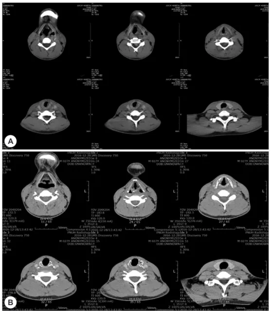
본 증례는 제 1 저자로서 27세의 남성이며, 언어치료 사로 근무하고 있으며 흡연 경험은 전혀 없었다. 2010 년 5월 5일, 축구 경기를 하던 중에 상대방의 머리에 의 해 좌측 경부 좌상을 입었으며, 당시 호흡 곤란, 무성증, 그리고 삼킴 통증을 호소하였다. 신체 검사 결과, 좌측 경부 압통 및 종창이 확인되었지만 염발음과 피하 기종 은 관찰되지 않았다. 후두경 검사상, 좌측 성대 및 피열 연골에 종창, 점막하 출혈이 있었으며, 운동성이 감소되 어있었지만, 우측 성대에서는 정상적인 움직임을 관찰 할 수 있었다. 수상 다음 날, 촬영한 CT 검사에서, 좌측 피 열 연골 및 성문하부에 종창이 관찰되었고, 좌측 윤상 연골궁의 전위골절이 관찰되었다(Fig. 1A).
본 증례는 Trone과 Schaefer가 제안한 후두 기관 손상 분류 중, 그룹 3에 해당되었다.6) 수상 후, 환자는 2주 동 안 항생제와 스테로이드를 복용하였고 증상이 호전되어
퇴원하였다. 수상 3주 후, 정상 발성이 가능하였지만, 종 종 발성 시, 음성 피로를 호소하였다. 2016년 12월, 환자 는 부산대학교병원 이비인후과에 내원하였고, 후두 내시 경 검사와 경부 CT 검사, 그리고 Multi-dimensional voice program(MDVP)와 PCquirer, 전기 성문도를 이용하여 음성 검사를 실시하였다. 후두 내시경 검사 시, 다기능 검사 시스템을 이용하여 후두 고속 비디오 내시경 검 사, 라인 스캐닝 디지털 카이모 그래피 검사, 평면 스캐 닝 디지털 카이모 그래피 검사, 그리고 후두 비디오 스 트로보스콥 검사를 시행하였다.5)
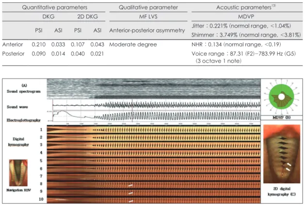
후두 고속 비디오 내시경 검사 및 후두 비디오 스트로 보스콥 검사에서는, 성대 전반부 및 후반부 위상의 비대 칭 정도를 주관적으로 평가하였다.7) 양측 성대의 진동 패턴을 평가하기 위해, 라인 스캐닝 디지털 카이모 그래 피 검사를 이용하여 좌-우측 강도 대칭 지수(amplitude asymmetry index, ASI)와 좌-우측 위상 대칭 지수 (phase symmetry index, PSI)를 정량적으로 분석하였다.8) 평 면 스캐닝 디지털 카이모 그래피 검사에서는 강덕훈 등 이 제시한 방법에 따라 좌-우측 성대의 PSI와 ASI를 측 정하였다.5)
후두 고속 비디오 내시경 검사에서 좌측 성대 전반부 에 성대 휨 현상과 점막 파동의 감소를 관찰할 수 있었고 (Fig. 2A), 전-후방 위상차를 관찰할 수 있었다(Fig. 2B).
라인 스캐닝 디지털 카이모 그래피 검사에서는 성대 전 반부 1/3에서 좌-우측 위상 및 강도 비대칭을 관찰할 수 있었지만, 후반부 2/3에서는 이러한 양상이 나타나지 않았다(Table 1, Fig. 3A). 이와 같은 양상은 평면 스캐 닝 디지털 카이모 그래피 검사에서도 관찰되었다(Table 1, Fig. 3B). 그러나 MDVP를 이용한 음성검사, 스펙트 로그램, 전기성문도 검사의 결과치는 정상인 것으로 나 타났다(Table 1, Fig. 3A, C). Axial CT 검사에서 좌측 윤 상 연골의 변형이 관찰되었다. 이러한 변화는 이전에 손 상 받았던 윤상 연골이 회복되어 가는 과정인 것으로 추정될 수 있다.
고 찰
본 증례는 경부의 좌상 이후 지속된 음성 피로를 주 증 상으로 내원하였다. 발생원인은 후두 외상으로 추정되며,
발생빈도는 전체 외상의 1% 미만에 해당하며, 인구 3만 명 중, 1명이 응급실에 방문한다고 한다.9,10) 후두 외상 은 기도를 막아 호흡 곤란 및 영구적인 음성 장애를 초 래할 수 있으므로, 치료 이전에 적절한 평가가 반드시 필요하다.
성대 진동을 평가하는 방법은 여러 가지가 있으나, 가
장 효과적인 방법은 후두고속 비디오 내시경 검사이다.
그러나, 후두 고속 비디오 내시경 검사는 촬영한 영상을 획득하는데 오랜 시간이 걸리고 획득한 영상의 용량이 매우 크기 때문에 임상에서 효과적으로 활용되지 못하 였다. 하지만, 이러한 단점이 해결되면 후두 비디오 스트 로보스콥 검사보다 더 빈번하게 사용될 수 있을 것으로
Fig. 1. Images of Neck CT at the time of neck trauma (A) and 6 years after trauma (B). The figure (A) showed dis- placed fracture of left lateral cricoid cartilage with soft tissue swelling at the level of glottis and subglottis resulting in distortion and narrowing of the left side subglottic larynx. The figure (B) showed deformity of left lateral cricoid carti- lage with markedly decreased extent of soft tissue swelling considered as healing process of previous injury of cricoid cartilage.
B
A
Table 1. Results of qualitative, quantitative and acoustic parameters
Quantitative parameters Qualitative parameter Acoustic parameters
13)DKG 2D DKG MF LVS MDVP
PSI ASI PSI ASI Anterior-posterior asymmetry Jitter : 0.221% (normal range, <1.04%) Shimmer : 3.749% (normal range, <3.81%) Anterior 0.210 0.033 0.107 0.043 Moderate degree NHR : 0.134 (normal range, <0.19) Posterior 0.090 0.014 0.040 0.021 Voice range : 87.31 (F2)-783.99 Hz (G5)
(3 octave 1 note)
Fig. 2. Sequential images of laryngeal high-speed videoendoscopy (A) and multi-frame laryngeal videostroboscopy (B) (F
0: 140 Hz). One cycle of laryngeal high-speed videoendoscopy was 11 frames, while one cycle of multi-frame laryngeal videostroboscopy at same frequency consists of 140 frames. Thinning and bowing change was noted on left vocal cord without mobility impairment. Anterior glottal gap and decreased mucosal wave was noted on left vocal cord in Figure (A), (B). Moderate anterior-posterior phase asymmetry was noted on Figure (B).
A B
Fig. 3. Multi-images of sound spectrogram, sound wave, electroglottography, line scanning digital kymography (A),
diagram of MDVP (B) and two-dimensional scanning digital kymography (C). The normal vibratory patterns were
noted in sound spectrogram, sound wave, and electroglottography. In line scanning digital kymography, the anteri-
or 1/3 of the left vocal fold began to move toward midline before the left vocal fold began (→), but the posterior 2/3
of vocal folds closed equally in line scanning digital kymography. In two-dimensional scanning digital kymography,
distinctive phase difference was noted on the anterior 1/3 of vocal fold, and the medial peak of left vocal cord was
sharper than that of right vocal cord. It suggested atrophic change of left anterior vocal fold (→).
생각된다. 후두 고속 비디오 내시경 영상을 원본 그래도 관찰하는 것은 쉽지 않기 때문에, 후처리 과정을 통하여 디지털 카이모 그래피,4) 성문 면적 파형(glottal area wave- form),11) 성문 폭 파형(glottal width waveform)12) 검사와 같은 방법들을 통해, 객관적으로 분석할 수 있게 되었다.
본 증례의 경우, 성대 진동과 관련된 생체 역학 정보를 획득할 수 있다.
본 증례의 경우, 좌측 성대의 전반부에서 성문틈이 발 생하고 점막파동이 감소되어있는 것을 확인할 수 있었 다. 또한, 성대 전반부 1/3의 경우에는 우측 성대가 좌측 성대보다 성문 중앙부로 먼저 움직이는 것을 확인하였 으며, 이는 좌-우측 성대 전반부 1/3에 위상차가 있다는 것을 의미한다. 우측 성대의 강도는 같은 위치의 좌측 성대의 강도보다 약한 것으로 나타났다. 성대 후반부 2/3의 경우에는, 양측 성대의 강도는 동일하였다. 전반 부 우측 성대의 medial peak 모양은 좌측 성대에 비해 모양이 둔한(blunt) 것으로 나타났다. 결과적으로 라인 스캐닝 디지털 카이모 그래피와 평면 스캐닝 디지털 카 이모 그래피를 통해 좌측 성대의 미세한 위축이 나타났 고, 이로 인해 나불거림(flagging appearance) 현상이 나 타나는 것을 확인할 수 있었다.
결 론
음성장애 환자에서는 대부분의 경우, 음성 및 전기 성 문 파형 검사 시, 간섭신호 (perturbation)가 증가하고, 후 두 비디오 스트로보스콥 검사에서는 주파수의 동기화 가 어려워서 성대 진동의 느린 동작을 관찰할 수 없는 경 우가 많다. 이로 인해 미세한 성대 진동의 변화를 감지 하기가 어렵다. 이런 경우에는 라인 스캐닝 디지털 카이 모 그래피와 평면 스캐닝 디지털 카이모 그래피를 병용 한 동시 다중 검사(simultaneous multi-modal exami- nation)가 도움을 줄 수 있을 것으로 사료된다.
중심 단어:후두 외상・라인 스캐닝 디지털 카이모그라 피・평면 스캐닝 디지털 카이모그라피.
이 논문은 부산대학교 기본연구지원사업(2년)에 의하여 연구되 었음.
REFERENCES
1) Hirose H. High-speed digital imaging of vocal fold vibra-
tion. Acta Oto-Laryngologica 1988;105(sup458):151-3.
2) Švec JG, Schutte HK. Videokymography: high-speed line
scanning of vocal fold vibration. J Voice 1996;10(2):201-5.
3) Wang S, Lee B, Lee J, Lim Y, Park Y, Park H. Development
of two-dimensional scanning videokymography for analy- sis of vocal fold vibration. J Korean Soc Laryngol Phoniatr Logop 2013;24(1):107-11.
4) Tigges M, Wittenberg T, Mergell P, Eysholdt U. Imaging
of vocal fold vibration by digital multi-plane kymography.
Computerized Medical Imaging and Graphics 1999;23(6):
323-30.
5) Kang DH, Wang SG, Park HJ, Lee JC, Jeon GR, Choi IS,
et al. Real-time simultaneous DKG and 2D DKG using high-speed digital camera. J Voice 2017;31(2):247:e1-7.
6) Trone TH, Schaefer SD, Carder HM. Blunt and penetrating
laryngeal trauma: a 13-year review. Otolaryngolo Head Neck Surg 1980;88(3):257-61.
7) Bonilha HS, Deliyski DD, Gerlach TT. Phase asymmetries
in normophonic speakers: visual judgments and objective findings. Am J Speech-Language Pathol 2008;17(4):367-76.
8) Qiu Q, Schutte HK, Gu L, Yu Q. An automatic method to
quantify the vibration properties of human vocal folds via videokymography. Folia Phoniatr Logop 2003;55(3):128-36.
9) Becker M, Leuchter I, Platon A, Becker CD, Dulguerov P, Varoquaux A. Imaging of laryngeal trauma. Eur J radiol
2014;83(1):142-54.
10) Schaefer SD. The acute management of external larynge-
al trauma: a 27-year experience. Arch Otolaryngol Head Neck Surg 1992;118(6):598-604.
11) Yan Y, Damrose E, Bless D. Functional analysis of voice
using simultaneous high-speed imaging and acoustic re- cordings. J Voice 2007;21(5):604-16.
12) Lohscheller J, Švec JG, Döllinger M. Vocal fold vibration
amplitude, open quotient, speed quotient and their variabil- ity along glottal length: Kymographic data from normal subjects. Logoped Phoniatr Vocol 2013;38(4):182-92.
13) Choi SH, Choi CH. The Characteristics of Voice Handicap
Index and Vocal Misuse and Overuse in Female Elemen- tary Teachers. J Kor Soc Speech Sci 2013;5(4):53-61.
14) Jang MJ, Lee YS, Wang SG, LEE BJ. Clinical application