pISSN 1738-3544 eISSN 2288-1662
Comparison of Methods for Measuring Histamine by ELISA and HPLC-MS Assay In Vitro
In Hee Lee 1 and Yoo Hyun Kim 2
1
Department of Microbiology, Dankook University, Cheonan 31116, Korea
2
Department of Clinical Laboratory Science, Wonkwang Health Science University, Iksan 54538, Korea
In Vitro에서 히스타민 측정 시 ELISA법과 HPLC-MS 분석법의 비교
이인희 1 , 김유현 2
1
단국대학교 미생물학과,
2원광보건대학교 임상병리학과
The measurement of histamine is to determine the degree of allergy because the allergic reaction can lead to the release of histamine. In general, the antigen-antibody reaction was quantified by measuring absorbance using a microplate reader. In this study, we compare the method using a general antigen-antibody reaction and the method using a high performance liquid chroma- tography mass spectrometer (HPLC-MS) of chemical analysis in the measurement of histamine secretion. The cell line used was RBL-2H3, an allergic reaction was induced by stimulation with C48/80 (compound 48/80). Allergy-induced cells degranulation rate was confirmed by measurement of -hexosaminidase and cytotoxicity was performed for the validity of the experiment. The quantitative determination of histamine showed a significant difference, since the quantitative limit of the measurement by the antigen-antibody reaction was 10.257 ppm while the quantitative limit of the measurement by HPLC-MS was 0.020 ppm. Measurement of histamine in allergic activity and anti-allergy tests showed that the HPLC-MS analysis rather than the analysis of the antigen-antibody reaction is a more precise and accurate test.
Keywords: Allergy, -hexosaminidase, Histamine, High performance liquid chromatography mass spectrometer
Corresponding author: Yoo Hyun Kim Department of Clinical Laboratory Science, Wonkwang Helath Science University, Iksan 54538, Korea
Tel: 82-63-840-1218 E-mail: yhkim@wu.ac.kr
This is an Open Access article distributed under the terms of the Creative Commons Attribution Non-Commercial License (http://creativecommons.org/licenses/by-nc/4.0) which permits unrestricted non-commercial use, distribution, and reproduction in any medium, provided the original work is properly cited.
Copyright © 2015 The Korean Society for Clinical Laboratory Science. All rights reserved.
Received: October 16, 2015 Revised 1st: November 6, 2015 Revised 2nd: November 7, 2015 Revised 3rd: November 9, 2015 Accepted: November 9, 2015
서 론
알러지는 일반적으로 반응을 보이지 않는 외부 자극에 대해 인 체의 면역 기전이 보통보다 과민한 반응을 유발시키는 일종의 항원- 항체반응이다(Jung 등, 1998; Park 등, 2007). 이러한 알러지 반응 은 5가지 유형이 있으며 그 중 대부분이 제 I형 알러지 반응에 속한 다(Roitt 등, 2001). 알러지성 질환을 유발시키는 외부물질이 유입 되면 비만세포는 탈 과립이 일어나고 이와 동시에 histamine을 방 출하여 혈중에 유리된다(Mosman 등, 1986; Cooper, 1994).
비만세포는 피부, 림프관 주위, 호흡기, 혈관 주위, 위장 관의 점 막, 뇌 등 전신에 분포되어 있으며 천식이나 알러지성 비염과 같은 반응을 매개하는 중요한 면역세포이다(Marshall, 2004). 비만세포 표면의 IgE (immunoglobulin E) 수용체에 결합된 IgE 항체와 특정 항원이 반응할 때 특이적 cross-linking으로 활성화 되면 비만세포 과립내의 histamine, proteoglycan, serine protease, carboxy- peptidase A, sulfatase, exoglycosidase 등의 화학적 매개체들과 특정 cytokine들이 방출되면서 알러지성 염증을 유발시킨다. 또한 화학적 매개체들이 세포로부터 지속적으로 생성되고 방출되면서
만성적 염증반응이 되기도 한다(Nadler와 Mattews, 2000).
Histamine은 indolamine (serotonine, histamine 등), cate- cholamine (dopamine, epinephrine 등), polyamine (spermine 등)과 같이 생체 내에 존재하는 생리활성 아민 이다. Histamine은 체내에서 histidine decarboxylase의 작용에 의하여 histidine으 로부터 생합성 되어 조직의 비만세포, 혈액의 호염기구의 과립 중 에 저장된다(Schayer, 1956). Histamine은 비만세포의 과립에서 가장 빠르게 방출되는 화학적 매개체로 점막표면에 대한 선 상피세 포의 분비 항진 작용, 기관지 평활근에 대한 수축작용, 말초혈관에 대한 투과성 항진과 확장작용 등을 유발시킨다(Petersen등, 1996;
Mekori와 Metcalfe, 2000). 이것은 생체의 항원-항체 반응에 의한 방어기전과 관련이 있는 것으로 알려져 있다. 이와 같이 알러지 반 응에 있어서 histamine은 필수적이고 중요한 역할을 하고 있으며 실험적으로도 알러지 반응의 정도를 측정하는 하나의 척도로 활용 되고 있다.
일반적으로 in vitro, in vivo 실험에서 방출된 histamine을 정량 할 때 항원-항체반응을 이용한 ELISA (enzyme-linked immuno- sorbent assay) 측정법을 많이 사용하고 있다. 그러나 실험에 사용 되는 시약의 제조법이 다양하고 복잡하기 때문에 대부분은 제품화 되어 있는 kit를 사용하고 있는 실정이다. 이러한 측정법은 정량적 인 분석법임에도 불구하고 사용되는 기기 및 실험자의 역량에 따라 결과에 많은 편차를 보이기도 하고, 많은 종류의 시약과 반응시간 이 필요하여 단순한 실험이라고 할 수는 없다. 이에 본 저자들은 복 잡하지 않고 정확도가 높은 histamine의 측정을 위한 연구를 진행 하였다. Histamine을 분비 하는 RBL-2H3 세포주를 C48/80 (compound 48/80)으로 자극하고 이때 방출되는 histamine의 정 량을 항원-항체반응을 이용한 ELISA kit와 분석 화학적인 방법으 로 HPLC-MS (high performance liquid chromatography mass spectrometer)를 이용하여 분석하였으며 그에 대한 차이점을 비 교하여 유의적인 차이를 확인하였다.
재료 및 방법
1. 세포배양 및 세포 독성능 평가
Histamine을 분비하는 RBL-2H3 세포주는 한국 세포주은행 (KTCC)에서 구입 하였으며, 37oC, 5% CO2 incubator (Pana- sonic, Tokyo, Japan)에서 10% FBS (fetal bovine serum, Gibco, Grand Island, NY, USA), 100 U/mL penicillin, 100 g/mL streptomycin (Gibco, Grand Island, NY, USA)이 포함된 DMEM (Dulbeco’s modified eagle’s medium, Gibco, Grand Island, NY, USA)에서 배양하였다. 본 실험에서 알러지가 유발됨의 정당
성을 확인하기 위하여 MTT [3-(4,5-dimethyl-thiazol-2-yl)- 2,5-diphenyltetrazolium bromide (Sigma-Aldrich Co., St.
Louis, USA)] assay로 세포독성을 측정하였다. RBL-2H3 세포 (2×10⁶ cells/mL)에 cetrizine (Sigma-Aldrich Co., St. Louis, USA) 을 10 L/mL의 농도로 처리하였다. 37oC, 5% CO2 조건에서 30분 배양한 후 C48/80 (Sigma-Aldrich Co., St. Louis, USA)을 1 mg/mL의 농도로 처리하여 20분간 동일조건에서 배양하였다. 각 well에 2 mg/mL 농도의 MTT 용액을 10 L씩 첨가하고 다시 4시간 동안 37oC incubator에서 배양하였다. 배지를 제거하고 200 L의 DMSO (dimethyl sulfoxide)로 well에 생성된 formazan 결정을 완 전히 용해시켜 microplate reader (Tecan, Zurich, Switzerland) 를 사용하여 640 nm에서 흡광도를 측정하였다. 세포독성은 시료 의 흡광도를 대조군의 흡광도에 대한 백분율로 나타내었다.
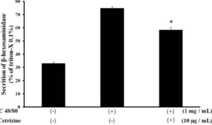
2. -hexosaminidase 방출량 측정
탈 과립 현상의 표지 물질인 -hexosaminidase의 방출량을 측 정함으로써 알러지 유발의 정도를 확인하였다(Schwartz 등, 1979). RBL-2H3 세포는 2×10⁶ cell/mL의 농도로 tyroid buffer (25 mM PIPES, 158 mM NaCl, 5 mM KCl, 0.4 mM MgCl2, 1 mM CaCl2, 5.6 mM glucose, pH7.2)로 3회 세척하였고 tyroid buffer 에 녹인 cetrizine 10 g/mL를 처리하였다. 30분간 37oC, 5% CO2
조건에서 배양한 후 탈 과립을 유도하기 위하여 1 mg/mL C48/80 을 처리하고 20분간 다시 배양하였다. 배양 후 4 oC에서 10분간 안 정화시켰으며 원심분리 후 상층액을 회수하였다. -hexosamini- dase의 기질인 p-nitrophenyl--acetyl-glucosaminide (Sigma- Aldrich Co., St. Louis, USA)를 1 mM의 농도로 처리하고 1시간 동 안 37oC에서 반응시킨 후, 2배수의 sodium bicarbonate (pH 10) 을 첨가하여 반응을 종료하였다. 반응 결과는 415 nm 파장의 microplate reader로 흡광도를 측정하였다.
-hexosaminidase release index
=(O.D at 415 nm of sample/O.D at 415 nm of positive control)×100
3. 항원-항체 반응을 이용한 histamine 방출량 측정
Histamine의 방출량을 항원-항체 반응으로 측정하기 위하여 시 판되고 있는 히스타민 정량 kit를 사용하였으며, 항원-항체반응법 에 의한 kit는 Cayman (Ann Arbor, MI, USA )사의 제품을 사용하 였다(Peters 등, 1984; Sydom 등, 1987; Naclerio 등, 1996).
-hexosaminidase 방출량 측정에서 준비된 동일농도의 샘플 (정상군, 대조군, cetrizine 처리군)의 상층액과 histamine 표준을
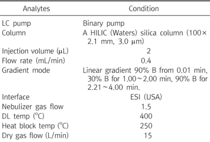
Table 1. HPLC-MS condition of histamine
Analytes Condition
LC pump Binary pump
Column A HILIC (Waters) silica column (100×
2.1 mm, 3.0 m)
Injection volume (L) 2
Flow rate (mL/min) 0.4
Gradient mode Linear gradient 90% B from 0.01 min, 30% B for 1.00∼2.00 min, 90% B for 2.21∼4.00 min.
Interface ESI (USA)
Nebulizer gas flow 1.5
DL temp (
oC) 400
Heat block temp (
oC) 250
Dry gas flow (L/min) 15
농도별(50, 10, 5, 1, 0.5 ppm)로 각각의 premix에 분주 후 voltexing 하였다. 잘 섞어진 각각의 용액을 microplate로 옮긴 다 음 415 nm 파장에서 흡광도를 측정하였다. 농도 별 histamine 표 준에서 얻은 흡광도 값으로 표준곡선을 3회 반복 실험하고 평균값 에 각 샘플의 측정 흡광도를 대입하여 측정치를 확인하였다.
4. HPLC 질량 분석기를 이용한 histamine 방출량 측정
Histamine의 방출량을 분석 화학적인 방법으로 측정하기 위하 여 HPLC 질량 분석기를 사용하였다. 항원-항체 반응을 이용한 histamine 방출량 측정에 사용한 동일한 샘플과 동일한 농도의 histamine (Sigma-Aldrich, St. Louis, USA) 표준(순도 98.00%)을 사용하였다. 실험에 사용된 HPLC 질량 분석기는 LC-20AD, SIL- 20AC, CBM-20A, CTO-20AC, LCMS-2020 (Shimadzu, Kyoto, Japan)이며, HPLC 용매인 acetonitrile (ACN), MeOH (J. T.
Baker, NJ, USA) HPLC용을 사용하였으며, 그 외의 시료 추출을 위 한 용매는 특급 시약을 사용 하였다.
질량 분석기의 분석을 위하여 전체적인 유속은 0.4 mL/min으 로 조정하였다. 이동상 A는 100 mM ammonium formate, pH 3.0 의 H2O이고, 이동상 B는 acetonitrile이였다. HPLC의 조건은 구배 용매 조성법(gradient mode)를 사용하였고, HPLC-MS는 elec- trospray ionization (ESI)로 112.2 m/z SIM(+)의 조건이었으며 HPLC의 분석 조건은 Table 1과 같다.
5. 검출한계(limit of detection) 및 정량한계(limit of quantification)
검출한계 및 정량한계를 측정하기 위하여 먼저 직선성을 검증하 였다. 다섯 가지 농도의 검체를 분석하여 얻은 값의 peak 면적비와 농도비의 관계를 알 수 있는 회귀분석법으로 표준곡선을 얻었다.
50, 10, 5, 1, 0.5 ppm으로 단계적으로 희석한 histamine 표준을
항원-항체 반응법을 이용한 kit와 HPLC-MS로 각각 분석하고 흡광 도와 peak 면적에 대한 표준 농도의 관계를 표시하는 표준곡선을 작성하였다. 3회 실험하여 평균값과 표준편차를 구하였고, 표준곡 선의 기울기와 표준편차를 통해 정량한계 및 검출한계를 측정하였 다. 검출한계는 시료에 포함되는 분석대상물의 검출이 가능한 최저 농도, 정량한계는 검체 중에 포함되어 있는 분석대상물의 정량이 가능한 최저농도로 식품의약처의 의약품 등 시험방법 validation 가이드라인의 계산법 세가지 중 반응의 표준편차와 검량선의 기울 기에 근거하는 방법으로 계산하였으며, 계산식에서 의미하는 LOD 는 검출한계, LOQ는 정량한계이고, 는 반응의 표준편차, S는 검 량선의 기울기를 의미한다(Moon 등, 2014).
LOD=3.3 * /S LOQ=10 * /S
6. 통계처리
각각의 실험결과에 대한 분석은 cetrizine을 투여한 실험군과 대 조군에 대해 t-test를 시행하여
p
<0.05 일 경우 유의성이 있다고 평가하였다.결 과
1. 세포독성 능 평가
RBL-2H3에 알러지를 유발시키기 위하여 처리한 C48/80군과 그에 대한 항 알러지 효과를 보기 위하여 처리한 cetrizine군에 대 하여 MTT assay를 통해 세포독성을 확인 하였다. 아무런 처리를 하 지 않은 정상군의 세포 생존율을 100±1.66%로 하였을 때, C48/80 1 mg/mL을 처리한 대조군은 95.53±3.56%, C48/80 1 mg/mL과 cetrizine 10 g/mL을 처리한 군은 92.67±4.31%로 유 의성 있는 차이는 없었다(Fig. 1).
2. -hexosaminidase 방출량 측정
-hexosaminidase 방출량은 triton-x로 RBL-2H3 세포주를 완전히 용해시켜 세포 자체가 가지고 있는 모든 -hexosamini- dase를 대비하여 방출된 양을 백분율(%)로 계산하였다. 어떤 처리 도 하지 않은 정상군은 33.02±0.93%, C48/80만을 처리한 군은 74.89±1.09%, 그리고 C48/80과 cetrizine을 모두 처리한 군은 58±1.80%로 cetrizine 처리시 유의한 감소를 보여 알러지에 대한 효능이 있다는 것을 확인하였다(Fig. 2).
Fig. 3. Effect of cetrizine on histamine release in C48/80-treated RBL-2H3 cells by antigen-antibody reaction. RBL-2H3 cells were treated with C48/80 (1 mg/mL) or cotreated with cetrizine (10
g/mL). The absorbance was measured at 640 nm using micro- plate reader. Data are the means±SD from triplicate experiments.
Fig. 1. Effect of C48/80 and cetrizine on cell viability of RBL-2H3 cell. Each column represents mean±S.D of 3 independent sam- ples. The absorbance was measured at 640 nm using microplate reader.
Fig. 2. Effect of C48/80 and cetrizine on -hexosaminidase release from RBL-2H3 cell. Each column represents mean±S.D of 3 inde- pendent samples. The absorbance was measured at 415 nm using microplate reader.
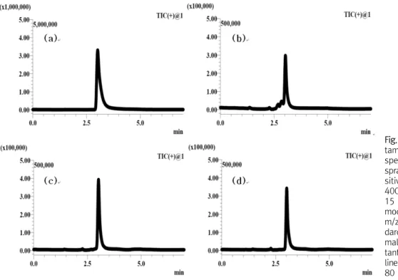
Fig. 4. Effect of cetrizine on histamine release in C48/80-treated RBL-2H3 cells by HPLC-MS of 112.2 m/z SIM(+). RBL-2H3 cells were treated with C48/80 (1 mg/mL) or cotreated with cetrizine (10 g/mL). Data are the means±SD from triplicate experiments.
3. 항원-항체 반응과 분석 화학적 방법을 이용한 histamine 방출량 측정
RBL-2H3 세포주에서 C48/80에 의하여 histamine이 어느 정 도 분비되었는지, 또 cetrizine이 어느 정도로 histamine의 분비를 억제하는지 확인하기 위하여 항원-항체 반응법을 이용한 kit와 HPLC-MS를 이용하여 histamine의 방출량을 측정하였다. 두 가지 측정법을 비교하기 위하여 모두 같은 샘플과 같은 histamine 표준 의 농도(50, 10, 5, 1, 0.5 ppm)를 사용하였다. 항원-항체 반응을 이용한 측정에서 정상군은 0.61±0.08 ppm, 대조군은 0.97±
0.13 ppm, cetrizine 처리군은 0.61±0.05 ppm으로 -hexosa- minidase 방출량과 비례하는 결과를 얻었다(Fig. 3). 분석 화학적 방법은 정상군 0.38±0.01 ppm, 대조군은 0.69±0.02 ppm, cetrizine 처리군은 0.55±0.01 ppm으로 비례하는 결과를 얻었으 며 수치상으로는 차이를 보였다(Fig. 4, 5).
4. 검출한계 및 정량한계
Histamine의 방출량을 측정하는 방법인 항원-항체 반응법과 HPLC-MS를 이용한 분석 화학적인 방법 각각의 정량한계를 측정 하였다. Histamine 분석법 각각을 3회씩 표준으로 표준곡선을 그 리고 기울기 값의 평균과 반응의 표준편차를 공식에 대입하여 계산 하였다. 검출한계는 항원-항체 반응법을 이용한 histamine측정에 서 3.385 ppm, HPLC-MS 분석에서 0.007 ppm 이었으며, 정량한 계는 항원-항체 반응법을 이용한 histamine측정에서 10.257 ppm 이었으며 HPLC-MS 분석에서 0.020 ppm 으로 현저히 낮은 수치를 나타내었다(Table 2).
고 찰
Histamine은 비만세포의 활성화에 대한 탈 과립(degranulation) 의 지표로써, histamine의 측정은 알러지 억제물질의 생물활성 측
Table 2. Detection limit and quantitation limit of respective mensuration
Mensuration S LOD (ppm) LOQ (ppm)
Antigen-antibody reaction 0.0057 0.0055±0.0003 3.385 10.2572
HPLC-MS 7538.1453 3722489.2379±55435.68 0.007 0.020
Abbreviation: , standard deviation of intercept; S, mean of slope; LOD, limit of detection; LOQ, limit of quantitation.
Fig. 5. HPLC-MS chromatogram of his- tamine standard, supernatant of re- spective RBL-2H3 cell line. Electro- spray ionization (ESI) interface in po- sitive mode, 250
oC DL temperature, 400
oC heat block temperature, and 15 L/min nitrogen gas. The single ion mode was used with the following m/z-values: 112.2. (a) Histamine stan- dard 10 ppm, (b) supernatant of nor- mal RBL-2H3 cell line, (c) superna- tant of C48/80-treated RBL-2H3 cell line, (d) supernatant of cotreated (C48/
80 and cetrizine) RBL-2H3 cell line.
정에 유용하게 사용되고 있다(Petersen, 1996; Mekori와 Matcalfe, 2000). 일반적으로 histamine을 정량 할 때는 항원-항체 반응법을 이용하여 발색되는 흡광도를 측정한다. 항원-항체 반응법에 의한 정량분석이 HPLC 분석보다 감도가 뛰어나다는 것도 보고되어 있 다(Muscarella 등, 2005). 그러나 Muscarella 등(2005)의 연구에 서 비교된 HPLC 분석법은 histamine을 유도체화 시킨 후 다 파장 검출기를 이용한 분석이었기 때문에 감도가 떨어진다. Histamine 을 유도체화 시키거나 발색하게 되면 검출하고자 하는 물질이 모두 유도체화 되었거나 발색이 되었다는 전제하에 분석을 진행하게 된 다. 하지만 질량 분석기를 사용하면 특정 반응이 아닌 물질 자체적 인 분석이 진행됨으로 정확도 및 정밀도가 증가하는 것은 당연하 다. 기존 유도체화를 통한 HPLC 분석법에서는 histamine에 대한 검출한계는 0.2-1.2 ppm, 정량한계는 0.4-2.4 ppm 이었다(Vida 등, 2011). 본 연구에서는 HPLC 질량 분석기를 이용하였으며 특정 분자량만을 검출하는 방법을 사용하였기 때문에 기존 보고보다 훨 씬 낮은 농도에서 검출한계를 보였다.
비만세포는 면역학적 자극원(항원, anti-IgE, lectin 등)과 약리
학적 복합물(ionophore A23187, C48/80 등), 보체(complement C3a와 C5a)와 같은 anaphylatoxin에 의하여 유도될 수 있다 (Roitt 등, 2001). 본 실험에서는 알러지를 유발시키기 위하여 RBL-2H3 세포주에 C48/80을 처리하였다(Yan 등, 2009; Choi 등, 2010; Kim과 Park, 2010). C48/80은 formaldehyde에 의하 여 연결된 phenethylamine의 혼합물이다. 이 C48/80이 비만세포 의 세포막에 작용하여 세포 밖에 있는 칼슘을 세포 안으로 유입시 켜 세포 내 유리칼슘을 증가시키며, 2차 신호전달자들인 cAMP (cyclic adenosine monophosphate)와 cGMP (cyclic guano- sine monophosphate) 양에 변화를 일으켜, 비만세포의 탈 과립과 히스타민을 유리시키는 비만세포 활성화 물질이다(Tasaka 등, 1991; Yoshii 등, 1991). 대표적인 알러지 치료제 중 2세대 항 히스 타민제로 levocetirizine과 cetirizine은 알러지성 비염, 두드러기 등의 질환 치료에 효과적이며(Baltes 등, 2001) 진정 또는 항 콜린 성 부작용이 적은 특징을 가지고 있다(Bartra 등, 2006). 본 연구에 서는 시중에서 가장 많이 사용되는 cetrizine을 항 알러지 효능의 대조약으로 사용하였다(Hasan 등, 2012).
아민 계열의 단백질인 histamine을 측정하는 것은 분석 화학적 으로 단순하지는 않다. 자체적으로 방출되는 UV파장이 없기 때문 에 일반적인 HPLC의 UV 검출기로는 분석이 되지 않는다. 이 경우 OPA (
-phthaldialdehyde, Yoshitake 등, 2003; Vida와 Paw, 2011), PITC (phenylisothiocyanate, Calull 등, 1991), NDA (2,3-naphthalene-dicarboxaldehyde, Kim 등, 2011) 등으로 유 도체화 시킨 후 HPLC 형광 검출기를 이용하여 분석하거나, ninhydrin과 반응시킨 후 UV 검출기로 검출하는 방법이 있으나 질 량 분석기를 이용한 직접적인 분석보다 감도가 떨어지는 것은 당연 하다.측정의 정밀성을 확인하는 방법은 검출한계와 정량한계 두 가지 가 있다. 검출한계는 정성적인 검출이 가능한가를 측정하는 방법이 며, 정량한계는 정량적인 검출이 가능한가를 측정하는 방법이다.
본 연구에서는 정확한 비교를 위하여 정량한계를 측정하였으며, 항 원-항체 반응법을 이용한 histamine 측정에서의 정량한계는 10.257 ppm 이었고 HPLC-MS로 분석한 정량한계는 0.02 ppm 으로 현저히 낮은 수치를 나타내었다. 이는 분석 화학적인 방법이 훨씬 낮은 농도의 histamine까지 검출이 가능하다는 것을 시사하 여 알러지 유발에 대한 histamine 관련 측정 실험에 있어서 비교적 정밀성 및 정확성이 높은 측정법이라는 것을 의미한다.
요 약
알러지 반응이 일어나면 histamine이 방출되기 때문에 hista- mine을 정량 측정함으로써 유발된 알러지의 정도를 확인 할 수 있 다. 일반적으로는 항원-항체반응으로 microplate reader를 이용 하여 흡광도를 측정하여 정량 한다. 본 연구에서는 histamine 방출 량을 측정함에 있어서 일반적인 항원-항체반응과 분석 화학적인 방법으로 HPLC-MS를 이용한 방법을 비교하였다. 세포주는 RBL- 2H3를 사용하였고, C48/80으로 자극시켜 알러지를 유발하였다.
유발된 알러지는 -hexosaminidase의 측정으로 탈 과립을 확인 하였으며 실험의 정당성을 위하여 세포독성 능을 확인하였다.
Histamine 정량에서 항원-항체반응에 의한 측정의 정량한계는 10.257 ppm이었고, HPLC-MS에 의한 정량한계는 0.020 ppm으 로 현저한 차이를 보였다. 알러지 활성 및 항 알러지 실험에 있어서 histamine의 측정은 HPLC-MS를 이용한 분석이 더 정밀하고 정확 한 실험인 것을 확인하였다.
Acknowledgements:
본 연구는 2015년도 원광보건대학교 연구지원에 의하여 수행된 연구임.
Funding:
NoneConflict of interest:
NoneReferences