Abstract
Early implant placement is one of the treatment options for postextraction rehabilitation in the anterior region. Implant placement to replace a single tooth in the esthetic zone is challenging to the clinician, particularly in sites with soft tissue or bone deficiency. This report describes an early implant placement in the esthetic zone, with hard tissue and soft tissue augmentation, providing favorable esthetic outcome on the facial gingival and interdental gingiva height. However, long-term follow up results should be studied.
Key Words: alveolar ridge augmentation, esthetics, single-tooth dental implant
전치부 심미영역에서 연조직 및 경조직 증대술을 동반한 조기 임플란트 식립
김유진, 임현창, 김민수, 박정철, 이중석, 정의원, 김창성, 조규성, 채중규, 최성호 연세대학교 치과대학 치주과학교실, 치주조직재생연구소
Early Implant Placement in the Esthetic Zone with Hard Tissue and Soft Tissue Augmentation
Yu-Jin Kim, Hyun-Chang Lim, Min-Soo Kim, Jung-Chul Park, Jung-Seok Lee, Ui-Won Jung, Chang-Sung Kim, Kyoo-Sung Cho, Jung-Kiu Chai, Seong-Ho Choi
Research Institute for Periodontal Regeneration, Department of Periodontology, College of Dentistry, Yonsei University, Seoul, Korea
Reprint requests: Seong-Ho Choi
Department of Periodontology, College of Dentistry, Yonsei University, 50, Yonsei-ro, Seodaemun-gu, Seoul 120-752, Korea
Tel: 82-2-2228-3189, Fax: 82-2-392-0398 E-mail: [email protected]
Received for publication: November 20, 2013 Accepted for publication: November 24, 2013
교신저자: 최성호
(120-752) 서울시 서대문구 연세로 50 연세대학교 치과대학 치주과학교실 Tel: 82-2-2228-3189, Fax: 82-2-392-0398 E-mail: [email protected]
원고접수일: 2013년 11월 20일 게재확정일: 2013년 11월 24일
Copyright © 2013. The Korean Academy of Oral & Maxillofacial Implantology
This is an Open Access article distributed under the terms of the Creative Commons Attribution Non-Commercial License (http://creativecommons.org/licenses/by-nc/3.0/) which permits unrestricted non-commercial use, distribution, and reproduction in any medium, provided the original work is properly cited.
한 적절한 두께의 순면 경조직의 회복은 전치부 수복에 있어서 매우 중요하다5.
연조직의 회복 또한 심미성에 있어 중요한 요소이다.
골 증대술 후 상방 연조직이 얇아지고 구강전정이 얕아 지는 것을 흔히 목격할 수 있으며 치아상실 후 원인에 따 라 연조직 소실이 심한 상황과 마주치기도 한다. 또한, 충분해 보이는 gingival outline을 가진 증례에서도 thin biotype의 경우 보철물 장착 후 금속 변연부가 노출되기 도 하여 치은의 biotype 변경 목적으로 연조직 이식술을 시행하기도 한다6.
조기식립을 시행한 본 증례에서는 최종적으로 심미적 인 전치부 임플란트 수복을 위하여 골유도 재생술과 연 조직 이식술을 동반하여 치조골 증대를 시행하였다.
II 증례보고
전신병력이 없는 두 환자에서 발치 후 2개월 이내에 조 기 임플란트를 식립하였다. 첫 번째 증례는 임플란트를 식립하면서 골유도 재생술을 시행 후 차폐막 노출이 발 생하여, 임플란트 이차수술 시 얇아진 연조직의 부피증 대를 위해서 상피하 결합조직 이식술을 시행한 증례이 다. 두 번째 증례는 thin biotype을 가지고 있는 환자로, 임플란트를 식립하면서 골유도 재생술 및 vascularized interpositional periosteal connective tissue (VIP-CT) graft technique7을 이용하여 치조제 증대술을 시행한 증 례이다.
1. 환자 1
1) 술 전 검사
20세 남자 환자가 상악 우측 중절치 부위의 심한 골소 실로 인해 임플란트 식립이 어렵다는 이유로 연세대학교
I 서론
단일 치아 상실 시 임플란트 수복 방법은 예지 성 있는 치료 방법으로 제시되고 있다. 상악 전치부 단일치 임플란트 수복은 Kemppainen 등1의 전향적 연구에서 성공률이 99%임이 보고되었으나, 발치 후 치유기간 동안 치조골의 흡수와 치간 치은의 소 실로 인한 연조직 및 경조직 이식술이 추가로 필요한 경 우가 많아 임상가에게는 도전적인 과제이다.
우선 치조제상에 임플란트를 식립하는 시기를 적절히 정하는 것이 매우 중요하다. 이는 임플란트 주위의 심미 도를 좌우하는 데 큰 영향을 끼치며, 특히 상악 전치부인 경우에 골 소실과 연조직 소실을 피할 수 없다면 최소화 하도록 치료시기를 선택해야한다. 임플란트의 식립 방식 은 발치 후 식립 시기에 따라 즉시식립, 조기식립, 지연 식립으로 구분된다2. 즉시식립을 대체할 방법으로는 전 치부 상실 후 빠른 수복을 위하여 발치 후 4~8주에 임플 란트를 식립하는 조기식립이 있다. 이 시기에는 임플란 트 식립 부위에 건전한 연조직을 얻을 수 있으며, 추후 예지성 있는 형태의 회복을 가능하게 한다. 반면 발치와 형태의 잔존으로 인해 임플란트의 적절한 식립과 고정이 어려운 경우가 있기도 하다.
장기적으로 심미적 안정성이 유지되려면 임플란트를 계획된 이상적인 위치에 심는 것이 중요하다. 협측으로 식립된 임플란트는 장기적인 심미성의 위험요소로 보고 되고 있다. 인접치아의 순면에서 연결하여 만든 가상선 보다 구개측으로 1 mm 이상 안쪽에 임플란트가 식립되 었을 때 그렇지 않았을 때와 비교하여 변연치은 퇴축이 3 배나 적다는 연구결과가 있으며3, Grunder4는 장기적인 심미성을 위해서는 적어도 2 mm 이상의 협측골의 존재 가 필수적이라고 보고한 바 있다. 골유도 재생술을 이용
치과대학병원 치주과에 의뢰되었다. 상악 우측 중절치는 신경치료를 받았던 병력이 있었던 치아로 치근 협측에 염증이 지속적으로 존재하여 2개월 전에 발치를 시행하 였다. 임상검사 및 콘빔 전산화단층촬영(cone-beam computed tomography)을 포함한 방사선 검사 결과 상악 우측 중절치부 발치와 근심 및 원심 치간골은 보존되어 있지만, 협측골 소실이 심한 saddle type 결손부를 보였 다. 전산화단층촬영 영상에서 모의 수술을 시행하였을 때, 약 10 mm의 열개형 결손부 발생이 예상되었다(Fig.
1).
2) 임플란트 식립과 골유도 재생술 시행
발치 후 2개월째에 임플란트 식립이 이루어졌다. 상악 우측 중절치의 치아 결손 부위에 연조직의 함몰된 부분이 포함되지 않도록 구개측에 치우쳐서 절개선을 긋고, 상 악 우측 측절치 원심측에 수직 절개를 하여 전층 판막을 거상하였다. 판막 거상 후 유지되고 있는 발치와의 협측 골 대부분이 소실되어 있음을 관찰할 수 있었다(Fig. 2).
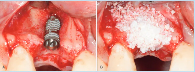
4.5×13 mm의 임플란트(GS III; Osstem Implant, Busan, Korea) 식립 후 길이 약 10 mm, 폭 약 4 mm의 열개를 관 찰하였다. 임플란트는 구개측과 치근단 쪽에서 고정을
Fig. 2. Intraoral photos after flap elevation. (A) Facial view: buccal bone defect is present. (B) Occlusal view: the interproximal bone level is intact.
Yu-Jin Kim et al. : Early Implant Placement in the Esthetic Zone with Hard Tissue and Soft Tissue Augmentation. Implantology 2013
Fig. 1. Intraoral photos and cone-beam computed tomography (CBCT) at first visit. (A) Facial view of maxillary right central incisor. (B) Occlusal view of maxillary right central incisor. (C) Preoperative CBCT images.
Yu-Jin Kim et al. : Early Implant Placement in the Esthetic Zone with Hard Tissue and Soft Tissue Augmentation. Implantology 2013
얻을 수 있었다. Bio-Oss® (Geistlich Biomaterials, Bern, Switzerland)를 임플란트 fixture 표면에 덮고, 그 상부를 Bio-gide® (Geistlich Biomaterials)로 이장하였다(Fig. 3).
감장 절개를 통하여 연조직의 일차의도 폐쇄를 이루었다 (Fig. 4).
3) 이차수술과 동시에 상피하 결합조직 이식술 시행 술 후 10일째 발사를 시행하였으며 술 전에 연조직 함
몰부위가 존재했던 부위 중심으로 차폐막 노출이 발생한 것을 관찰할 수 있었다. 매일 소독을 시행한 결과 3주 후 연조직 피개가 완전히 이루어졌으나, 연조직의 두께는 얇아져 있었다(Fig. 5). 술 후 5개월째에 이차수술을 시 행하였다. 얇아진 연조직의 부피증대를 위해 구개측에서 상피하 결합조직을 채득하여 협측에 이식하였다(Fig. 6).
Fig. 4. Intraoral photos after flap closure. Primary intension without tension was achieved. (A) Facial view. (B) Occlusal view.
Yu-Jin Kim et al. : Early Implant Placement in the Esthetic Zone with Hard Tissue and Soft Tissue Augmentation. Implantology 2013
Fig. 3. Intraoral photos at implant placement and guied bone regeneration. (A) Placement of the implant resulted in a buccal dehiscence. (B) Peri-implant dehiscence was corrected with guided bone regeneration techniques.
Yu-Jin Kim et al. : Early Implant Placement in the Esthetic Zone with Hard Tissue and Soft Tissue Augmentation. Implantology 2013
4) 술 후 평가
임플란트 이차 수술 후 임시 보철물을 사용하여 3개월 동안 단계적으로 연조직 형성(tissue molding)을 시행한 후 최종 보철물을 장착하였다. 초기에는 근심측, 원심측 치간유두 형성이 다소 부족하였으나, 5개월 후 black triangle이 사라진 것을 관찰할 수 있었다(Fig. 7).
2. 환자 2
1) 술 전 검사
50세 여자 환자가 재근관 치료를 받은 상악 좌측 측절 치의 치근 수평파절이 발견되어 치주과에 발치 의뢰되었 다. 상악전치부 전반적으로 짧은 임상 치관 소견이 있었 다. 방사선 검사상 상악 좌측 중절치 원심측 골의 수직적 소실이 관찰되었다(Fig. 8).
Fig. 6. Intraoral photos at second surgery with connective tissue graft. (A) Soft tissue depression was observed on the buccal gingiva at site #11. (B) The subepithelial connective tissue graft was placed to compensate for the soft tissue deficiency.
Yu-Jin Kim et al. : Early Implant Placement in the Esthetic Zone with Hard Tissue and Soft Tissue Augmentation. Implantology 2013
Fig. 5. Intraoral photos during the follow-up period. (A) The augmented site was exposed during the healing period. (B) After 3 weeks, soft tissue closure was achieved.
Yu-Jin Kim et al. : Early Implant Placement in the Esthetic Zone with Hard Tissue and Soft Tissue Augmentation. Implantology 2013
2) 임플란트 식립과 동시에 연조직, 경조직 증대술 시행
발치 후 6주째에 임플란트를 식립하였다. 상악 좌측 측 절치의 치아 결손부위에 발치와가 포함되지 않도록 구개 측에 치우쳐서 절개선을 긋고, 상악 좌측 견치 원심측에 수직 절개를 하여 전층 판막을 거상하였다. 연조직의 두 께가 1 mm 정도로 thin biotype임을 확인할 수 있었다.
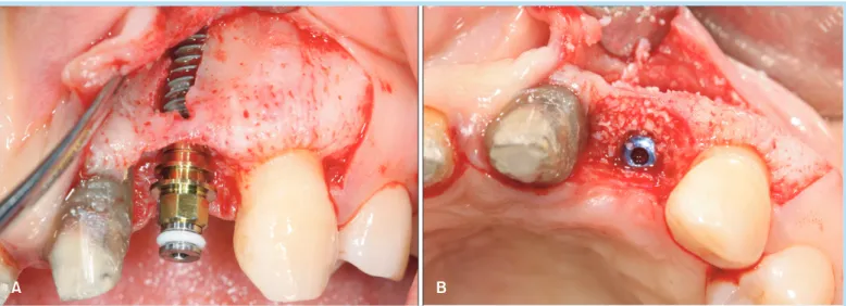
3.5×10 mm의 임플란트(GS III; Osstem) 식립 후 치조골 천공결손(fenestration defect) 및 교합면에서 임플란트 협측으로 간극 결손부(gap defect)가 2 mm 형성되었고, 결손부에 Bio-Oss®를 이식(Fig. 9)하고 Bio-gide®로 이 장하였다. Thin biotype의 개선과 일차의도 폐쇄를 위해 동측 구개부에서 VIP-CT7를 형성하여 협측 판막 쪽으로 고정하였다(Figs. 10, 11).
Fig. 7. Intraoral photos and periapical radiograph after final prosthesis. (A) Immediately after delivery. (B) The complete papilla fill is observed at 5 months after delivery.
Yu-Jin Kim et al. : Early Implant Placement in the Esthetic Zone with Hard Tissue and Soft Tissue Augmentation. Implantology 2013
Fig. 8. Intraoral photos after old restoration removal at first visit. (A) Facial view of maxillary left lateral incisor. (B) Occlusal view fo maxillary left lateral incisor.
Yu-Jin Kim et al. : Early Implant Placement in the Esthetic Zone with Hard Tissue and Soft Tissue Augmentation. Implantology 2013
3) 이차수술과 동시에 임상 치관 연장술 시행 환자는 상악 우측 중절치 및 좌측 중절치에 임시 보철 물을 착용하고 있는 상태이며 altered passive eruption 으로 짧은 임상치관을 보였다. 임플란트 일차 수술 시 연
조직 증강이 이루어 졌음에도 불구하고, 상악 좌측 중절 치의 원심측 골 소실로 인해 상악 좌측 측절치 근심부위 연조직은 부족한 상태였다. 상악 좌측 중절치 원심면의 연조직 양의 부족을 해소하고 상악 우측 중절치 및 좌측 Fig. 9. Intraoral photos at implant placement and guied bone regeneration. (A) Placement of the implant resulted in a buccal fenestration and gap defect. (B) The gap around the implant fixture was filled using guided bone regeneration techniques.
Yu-Jin Kim et al. : Early Implant Placement in the Esthetic Zone with Hard Tissue and Soft Tissue Augmentation. Implantology 2013
Fig. 10. Intraoral photos at soft tissue augmentation using vascularized interpositional periosteal connective tissue graft. A soft tissue graft was placed in effort to convert the biotype of gingiva at site #22 from thin to thick. (A) The palatal donor site preparation began by extending the incision horizontally to the second premolar. (B) The pediculated graft was elevated from the bone. (C) The connective tissue pedicle was rotated over the the surgical site covering with the collagen membrane and the labial gingival flap.
Yu-Jin Kim et al. : Early Implant Placement in the Esthetic Zone with Hard Tissue and Soft Tissue Augmentation. Implantology 2013
중절치의 심미성 개선을 위해 상악 좌측 측절치 임플란 트 부위에 이차수술과 함께 상악전치부의 심미적 임상 치관 연장술을 시행하였다(Fig. 12).
4) 술 후 평가
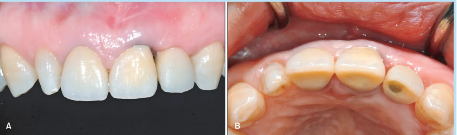
현재 테세라(TesceraTM; Bisco, Schaumburg, IL, USA) indirect composite provisional restoration을 제작하여 장착한 상태이며 2개월까지 임상관찰을 시행하였다. 향 후 provisional restoration을 수정하여 연조직 형성(tis- sue molding)을 시행한 후 최종 보철물을 장착할 계획이 다. 술 후 초진에 비하여 심미적으로 개선되었으나, 상악 좌측 측절치 임플란트의 근심측 치간유두 형성은 부족함 을 보였다(Fig. 13).
III 총괄 및 고찰
본 증례들은 전치부에서 심미성을 얻기 위해 조기 임플 란트 식립을 하면서 경조직, 연조직 재건을 시행한 보고 이다. 임플란트의 심미성을 평가하기 위한 척도로 여러 가지 기준이 제시되어 왔다. Jemt8의 papilla index와 Fig. 11. Intraoral photos after flap closure. Primary intension without tension was achieved. (A) Facial view. (B) Occlusal view.
Yu-Jin Kim et al. : Early Implant Placement in the Esthetic Zone with Hard Tissue and Soft Tissue Augmentation. Implantology 2013
Fig. 12. Intraoral photos at second surgery. Esthetic clinical crown lengthening procedure was performed simultaneously during second surgery at site #22.
Yu-Jin Kim et al. : Early Implant Placement in the Esthetic Zone with Hard Tissue and Soft Tissue Augmentation. Implantology 2013
Belser 등9의 index를 사용하여 임플란트 주변 연조직과 임플란트 보철물의 심미적인 평가를 시행할 수 있었다.
Papillae index는 두 치아의 labial gingiva를 연결하는 선, papillary tip, 그리고 이 두 선을 이등분하는 선을 기 준으로 분류하며 0: no papilla, 1: less than half of the height of the papilla, 2: at least half, 3: papilla fills up the entire proximal space, 4: hyperplastic의 기준으로 평가되었다. Belser 등9의 index는 pink esthetic score (PES)와 white esthetic score (WES)로 나누어 평가하였 는데, 각각 평가 점수는 10점을 만점으로 하였고 심미적 으로 받아들일 수 있는 최소 점수를 6점이라고 하였다.
PES는 mesial papilla, distal papilla, curvature of facial
mucosa, level of facial mucosa, root convexity/soft tis- sue color and texture의 5가지 요소를 평가하였다.
Papilla의 평가는 0 (absent), 1 (incomplete), 2 (com- plete)로 수치화하였다. 나머지 3개의 변수는 상악 좌측 중절치와 비교하여 불일치 정도에 따라 0 (major), 1 (minor), 2 (none)로 수치화하였다. WES 역시 tooth form, tooth outline and volume, color (hue and value), surface texture, translucency 등 5가지 요소를 평가하였 다. 상악 좌측 중절치와 비교하여 불일치 정도에 따라 0 (major), 1 (minor), 2 (none)로 수치화하였다.
상기 기준에 의거하여 심미 평가를 시행해 보았을때 첫 번째 증례의 경우는 임플란트 보철물의 근심측, 원심 Fig. 13. Intraoral photos after provisional prosthesis. Two month after delivery of provisional restoration. (A) Facial view. (B) Occlusal view.
Yu-Jin Kim et al. : Early Implant Placement in the Esthetic Zone with Hard Tissue and Soft Tissue Augmentation. Implantology 2013
Table 1. Evaluation of the outcome using esthetic parameters (Case 1)
Pink esthetic score White esthetic score Mesial papilla
Distal papilla
Curvature of facial mucosa Level of facial mucosa Soft tissue color and texture Total
2 2 1 1 2 8
Tooth form
Tooth volume and outline Color (hue/value)
Surface texture Translucency Total
2 2 2 2 2 10
Yu-Jin Kim et al. : Early Implant Placement in the Esthetic Zone with Hard Tissue and Soft Tissue Augmentation. Implantology 2013
측 치간유두의 재건이 심미적으로 이루어져 있어 Jemt8 의 papilla index 기준에 의해 근원심 모두 index 3을 기 록할 수 있다. Belser 등9의 index에 의해 평가해 보면 PES는 8점, WES는 10점으로 평가해 볼수 있다(Table 1).
이는 PES와 WES를 평가 기준으로 한 연구5에 비해 높은 수치이고 심미적으로 성공적인 임플란트 보철 수복이라 고 평가할 수 있겠다. 또한 상악 우측 중절치 소실로 수 복된 임플란트 보철물의 치은 변연이 인접 자연치보다 오히려 심미적으로 판단된다. 향후 상악 좌측 중절치 및 측절치의 치관연장술을 통하여 전치부의 심미성을 보다 개선할 수 있을 것이다. 두 번째 증례의 경우 임플란트 보철물의 근심측 치간유두 형태가 연조직 증대술과 인접 자연치의 치관연장술을 통하여 술 전에 비해 개선되었음 을 확인하였다. Papilla index에 따라 근심 치간유두는 index 1, 원심 치간유두는 index 2로 평가해 볼 수 있다.
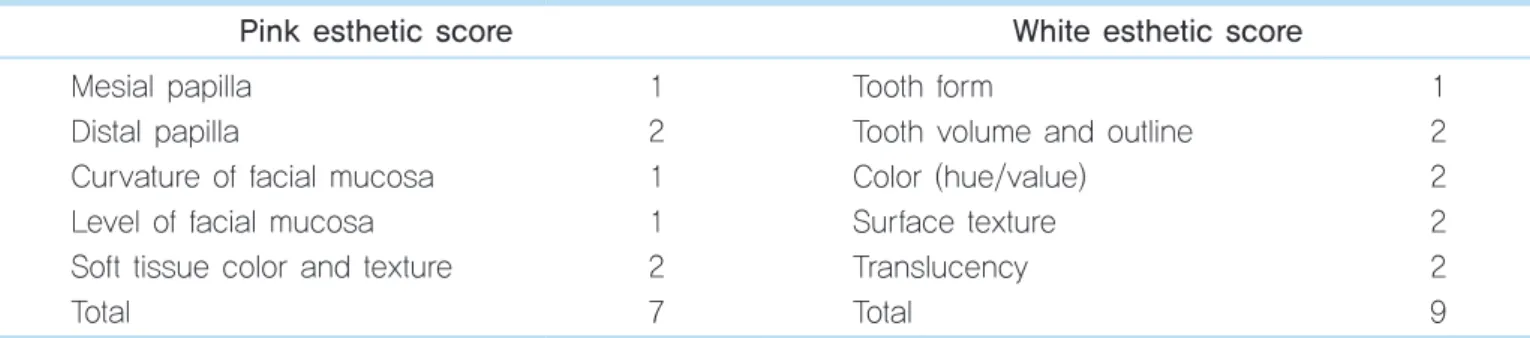
Belser 등9의 index에 의해 평가해 보면 PES는 7점, WES 는 9점으로 평가해 볼수 있다(Table 2). 초진과 비교하였 을 때 심미적으로 향상된 결과를 얻을 수 있었다.
전치부 임플란트 보철 시 장기적으로 심미적 안정성이 유지되려면 임플란트 주변의 순면과 치간부 연조직에 대 한 고려가 필수적이다. 첫 번째로 순면 연조직의 형성은 임플란트의 협설측 식립 위치와 연관이 깊은데, 첫 번째 증례에서는 심한 치조골 결손으로 인하여 임플란트가 다 소 협측에 식립(Fig. 6B)되었으며 약 10 mm의 열개형 결
손부에 많은 양의 골이식을 시행하였다. 차폐막 노출이 발생하여 얇아진 연조직과 임플란트 순면 연조직의 부피 를 증가시키기 위해 이차수술 시 연조직 이식술을 추가 로 시행하게 되었다. 이 증례에서는 단기적으로 좋은 심 미성을 얻었지만, 인접치아의 순면을 이은 가상선과 비 교해 보았을 때 전반적인 bone housing에서 협측으로 위 치되었기 때문에 장기적으로 추적검사가 필요할 것으로 보인다. 두 번째 증례에서는 thin biotype으로 장기적인 순면 연조직 유지를 위해 VIP-CT를 임플란트 식립과 함 께 시행하여 thick biotype으로 biotype을 전환할 수 있었 다. 이 증례의 경우 인접치아의 순면에서 연결하여 만든 가상선보다 구개측으로 1 mm 이상 안쪽에 임플란트가 식립되어 임플란트 협측으로 2 mm 이상으로 협측골을 확보할 수 있었기 때문에 장기적인 심미성의 측면에서 유리할 것으로 생각된다(Fig. 9B). 두 번째로 인접치아가 자연치인 단일치아 임플란트 수복에서 치간부 연조직의 형성에는 인접 자연치의 치간골 위치가 중요하다4. 첫 번 째 증례에서 협측골의 소실에도 불구하고 근원심 치간골 은 잘 유지가 되어 보철물 주변에 치간유두가 심미적으 로 형성될 수 있었다. 두 번째 증례에서는 상악 좌측 중 절치의 원심 치조골 결손이 존재하여, 짧은 임상치관을 수정하기 위해 치관연장술을 시행하였음에도 완전한 치 간유두 형태의 회복은 어려웠다.
Table 2. Evaluation of the outcome using esthetic parameters (Case 2)
Pink esthetic score White esthetic score Mesial papilla
Distal papilla
Curvature of facial mucosa Level of facial mucosa Soft tissue color and texture Total
1 2 1 1 2 7
Tooth form
Tooth volume and outline Color (hue/value)
Surface texture Translucency Total
1 2 2 2 2 9
Yu-Jin Kim et al. : Early Implant Placement in the Esthetic Zone with Hard Tissue and Soft Tissue Augmentation. Implantology 2013
IV 결론
상악 전치부 단일 치아 임플란트 수복에서 장기적으로 심미적인 안정성을 확보하기 위해서 순면 변연치은과 치 간 조직을 잘 형성해 주는 것이 중요하다. 지연 임플란트 식립을 하면서 경조직, 연조직 재건을 통하여 단기적으 로는 심미적으로 만족할 만한 결과를 얻을 수 있었다. 장 기간의 추적 검사를 통하여 재평가가 필요하다.
참고문헌
1. Kemppainen P, Eskola S, Ylipaavalniemi P. A comparative prospective clinical study of two single-tooth implants: a preliminary report of 102 implants. J Prosthet Dent. 1997; 77: 382-387.
2. Hämmerle CH, Chen ST, Wilson TG Jr. Consensus statements and recommended clinical procedures regarding the placement of implants
in extraction sockets. Int J Oral Maxillofac Implants. 2004; 19(Suppl):
26-28.
3. Evans CD, Chen ST. Esthetic outcomes of immediate implant placements. Clin Oral Implants Res. 2008; 19: 73-80.
4. Grunder U. Stability of the mucosal topography around single-tooth implants and adjacent teeth: 1-year results. Int J Periodontics Restorative Dent. 2000; 20: 11-17.
5. Buser D, Halbritter S, Hart C, et al. Early implant placement with simultaneous guided bone regeneration following single-tooth extraction in the esthetic zone: 12-month results of a prospective study with 20 consecutive patients. J Periodontol. 2009; 80: 152-162.
6. Fu JH, Lee A, Wang HL. Influence of tissue biotype on implant esthetics. Int J Oral Maxillofac Implants. 2011; 26: 499-508.
7. Nemcovsky CE, Artzi Z, Moses O. Rotated split palatal flap for soft tissue primary coverage over extraction sites with immediate implant placement. Description of the surgical procedure and clinical results. J Periodontol. 1999; 70: 926-934.
8. Jemt T. Restoring the gingival contour by means of provisional resin crowns after single-implant treatment. Int J Periodontics Restorative Dent. 1999; 19: 20-29.
9. Belser UC, Grütter L, Vailati F, et al. Outcome evaluation of early placed maxillary anterior single-tooth implants using objective esthetic criteria:
a cross-sectional, retrospective study in 45 patients with a 2- to 4-year follow-up using pink and white esthetic scores. J Periodontol. 2009; 80:
140-151.