Ameloblastoma is the second most common odontoge- nic tumor.1 Ameloblastoma is a true neoplasm which is described as tumor that is usually unicentric, nonfunction- al, intermittent in growth, anatomically benign and clini- cally persistent.2 Ameloblastoma is commonly a central lesion but rarely a peripheral epulis-like lesion.1
Based mainly on the clinical behaviour and prognosis, three types of ameloblastoma can presently be distinguish- ed: (1) the ‘conventional or classical’, intraosseous, solid or multicystic ameloblastoma, (2) the unicystic amelobla- stoma, and (3) the peripheral ameloblastoma.3Recent stud- ies could well indicate that desmoplastic ameloblastoma may be classified as a fourth subtype of ameloblastomas because of its biological behaviour, radiographic appear- ance, and unique histology.3Several causative factors have been proposed, including (1) nonspecific irritating factors such as extraction, caries, trauma, infection, inflammation, or tooth eruption; (2) nutritional deficit disorders, and (3) viral pathogenesis.4
Among these types, intraosseous ameloblastoma is the
most common and simple type encountered. Clinico-radio- graphically, intraosseous ameloblastoma presents as a painless swelling or slow expansion of the jaws, and it is described as multilocular expansile radiolucency that occurs most frequently in mandibular molar/ramus area.
Peripheral ameloblastoma, a very rare pattern of amelo- blastoma, generally occurs in the gingiva, and there is no bony involvement. It presents as a painless, sessile, firm, and exophytic growth that is usually relatively smooth or granular.5
Various cases of intraosseous ameloblastoma have been reported with the usual clinical presentation described above. In 1991, Kuru6reported the first case of intraosse- ous ameloblastoma manifesting as a peripheral (exophytic) lesion. Later, two similar cases were reported by Tongdee and Ganggakavin7and Stevenson and Austin8respectively.
As for our knowledge, this could be the fourth such a case to be reported in the English literature.
Case Report
A 45-year-old male patient reported with a gingival growth on the right anterior region of the mandible for one year and also complained of pain in the same region for 6 to 7 months. The exophytic lesion was small initially
Intraosseous ameloblastoma masquerading as exophytic growth: a case report
Sanjay CJ, Chaya M David, Rachna Kaul*, Ramnarayan BK, Prashanth Ramachandra
Department of Oral Medicine and Radiology, Dayananda Sagar College of Dental Sciences, Bangalore, India
*Department of Oral Medicine and Radiology, Dr Shyamala Reddy Dental College and Hospital, Bangalore, India ABSTRACT
Intraosseous ameloblastoma is the most common and simple type of ameloblastoma prevalent among odontogenic tumors. Clinico-radiographically intraosseous ameloblastoma presents as slow, painless swelling or expansion of the jaws and described as multilocular expansile radiolucency that occurs most frequently in mandibular molar/ramus area. This article describes a case of follicular ameloblastoma involving 45 year old male which is different from the usual presentation, which includes-exophytic growth, different location and without expansion of the cortex. (Imaging Sci Dent 2011; 41 : 89-93)
KEY WORDS : Ameloblastoma; Diagnosis; Case Reports
Received January 19, 2011; Revised March 16, 2011; Accepted March 25, 2011 Correspondence to : Dr. Sanjay CJ
Department of Oral Medicine and Radiology, Dayananda Sagar College of Dental Sciences, Shavige Malleshwara Hills, Kumaraswamy Layout, Bangalore 560 078, India
Tel) 91-9845038140, E-mail) sanjay_cj@yahoo.co.in
Copyright ⓒ 2011 by Korean Academy of Oral and Maxillofacial Radiology
This is an Open Access article distributed under the terms of the Creative Commons Attribution Non-Commercial License (http://creativecommons.org/licenses/by-nc/3.0) which permits unrestricted non-commercial use, distribution, and reproduction in any medium, provided the original work is properly cited.
Imaging Science in Dentistry∙pISSN 2233-7822 eISSN 2233-7830
and increased gradually to the current size displacing the adjacent teeth. He had mild and intermittent pain. He had visited a dentist with the same complaint 7 months before.
His general physical status was normal. Extraorally, a dif- fuse swelling measuring around 1.5×1.5 cm in size was seen on the right lower portion of the face. The skin over the swelling was normal and was tender on palpation.
Intraoral examination revealed an exophytic lesion on
the right mandible (Fig. 1A) at the interdental gingiva between canine and the first premolar, which was roughly circular, measuring about 3.5×3.5 cm in size, extended labially from the attached gingiva to the lingual sulcus of the right mandibular incisors, canine, and premolar. The surface of the lesion (Fig. 1B) was pebbled and rough with indentations of the opposing teeth with mesially displaced canine. Color of the lesion was normal as that of the sur- rounding mucosa and firm in consistency. The associated teeth were vital but tender, and grade I mobility was pre- sent. No cortical expansion was found.
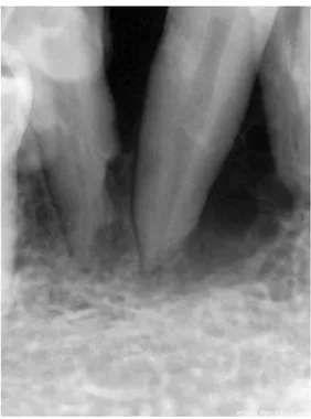
The periapical digital radiograph (Fig. 2) showed the presence of characteristic multilocular soap bubble appear- ance in the region of canine and first premolar, and knife
Fig. 1.Clinical photographs show an exophytic growth at the inter-dental area in right mandible between canine and first premolar extend- ing labially (A) from the attached gingiva of the teeth to the lingual sulcus of incisors, canine, and first premolar teeth (B).
Fig. 2.Intraoral periapical radiograph shows a presence of charac- teristic soap bubble appearance in the region of the right mandibular canine and first premolar, and knife edge root resorption pattern in relation to the first premolar.
Fig. 3.Panoramic radiograph shows a radiolucent area measuring 4×4 cm in diameter extending from the distal aspect of the right lateral incisor to the mesial aspect of the second premolar.
A B
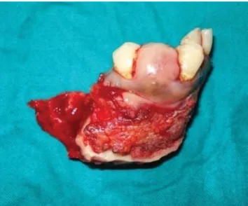
edge shaped root resorption pattern in relation to the first premolar. The panoramic radiograph (Fig. 3) showed radi- olucency measuring 4×4 cm in diameter extending from the distal aspect of lateral incisor to the mesial aspect of second premolar. The occlusal radiograph (Fig. 4) showed no expansion of the cortex. Blood investigations were within normal limits and serum chemistry showed no sig- nificant changes. The lesion was surgically removed (Fig.
5) and was subjected for microscopic examination (Fig.
6) which revealed normal stratified squamous epithelium
which was parakeratinized. Connective tissue showed follicles which were arranged in various sizes and shapes.
The follicles were lined by a single layer of tall columnar ameloblast-like cells and little cystic degeneration. Conti- nuity of the tumor cells with epithelium was not evident.
These features were suggestive of amelobalstoma (follicu- lar type).
Discussion
Ameloblastoma usually manifests as an intraosseous or central lesion or rarely as a peripheral epulis-like lesion.1 The lesion may arise most commonly from cell rests of enamel organ, epithelium of odontogenic cysts, distur- bances of the developing enamel organ, and basal cells of oral epithelium. Clinico-radiographically, ameloblastoma can be divided into 3 distinct patterns: (1) the conventional solid/multicystic (86% of all cases), (2) the unicystic (13%
of all cases), and (3) the peripheral (1% of all cases) (extra- osseous).2
Various histologic forms have been described which include follicular, plexiform, acanthomatous, granular cell, desmoplastic, and basal cell patterns.2 Follicular ameloblastoma presents as a painless swelling or slow expansion of the jaws, and it is described as multilocular expansile radiolucency that occurs most frequently in mandibular molar/ramus area.9 Desmoplastic variant
Fig. 4. Occlusal radiograph shows no cortical expansion.
Fig. 5.Gross specimen after surgical removal.
Fig. 6. Histopathological examination demonstrates connective tissue with numerous follicles lined by single layer of tall columnar ameloblast-like cells (H&E stain, 40×).
occurs more frequently in the anterior mandibular region10 and presents a mixed radiopaque and radiolucent appear- ance. Unicystic ameloblastoma is usually seen in younger age and most commonly associated with pericoronal radiolucency with unerupted 3rd molar (38%).11
In this particular case, the clinical presentation of the follicular ameloblastoma was different from the usual presentation, which included exophytic growth, uncommon location, and absence of expansion of the cortex. The lesion had started intraosseously, however eventually per- forated to involve the soft tissue. Clinically differential diagnosis of this exophytic growth included peripheral ameloblastoma, peripheral odontogenic fibroma, peripheral giant cell granuloma, and other peripheral hyperplastic swellings superficial to the alveolar ridge.9 The final diagnosis was made by the clinical, radiological, and his- topathological features after the complete excision of the lesion.
Kuru first reported an intraosseous ameloblastoma hav- ing penetrated through the alveolar bone, fused with the oral epithelium, and eventually presenting itself clinically as a ‘peripheral lesion’.6Our case had the similar clinical and radiological features of the previously reported case by Kuru but with no expansion of the cortex. Two other sim- ilar cases have been reported by Tongdee and Gangga- kavin7in 1978 and Stevenson and Austin8in 1990, res- pectively. The exophytic growth is also a characteristic finding of the peripheral (extraosseous) ameloblastoma which is very rare. Radiographically, or at surgery, a super- ficial erosion of the bone or a superficial bony depression -known as cupping or saucerization- may be noticed, a finding that is thought to be due to pressure resorption, in contrast to resorption caused by neoplastic invasion.5The diagnostic criteria of peripheral ameloblastoma include the origin from the overlying epithelium, presence of odontogenic epithelium islands in the lesion, and lack of potential to bone infiltration.12 Also, peripheral odonto- genic fibroma and peripheral giant-cell granuloma share the same clinical characteristic of exophytic growth most commonly at mandibular premolar region, despite the histopathological difference. However, radiographically, both of them create depression/resorption in the underly- ing bone very rarely.13
Most oral surgeons and pathologists unquestionably consider peripheral ameloblastoma to be a nonaggressive lesion without actual infiltration into the underlying bone.14 One of the main problems regarding peripheral ameloblas- toma is its possible origin. The two main theories are the following: (1) origin from the extra osseous epithelial
remnants of dental lamina and its organ derivatives within the underlying connective tissue; (2) origin from the basal cell layer of the oral mucosa, which is believed to have odontogenic potential.15 Those lesions that are entirely separated from the overlying surface epithelium probably arise from the odontogenic remnants, however this hypo- thesis can be questioned if there is continuity between the tumor and the surface epithelium.
In conclusion, even though the incidence of intraosseous ameloblastoma manifesting as exophytic lesion is very rare, it may have to be considered in the differential diag- nosis of exophytic lesions at the region of mandibular canine premolar region along with peripheral ameloblas- toma, peripheral odontogenic fibroma, peripheral giant cell granuloma, and rarely peripheral hyperplastic swellings superficial to the alveolar ridge.
References
1. Langlais RP, Langland OE, Nortje CJ. Multilocular radioluc- encies. In: Diagnostic imaging of jaws; 1st ed. Williams &
Wilkins Co; 1995. p. 327-84.
2. Rajendran R. Cyst and tumors of odntogenic origin. In: Rajen- dran R, Sivapathasundharam B. Shafer’s text book of oral pathology. 6th ed. Noida: Elsevier; 2009. p. 254-308.
3. Philipsen HP, Reichart PA. Unicystic ameloblastoma. A review of 193 cases from the literature. Oral Oncol 1998; 34 : 317- 25.
4. Kim SG, Jang HS. Ameloblastoma: a clinical, radiographic, and histopathologic analysis of 71 cases. Oral Surg Oral Med Oral Pathol Oral Radiol Endod 2001; 91 : 649-53.
5. Philipsen HP, Reichart PA, Nikai H, Takata T, Kudo Y. Periph- eral ameloblastoma: biological profile based on 160 cases from the literature. Oral Oncol 2001; 37 : 17-27.
6. Kuru H. Ueber das Adamantinom. Zentralbl Allg Pathol 1911;
22 : 291-5.
7. Tongdee C, Ganggavakin S. Peripheral ameloblastoma (report of a case and review literature). J Dent Assoc Thai 1978; 28 : 31-8.
8. Stevenson AR, Austin BW. A case of ameloblastoma presenting as an exophytic gingival lesion. J Periodontol 1990; 61 : 378- 81.
9. Ueno S, Nakamura S, Mushimoto K, Shirasu R. A clinicopa- thologic study of ameloblastoma. J Oral Maxillofac Surg 1986;
44 : 361-5.
10. Waldron CA, el-Mofty SK. A histopathologic study of 116 ameloblastomas with special reference to the desmoplastic variant. Oral Surg Oral Med Oral Pathol 1987; 63 : 441-51.
11. Eversole LR, Leider AS, Strub D. Radiographic characteristics of cystogenic ameloblastoma. Oral Surg Oral Med Oral Pathol 1984; 57 : 572-7.
12. Martelli-Júnior H, Souza LN, Santos LA, Melo-Filho MR, De Paula AM. Peripheral ameloblastoma: a case report. Oral Surg Oral Med Oral Pathol Oral Radiol Endod 2005; 99 : E31-3.
13. de Villiers-Slabbert H, Altini M. Peripheral odontogenic fibro-
ma: a clinicopathologic study. Oral Surg Oral Med Oral Pathol 1991; 72 : 86-90.
14. Ide F, Kusama K, Tanaka A, Sakashita H. Peripheral amelobla- stoma is not a hamartoma but rather more of a neoplasm. Oral
Oncol 2002; 38 : 318-20.
15. Yamamoto T, Ueta E, Yoneda K, Osaki T. Peripheral amelob- lastoma: case report with immunohistochemical investigation.
J Oral Maxillofac Surg 1990; 48 : 197-200.