Received on June 28, 2014. Revised on July 22, 2014. Accepted on July 28, 2014.
CC This is an open access article distributed under the terms of the Creative Commons Attribution Non-Commercial License (http://creativecommons.org/licenses/by-nc/3.0) which permits unrestricted non-commercial use, distribu- tion, and reproduction in any medium, provided the original work is properly cited.
*Corresponding Author. Seung-Hyo Lee, Cellular Immunology Laboratory, Graduate School of Medical Science and Engineering, Korea Advanced Institute of Science and Technology, 291 Daehak-ro, Yuseong-gu, Daejeon, Korea. Tel:
82-42-350-4235; Fax: 82-42-350-4240; E-mail: sl131345@kaist.ac.kr
Abbreviations: LV, lymphatic vessel; LEC, lymphatic endothelial cell; DC, dendritic cell; LN, lymph node; VEGF, vascular endothelial growth factor; LT, lymphotoxin; IL, interleukin; HGF, hepatocyte growth factor; FGF, fibroblast growth factor;
IFN, interferon; TGF, transforming growth factor
Interplay between Inflammatory Responses and Lymphatic Vessels
Kihyuk Shin1,2 and Seung-Hyo Lee2*
1Department of Medicine, Pusan National University Hospital, Busan 602-739, 2Graduate School of Medical Science and Engineering, and Biomedical Research Center, and KAIST Institute for the BioCentury, Korea Advanced Institute of Science and Technology (KAIST), Daejeon 305-701, Korea
Lymphatic vessels are routes for leukocyte migration and flu- id drainage. In addition to their passive roles in migration of leukocytes, increasing evidence indicates their active roles in immune regulation. Tissue inflammation rapidly induces lym- phatic endothelial cell proliferation and chemokine pro- duction, thereby resulting in lymphangiogenesis. Further- more, lymphatic endothelial cells induce T cell tolerance through various mechanisms. In this review, we focus on the current knowledge on how inflammatory cytokines affect lymphangiogenesis and the roles of lymphatic vessels in modulating immune responses.
[Immune Network 2014;14(4):182-186]
Keywords: Lymphangiogenesis, Lymphatic vessels, Inflam- mation, Immune responses
INTRODUCTION
Lymphatic vessels (LVs) exist in most vascularized organs (1).
LVs contribute to fluid homeostasis by absorbing tissue fluid and draining it into the venous circulation (2-4). In addition to fluid homeostasis, LVs are also important for immune sur- veillance (5). In contrast to blood vessels, lymphatic capil- laries are connected by monolayers of lymphatic endothelial cells (LECs) and discontinuous basement membrane; thus,
dendritic cells (DCs) can migrate into LVs (6,7). Furthermore, LVs in lymph nodes (LNs) and peripheral tissues are highly plastic; hence, LVs undergo proliferation or remodeling dur- ing various pathological conditions such as inflammation (8-11).
Recent studies have revealed that lymphangiogenesis dur- ing inflammation influences the extent of inflammation and immune responses by modulating leukocyte migration and in- ducing T cell tolerance. In this review, we will describe the inflammatory mediators that affect lymphangiogenesis and the tolerogenic roles of LVs during immune responses.
FACTORS AFFECTING LYMPHANGIOGENESIS DURING INFLAMMATION
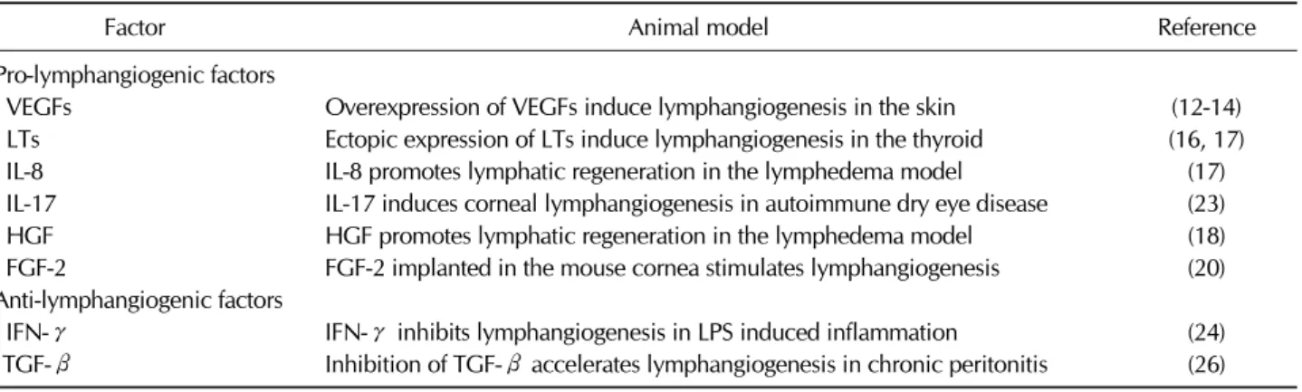
Several factors regulate lymphangiogenesis (Table I). However, here, we focus only on those factors that have been inves- tigated both in vivo and in vitro. Of them, vascular endothe- lial growth factors (VEGFs), mainly produced by macro- phages are the most well-known and well-studied pro-lym- phangiogenic factors. VEGF-C/D and VEGF-A have been re- ported to induce lymphangiogenesis in various inflammatory models (12-14). Blocking the VEGF receptor tyrosine kinase inhibits proliferation and tube formation of umbilical vein en- dothelial cells and LECs in vitro. Furthermore, in a transgenic
Table I. Inflammatory mediators which have pro- and anti-lymphangiogenic activity
Factor Animal model Reference
Pro-lymphangiogenic factors
VEGFs Overexpression of VEGFs induce lymphangiogenesis in the skin (12-14)
LTs Ectopic expression of LTs induce lymphangiogenesis in the thyroid (16, 17)
IL-8 IL-8 promotes lymphatic regeneration in the lymphedema model (17)
IL-17 IL-17 induces corneal lymphangiogenesis in autoimmune dry eye disease (23)
HGF HGF promotes lymphatic regeneration in the lymphedema model (18)
FGF-2 FGF-2 implanted in the mouse cornea stimulates lymphangiogenesis (20)
Anti-lymphangiogenic factors
IFN-γ IFN-γ inhibits lymphangiogenesis in LPS induced inflammation (24)
TGF-β Inhibition of TGF-β accelerates lymphangiogenesis in chronic peritonitis (26)
Figure 1. The lymphangiogenic balance between pro-lymphangioge- nic factors and anti-lymphangiogenic factors regulates lymphatic vas- cular homeostasis. Lymphangiogenesis under pathophysiological conditions is associated with increased pro-lymphangiogenic factors and/or decreased anti-lymphangiogenic factors.
mouse model of psoriasis, treatment with a VEGF receptor tyrosine-kinase inhibitor reduces the number of LVs in the skin (15).
In addition to VEGFs, lymphotoxin (LT)-α and LTα1β2 are involved in lymphangiogenesis during infection and lymphoid organ formation. Ectopic expression of LTα promotes the de- velopment of LVs within tertiary lymphoid organs. In addi- tion, genetic deletion of LTα1β2 or LTα abrogates develop- ment of LVs in the inflamed thyroid (16,17).
Furthermore, interleukin (IL)-8 and hepatocyte growth fac- tor (HGF) promote proliferation and tube formation of LECs without activating VEGF signaling in vitro. IL-8 and HGF pro- mote formation of LVs and improve amelioration of lymphe- dema in an experimental lymphedema model (18,19).
Fibroblast growth factor (FGF)-2 also exhibits pro-lym-
phangiogenic activity in the mouse cornea (20). Moreover, FGF-2 induces lymphangiogenesis via both direct and indirect effects. FGF-2 binds LECs and induces proliferation and mi- gration in vitro (21). Furthermore, recent findings suggest that FGF-2 interacts with VEGF-C to induce additional pro-lym- phangiogenic activity (22).
Similarly, IL-17 from type 17 T helper (Th17) cells induces lymphangiogenesis in an autoimmune dry eye disease model.
IL-17 directly promotes growth of LVs by inducing increased expression of VEGF-D and proliferation of LECs in vitro. Furthermore, in vivo blockade of IL-17 in a model of Th17 dominant autoimmune ocular disease results in a reduction in corneal lymphangiogenesis (23).
By contrast, some inflammatory cytokines have been re- ported to show anti-lymphangiogenic activity. Interferon (IFN)-γ, which is mainly produced by Th1 cells, inhibits LV formation in an LPS-induced inflammation model. Notably, resolution of increased LVs is dependent on IFN-γ in this model. Furthermore, IFN-γ production by T cells suppresses lymphatic-specific genes in LECs and causes reduction of LV formation in vitro (24).
In addition, in thioglycollate-induced peritonitis and in a lymphedema model, inhibition of transforming growth factor (TGF)-β promotes LV formation (25). Expression of LEC markers, including LYVE-1 and Prox1, is inhibited by TGF-β but is enhanced by a TGF-β type I receptor inhibitor (26).
Therefore, the balance of pro- and anti-lymphangiogenic factors could determine the nature of LVs in various in- flammatory conditions (Fig. 1). LVs are involved in immune responses; thus, these results imply that controlling these fac- tors could be a good tool to control lymphangiogenesis and immune responses.
CROSSTALK BETWEEN IMMUNE CELLS AND LYMPHATIC VESSELS
LVs provide pathways for DCs and lymphocyte migration to LNs, thus facilitating inflammation or possible immune toler- ance (7,27). For leukocyte trafficking, adhesion molecules like intercellular adhesion molecule (ICAM)-1 and vascular cell adhesion molecule (VCAM)-1 are induced on LECs.
Moreover, various chemokines are produced by LECs during inflammation (28,29). Of them, CCL21, which binds to CC-chemokine receptor 7 (CCR7) expressed on DCs and T cells, is the primary determinant of leukocyte migration to LNs (30-32). CXCL12 also enhances the migration of CXCR4- expressing leukocytes (33). Furthermore, CX3CL1 was re- ported to enhance DC transmigration across LECs to LNs (34).
Therefore, although CCL21 has a major role in DC migration during inflammation, the expression of other LEC adhesion molecules and chemokines also contributes to migration (29).
In addition to the ability of LECs to control leukocyte traf- ficking through adhesion molecules, LECs control immune re- sponses via their functions as professional antigen presenting cells (APCs). LECs in LNs constitutively express major histo- compatibility complex (MHC)-II molecules. Moreover, they endocytose and cross-present antigens in context with MHC-I molecules to T cells (35-37). However, LECs do not express co-stimulatory molecules such as CD80, CD86, OX40L, or 4-1BBL, thus fail to induce T cell activation and proliferation (38,39). Although the lack of co-stimulatory molecules on LECs implies immunological tolerance, further study is need- ed to elucidate the exact roles of LECs in CD4 T cell activation.
Recently, it was reported that LECs affect not only CD4 T cell tolerance but also CD8 T cell tolerance. LECs express pe- ripheral tissue antigens (PTA) that are restricted to specific tis- sues such as skin, gut, pancreas, and central nervous system (40-44). Furthermore, LECs induce deletion or abortive pro- liferation of PTA-specific CD8 T cells through a lack of co-stimulation or engagement with inhibitory molecules such as PD-L1 (38,45-48). Other co-stimulatory molecules such as HVEM or CD48 are constitutively expressed; thus, their ex- pression suggests that LECs may have immune regulatory roles in steady state as well as in inflammatory conditions (49).
CONCLUSION
So far, we have discussed inflammation, LV formation, and
immune regulation. Different pro- and anti-lymphangiogenic factors are produced by different inflammatory stimuli, and their overall effects determine the extent of lymphangio- genesis. Furthermore, although LECs promote leukocyte mi- gration and enhance immune responses, LECs also attenuate T cell-mediated immune responses by mediating tolerance.
LECs attract lymphocytes and DCs through cell adhesion mol- ecules and chemokines. In addition, LECs induce CD8 T cell tolerance through PD-L1 and lack of co-stimulatory mole- cules. However, mechanisms of CD4 T cell tolerance and roles of other inhibitory molecules on LECs need to be investigated. Finally, controlling lymphangiogenesis could be a novel therapeutic strategy to regulate autoimmunity, en- hance tumor immunotherapy, and reduce transplantation rejection.
ACKNOWLEDGEMENTS
This study is supported by a grant of the Korea Healthcare Technology R&D Project, Ministry for Health, Welfare and Family Affairs, Republic of Korea (A121145).
CONFLICTS OF INTEREST
The authors have no financial conflict of interest.
REFERENCES
1. Adams, R. H., and K. Alitalo. 2007. Molecular regulation of angiogenesis and lymphangiogenesis. Nat. Rev. Mol. Cell Biol. 8: 464-478.
2. Oliver, G. 2004. Lymphatic vasculature development. Nat.
Rev. Immunol. 4: 35-45.
3. Oliver, G., and K. Alitalo. 2005. The lymphatic vasculature:
recent progress and paradigms. Annu. Rev. Cell Dev. Biol.
21: 457-483.
4. Cueni, L. N., and M. Detmar. 2006. New insights into the molecular control of the lymphatic vascular system and its role in disease. J. Iinvest. Dermatol. 126: 2167-2177.
5. Cueni, L. N., and M. Detmar. 2008. The lymphatic system in health and disease. Lymphat. Res. Biol. 6: 109-122.
6. Schulte-Merker, S., A. Sabine, and T. V. Petrova. 2011.
Lymphatic vascular morphogenesis in development, physiol- ogy, and disease. J. Cell Biol. 193: 607-618.
7. Pflicke, H., and M. Sixt. 2009. Preformed portals facilitate dendritic cell entry into afferent lymphatic vessels. J. Exp.
Med. 206: 2925-2935.
8. Alitalo, K., T. Tammela, and T. V. Petrova. 2005. Lymphang- iogenesis in development and human disease. Nature 438:
946-953.
9. He, Y., I. Rajantie, K. Pajusola, M. Jeltsch, T. Holopainen,
S. Yla-Herttuala, T. Harding, K. Jooss, T. Takahashi, and K.
Alitalo. 2005. Vascular endothelial cell growth factor receptor 3-mediated activation of lymphatic endothelium is crucial for tumor cell entry and spread via lymphatic vessels. Cancer Res. 65: 4739-4746.
10. Achen, M. G., and S. A. Stacker. 2006. Tumor lymphangio- genesis and metastatic spread-new players begin to emerge.
Int. J. Cancer 119: 1755-1760.
11. Kataru, R. P., K. Jung, C. Jang, H. Yang, R. A. Schwendener, J. E. Baik, S. H. Han, K. Alitalo, and G. Y. Koh. 2009. Critical role of CD11b+ macrophages and VEGF in inflammatory lym- phangiogenesis, antigen clearance, and inflammation resolu- tion. Blood 113: 5650-5659.
12. Jeltsch, M., A. Kaipainen, V. Joukov, X. Meng, M. Lakso, H.
Rauvala, M. Swartz, D. Fukumura, R. K. Jain, and K. Alitalo.
1997. Hyperplasia of lymphatic vessels in VEGF-C transgenic mice. Science 276: 1423-1425.
13. Makinen, T., T. Veikkola, S. Mustjoki, T. Karpanen, B.
Catimel, E. C. Nice, L. Wise, A. Mercer, H. Kowalski, D.
Kerjaschki, S. A. Stacker, M. G. Achen, and K. Alitalo. 2001.
Isolated lymphatic endothelial cells transduce growth, survival and migratory signals via the VEGF-C/D receptor VEGFR-3.
EMBO J. 20: 4762-4773.
14. Wirzenius, M., T. Tammela, M. Uutela, Y. He, T. Odorisio, G. Zambruno, J. A. Nagy, H. F. Dvorak, S. Yla-Herttuala, M.
Shibuya, and K. Alitalo. 2007. Distinct vascular endothelial growth factor signals for lymphatic vessel enlargement and sprouting. J. Exp. Med. 204: 1431-1440.
15. Halin, C., H. Fahrngruber, J. G. Meingassner, G. Bold, A.
Littlewood-Evans, A. Stuetz, and M. Detmar. 2008. Inhibition of chronic and acute skin inflammation by treatment with a vascular endothelial growth factor receptor tyrosine kinase inhibitor. Am. J. Pathol. 173: 265-277.
16. Furtado, G. C., T. Marinkovic, A. P. Martin, A. Garin, B.
Hoch, W. Hubner, B. K. Chen, E. Genden, M. Skobe, and S. A. Lira. 2007. Lymphotoxin beta receptor signaling is re- quired for inflammatory lymphangiogenesis in the thyroid.
Proc. Natl. Acad. Sci. U. S. A. 104: 5026-5031.
17. Mounzer, R. H., O. S. Svendsen, P. Baluk, C. M. Bergman, T. P. Padera, H. Wiig, R. K. Jain, D. M. McDonald, and N.
H. Ruddle. 2010. Lymphotoxin-alpha contributes to lym- phangiogenesis. Blood 116: 2173-2182.
18. Choi, I., Y. S. Lee, H. K. Chung, D. Choi, T. Ecoiffier, H.
N. Lee, K. E. Kim, S. Lee, E. K. Park, Y. S. Maeng, N. Y.
Kim, R. D. Ladner, N. A. Petasis, C. J. Koh, L. Chen, H. J.
Lenz, and Y. K. Hong. 2013. Interleukin-8 reduces post-surgi- cal lymphedema formation by promoting lymphatic vessel regeneration. Angiogenesis 16: 29-44.
19. Saito, Y., H. Nakagami, R. Morishita, Y. Takami, Y. Kikuchi, H. Hayashi, T. Nishikawa, K. Tamai, N. Azuma, T. Sasajima, and Y. Kaneda. 2006. Transfection of human hepatocyte growth factor gene ameliorates secondary lymphedema via promotion of lymphangiogenesis. Circulation 114: 1177-1184.
20. Platonova, N., G. Miquel, B. Regenfuss, S. Taouji, C.
Cursiefen, E. Chevet, and A. Bikfalvi. 2013. Evidence for the interaction of fibroblast growth factor-2 with the lymphatic endothelial cell marker LYVE-1. Blood 121: 1229-1237.
21. Chang, L. K., G. Garcia-Cardena, F. Farnebo, M. Fannon, E.
J. Chen, C. Butterfield, M. A. Moses, R. C. Mulligan, J.
Folkman, and A. Kaipainen. 2004. Dose-dependent response of FGF-2 for lymphangiogenesis. Proc. Natl. Acad. Sci.U. S.
A. 101: 11658-11663.
22. Cao, R., H. Ji, N. Feng, Y. Zhang, X. Yang, P. Andersson, Y. Sun, K. Tritsaris, A. J. Hansen, S. Dissing, and Y. Cao.
2012. Collaborative interplay between FGF-2 and VEGF-C promotes lymphangiogenesis and metastasis. Proc. Natl.
Acad. Sci.U. S. A. 109: 15894-15899.
23. Chauhan, S. K., Y. Jin, S. Goyal, H. S. Lee, T. A. Fuchsluger, H. K. Lee, and R. Dana. 2011. A novel pro-lymphangiogenic function for Th17/IL-17. Blood 118: 4630-4634.
24. Kataru, R. P., H. Kim, C. Jang, D. K. Choi, B. I. Koh, M.
Kim, S. Gollamudi, Y. K. Kim, S. H. Lee, and G. Y. Koh.
2011. T lymphocytes negatively regulate lymph node lym- phatic vessel formation. Immunity 34: 96-107.
25. Avraham, T., S. Daluvoy, J. Zampell, A. Yan, Y. S. Haviv, S. G. Rockson, and B. J. Mehrara. 2010. Blockade of trans- forming growth factor-beta1 accelerates lymphatic re- generation during wound repair. Am. J. Pathol. 177:
3202-3214.
26. Oka, M., C. Iwata, H. I. Suzuki, K. Kiyono, Y. Morishita, T.
Watabe, A. Komuro, M. R. Kano, and K. Miyazono. 2008.
Inhibition of endogenous TGF-beta signaling enhances lymphangiogenesis. Blood 111: 4571-4579.
27. Lammermann, T., B. L. Bader, S. J. Monkley, T. Worbs, R.
Wedlich-Soldner, K. Hirsch, M. Keller, R. Forster, D. R.
Critchley, R. Fassler, and M. Sixt. 2008. Rapid leukocyte mi- gration by integrin-independent flowing and squeezing.
Nature 453: 51-55.
28. Johnson, L. A., S. Clasper, A. P. Holt, P. F. Lalor, D. Baban, and D. G. Jackson. 2006. An inflammation-induced mecha- nism for leukocyte transmigration across lymphatic vessel endothelium. J. Exp. Med. 203: 2763-2777.
29. Vigl, B., D. Aebischer, M. Nitschke, M. Iolyeva, T. Rothlin, O. Antsiferova, and C. Halin. 2011. Tissue inflammation mod- ulates gene expression of lymphatic endothelial cells and dendritic cell migration in a stimulus-dependent manner.
Blood 118: 205-215.
30. Forster, R., A. Braun, and T. Worbs. 2012. Lymph node hom- ing of T cells and dendritic cells via afferent lymphatics.
Trends Immunol. 33: 271-280.
31. Issa, A., T. X. Le, A. N. Shoushtari, J. D. Shields, and M.
A. Swartz. 2009. Vascular endothelial growth factor-C and C-C chemokine receptor 7 in tumor cell-lymphatic cross-talk promote invasive phenotype. Cancer Res. 69: 349-357.
32. Martln-Fontecha, A., S. Sebastiani, U. E. Hopken, M.
Uguccioni, M. Lipp, A. Lanzavecchia, and F. Sallusto. 2003.
Regulation of dendritic cell migration to the draining lymph node: impact on T lymphocyte traffic and priming. J. Exp.
Med. 198: 615-621.
33. Kabashima, K., N. Shiraishi, K. Sugita, T. Mori, A. Onoue, M. Kobayashi, J. Sakabe, R. Yoshiki, H. Tamamura, N. Fujii, K. Inaba, and Y. Tokura. 2007. CXCL12-CXCR4 engagement is required for migration of cutaneous dendritic cells. Am. J.
Pathol. 171: 1249-1257.
34. Johnson, L. A., and D. G. Jackson. 2013. The chemokine CX3CL1 promotes trafficking of dendritic cells through in-
flamed lymphatics. J. Cell Sci. 126: 5259-5270.
35. Amatschek, S., E. Kriehuber, W. Bauer, B. Reininger, P.
Meraner, A. Wolpl, N. Schweifer, C. Haslinger, G. Stingl, and D. Maurer. 2007. Blood and lymphatic endothelial cell-specif- ic differentiation programs are stringently controlled by the tissue environment. Blood 109: 4777-4785.
36. Tripp, C. H., B. Haid, V. Flacher, M. Sixt, H. Peter, J. Farkas, R. Gschwentner, L. Sorokin, N. Romani, and P. Stoitzner.
2008. The lymph vessel network in mouse skin visualised with antibodies against the hyaluronan receptor LYVE-1.
Immunobiology 213: 715-728.
37. Lund, A. W., F. V. Duraes, S. Hirosue, V. R. Raghavan, C.
Nembrini, S. N. Thomas, A. Issa, S. Hugues, and M. A.
Swartz. 2012. VEGF-C promotes immune tolerance in B16 melanomas and cross-presentation of tumor antigen by lymph node lymphatics. Cell Rep. 1: 191-199.
38. Tewalt, E. F., J. N. Cohen, S. J. Rouhani, C. J. Guidi, H.
Qiao, S. P. Fahl, M. R. Conaway, T. P. Bender, K. S. Tung, A. T. Vella, A. J. Adler, L. Chen, and V. H. Engelhard. 2012.
Lymphatic endothelial cells induce tolerance via PD-L1 and lack of costimulation leading to high-level PD-1 expression on CD8 T cells. Blood 120: 4772-4782.
39. Norder, M., M. G. Gutierrez, S. Zicari, E. Cervi, A. Caruso, and C. A. Guzman. 2012. Lymph node-derived lymphatic en- dothelial cells express functional costimulatory molecules and impair dendritic cell-induced allogenic T-cell proliferation.
FASEB J. 26: 2835-2846.
40. Lee, J. W., M. Epardaud, J. Sun, J. E. Becker, A. C. Cheng, A. R. Yonekura, J. K. Heath, and S. J. Turley. 2007.
Peripheral antigen display by lymph node stroma promotes T cell tolerance to intestinal self. Nat. Immunol. 8: 181-190.
41. Nichols, L. A., Y. Chen, T. A. Colella, C. L. Bennett, B. E.
Clausen, and V. H. Engelhard. 2007. Deletional self-tolerance to a melanocyte/melanoma antigen derived from tyrosinase is mediated by a radio-resistant cell in peripheral and mesen- teric lymph nodes. J. Immunol. 179: 993-1003.
42. Gardner, J. M., J. J. Devoss, R. S. Friedman, D. J. Wong,
Y. X. Tan, X. Zhou, K. P. Johannes, M. A. Su, H. Y. Chang, M. F. Krummel, and M. S. Anderson. 2008. Deletional toler- ance mediated by extrathymic Aire-expressing cells. Science 321: 843-847.
43. Cohen, J. N., C. J. Guidi, E. F. Tewalt, H. Qiao, S. J.
Rouhani, A. Ruddell, A. G. Farr, K. S. Tung, and V. H.
Engelhard. 2010. Lymph node-resident lymphatic endothelial cells mediate peripheral tolerance via Aire-independent direct antigen presentation. J. Exp. Med. 207: 681-688.
44. Fletcher, A. L., V. Lukacs-Kornek, E. D. Reynoso, S. E.
Pinner, A. Bellemare-Pelletier, M. S. Curry, A. R. Collier, R.
L. Boyd, and S. J. Turley. 2010. Lymph node fibroblastic re- ticular cells directly present peripheral tissue antigen under steady-state and inflammatory conditions. J. Exp. Med. 207:
689-697.
45. Harding, F. A., J. G. McArthur, J. A. Gross, D. H. Raulet, and J. P. Allison. 1992. CD28-mediated signalling co-stim- ulates murine T cells and prevents induction of anergy in T-cell clones. Nature 356: 607-609.
46. Hawiger, D., K. Inaba, Y. Dorsett, M. Guo, K. Mahnke, M.
Rivera, J. V. Ravetch, R. M. Steinman, and M. C.
Nussenzweig. 2001. Dendritic cells induce peripheral T cell unresponsiveness under steady state conditions in vivo. J.
Exp. Med. 194: 769-779.
47. Hernandez, J., S. Aung, K. Marquardt, and L. A. Sherman.
2002. Uncoupling of proliferative potential and gain of effec- tor function by CD8(+) T cells responding to self-antigens.
J. Exp. Med. 196: 323-333.
48. Martin-Orozco, N., Y. H. Wang, H. Yagita, and C. Dong.
2006. Cutting Edge: Programmed death (PD) ligand-1/PD-1 interaction is required for CD8+ T cell tolerance to tissue antigens. J. Immunol. 177: 8291-8295.
49. Liu, X., M. Alexiou, N. Martin-Orozco, Y. Chung, R. I.
Nurieva, L. Ma, Q. Tian, G. Kollias, S. Lu, D. Graf, and C.
Dong. 2009. Cutting edge: A critical role of B and T lympho- cyte attenuator in peripheral T cell tolerance induction. J.
Immunol. 182: 4516-4520.