Skeletal Fragility in Type 2 Diabetes Mellitus
Jakob Starup-Linde1,2, Katrine Hygum1, Bente Lomholt Langdahl1
1Department of Endocrinology and Internal Medicine, Aarhus University Hospital, Aarhus; 2Steno Diabetes Center North Jutland, Aalborg University Hospital, Aalborg, Denmark
Type 2 diabetes (T2D) is associated with an increased risk of fracture, which has been reported in several epidemiological studies.
However, bone mineral density in T2D is increased and underestimates the fracture risk. Common risk factors for fracture do not fully explain the increased fracture risk observed in patients with T2D. We propose that the pathogenesis of increased fracture risk in T2D is due to low bone turnover caused by osteocyte dysfunction resulting in bone microcracks and fractures. Increased levels of sclerostin may mediate the low bone turnover and may be a novel marker of increased fracture risk, although further research is needed. An impaired incretin response in T2D may also affect bone turnover. Accumulation of advanced glycosylation endproducts may also impair bone strength. Concerning antidiabetic medication, the glitazones are detrimental to bone health and associated with increased fracture risk, and the sulphonylureas may increase fracture risk by causing hypoglycemia. So far, the results on the effect of other antidiabetics are ambiguous. No specific guideline for the management of bone disease in T2D is available and current evi- dence on the effects of antiosteoporotic medication in T2D is sparse. The aim of this review is to collate current evidence of the pathogenesis, detection and treatment of diabetic bone disease.
Keywords: Diabetes mellitus, type 2; Fracture; Bone remodeling; Antidiabetics; Sclerostin
INTRODUCTION
Type 2 diabetes (T2D) is a prevalent disease currently affecting 420 million individuals worldwide with an expected increase to 629 million in the year 2045 [1]. Thus, prevention and interven- tion of complications is important to reduce morbidity and so- cioeconomic costs. A recently discovered complication of T2D is an increased risk of fractures, so-called diabetic bone disease.
A number of previous meta-analyses have reported an increased risk of hip fractures in T2D of 1.4- to 1.7-fold [2,3]. More recent studies support this as both and Holm et al. [4] and Leslie et al.
[5] report a 1.8 fold increase in hip fractures in T2D. However, not all studies report large increases in hip fracture risk. The study by Hothersall et al. [6] found merely a 1.05-fold increase
in hip fracture risk in women with T2D compared to the general population and no increased hip fracture risk in men with T2D.
A few additional studies reported no increased hip fracture risk in patients with T2D, but the number of patients with T2D are limited in these studies (ranging between 216 and 583) and may thus not have sufficient statistical power to detect hip fracture differences [7-9]. Besides hip fractures, the risk of low energy vertebral fracture is reported to be borderline significantly in- creased by 1.2-fold in a recent meta-analysis [10]. This finding is supported by the study by Napoli et al. [11] who reported a borderline significantly increased risk of vertebral fractures in patients with T2D after adjusting for bone mineral density (BMD). Furthermore, both fragility fractures and so-called low bone mass-related fractures are increased by 1.2-fold in patients
Received: 17 August 2018, Revised: 22 August 2018, Accepted: 29 August 2018 Corresponding author: Bente Lomholt Langdahl
Department of Endocrinology and Internal Medicine, Aarhus University Hospital, Palle Juul Jensens Blvd 99, DK-8200 Aarhus N, Denmark
Tel: +45-78467678, Fax: +45-89467659, E-mail: bente.langdahl@aarhus.rm.dk
Copyright © 2018 Korean Endocrine Society
This is an Open Access article distributed under the terms of the Creative Com- mons Attribution Non-Commercial License (http://creativecommons.org/
licenses/by-nc/4.0/) which permits unrestricted non-commercial use, distribu- tion, and reproduction in any medium, provided the original work is properly cited.
with T2D [10,12]. Patients with type 1 diabetes (T1D) may be included in some of the analyses if the diagnosis of T2D is based on information from registries. Thus, fracture risk may be overestimated and interpretation of the results should be conser- vative. However, limitations aside, T2D is associated with an increased risk of fractures—especially hip fractures. In a cross- sectional study, osteoporotic fractures impaired quality of life as much as other complications to diabetes [13]. Both hip fractures and vertebral fractures are related to increased mortality [14,15].
Furthermore, the socioeconomic expenses related to fragility fractures in the European Union are major with an estimated ex- pense of 37 billion euro in 2010 together with a significant loss of quality-adjusted life years [16]. As the population attributable risk of fractures will increase with the expected increasing prev- alence of T2D [17], prevention of fractures is urgently needed in T2D. The aim of this review is to collate evidence of the pathogenesis, detection, and treatment of diabetic bone disease.
LOW BONE TURNOVER IN DIABETIC BONE DISEASE
The increased fracture risk in patients with T2D may be due to a decreased bone quality. Bone quality is a compound entity which includes bone mass, bone turnover and bone material properties. However, in meta-analyses of BMD in patients with T2D compared to controls, BMD is increased and would thus mediate a relatively decreased fracture risk [3,18]. Hence, other mechanisms to the increased fracture risk in T2D apply than de- creased BMD. As stated previously, bone material properties may be impaired. Few studies have investigated bone tissue bi- opsies from patients with T2D. Krakauer et al. [19] investigated five bone tissue biopsies and Manavalan et al. [20] four bone tissue biopsies from patients with T2D and both found evidence of low bone turnover in T2D. In an animal study, rats with T2D show evidence of decreased biomechanical efficiency and duc- tility of bone, which increase bone fragility at a given bone mass [21]. In another animal study, mice with T2D exhibited in- creased collagen maturity and bone mineral content compared to controls which is consistent with a reduced bone turnover and accumulation of old bone in patients with T2D [22]. It is sug- gested that advanced glycosylation endproducts (AGEs) pro- duced in response to hyperglycemia are incorporated in the bone structure and reduce bone strength. Furthermore, AGEs may damage bone cells and alter bone turnover [23]. However, in a study assessing femoral neck biopsies from patients undergoing hip replacement, the AGE-content in neither bone nor serum
was increased in patients with T2D compared with controls [24].
A meta-analysis from our group reports decreased bone turn- over in patients with T2D based on bone turnover markers [25].
Levels of circulating bone resorption markers (tartrate resistant acid phosphatase and C-terminal cross-linked telopeptide of type-I collagen [CTX]) and bone formation markers (pro-colla- gen type I N-terminal propeptide [PINP] and osteocalcin) were lower in patients with T2D compared to controls, whereas cir- culating levels of the mineralization marker; bone specific alka- line phosphatase were similar to controls [25]. These findings suggest a relative hypermineralization in patients with T2D which could explain the higher BMD [26]. Low levels of bone turnover markers in T2D has also been reported in other meta- analysis [27,28]. Bone turnover markers are lower in patients with T2D compared with patients with T1D [29]; thus, the low bone turnover-state may be specific to T2D. In our meta-analy- sis we also found increased levels of circulating sclerostin in pa- tients with T2D compared with controls [25]. Circulating levels of sclerostin have previously been associated with low BMD and also correlated negatively with circulating levels of PINP in patients with T2D [30]. Sclerostin is produced by the osteocytes and regulate bone turnover by being an antagonist of the Wnt pathway [31]. The osteocytes respond to mechanical loading by reducing sclerostin production [32]. In T2D higher circulating levels of sclerostin were present in women with less than 2 hours walking activity daily compared to more walking activity [33]; thus, suggesting osteocytic dysfunction due to relative im- mobilization. Furthermore, when osteocyte-like cell lines are exposed to hyperglycemia, the sclerostin expression is increased [34,35]. Taken together, osteocytic dysfunction may result in high levels of sclerostin and low bone turnover in patients with T2D. It has been suggested that the low bone turnover in T2D cause microcracks similar to what is observed in bisphospho- nate therapy, thereby increasing fracture risk [36]. Bone materi- al strength (BMS) index measured by micro-indentation is thought to reflect the resistance to microcrack generation [37].
In patients with T2D BMS is decreased compared to controls [38,39]. The increased circulating sclerostin levels seem to be specific to T2D as it is not elevated in adult late autoimmune di- abetes or in patients with T1D [40,41].
Hyperglycemia may directly influence osteoclasts and osteo- blasts besides affecting the osteocytes. In vitro studies have shown that serum from patients with T2D inhibited the differen- tiation of mesenchymal stem cells to osteoblasts [42] and ex- posing human osteoblast-like cells to hyperglycemia for 7 and 14 days increased the matrix calcification with low quality min-
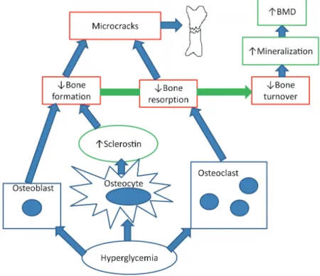
eral. However, the effect of hyperglycemia on osteoblasts in in vitro studies vary as osteocalcin production was decreased in some studies [43-46] and increased in other studies [47,48]. In vitro studies of osteoclasts have demonstrated that hyperglyce- mia decreased osteoclast number, osteoclastogenesis, and osteo- clast activity and decreased pit resorption has also been reported [49-51]. The effects of hyperglycemia on osteoblasts and osteo- clasts lead to hypermineralization and impaired resorption of mineralized matrix, which may contribute to the diabetic bone disease. Fig. 1 illustrates the hypothesis of ostecyte dysfunction and hypermineralization in T2D. Besides hyperglycemia and osteocytic dysfunction, insulin resistance has been correlated with low levels of bone turnover markers in patients with T2D [52] and the metabolic syndrome [53]. Furthermore, insulin re- sistance is correlated with decreased bone strength at the femo- ral neck measured by quantitative computed tomography (QCT) in healthy individuals in two studies [54,55].
An impaired incretin response may also contribute to bone disease in diabetes. The incretins glucose-dependent insulino- tropic peptide (GIP), glucagon-like peptide 1 (GLP-1), and GLP-2 are gastrointestinal hormones that are secreted postpran- dially with numerous beneficial effects on glucose metabolism.
It is thought that incretins also mediate the reduction in bone turnover observed after feeding [56] as part of a gut-bone-axis.
Receptors for GIP [57], GLP-1, and GLP-2 have been described in osteoblastic cell lines [58], GLP-1-receptors also in mature osteoblasts [59] and GIP-receptors in osteoclasts [60]. In pa- tients with T2D, the effect of feeding on bone resorption is re- duced compared with non-diabetes patients [61], possibly by an impaired GLP-1-response [62]. Further studies are warranted to investigate the role of incretins in bone turnover in T2D.
BONE MICROARCHITECTURE IN DIABETIC BONE DISEASE
The changes in bone turnover may be reflected in the microar- chitecture of the bone. However, a cross-sectional study with patients undergoing hip replacement surgery where samples were obtained from the femoral neck, no difference in cortical porosity or trabecular microstructure measured by microcom- puted tomography was present compared to controls [24]. A limitation to the study was that only 20 patients with T2D where included. Non-invasive techniques may also be used to investi- gate the bone microarchitecture in T2D. Trabecular bone score
Fig. 1. The hypothesis of osteocyte dysfunction and hypermineralization. Hyperglycemia decreases bone resorption by inhibiting the osteo- clast and decreases bone formation directly by inhibiting the osteoblast and indirectly by increasing sclerostin production by the osteocytes.
The reduced bone turnover leads to microcracks and bone fractures. Furthermore, the hyperglycemia triggers hypermineralization in the bone causing high bone mineral density (BMD).
(TBS) is a dual-energy X-ray absorptiometry (DXA)-derived variable which is thought to provide information about microar- chitecture of the vertebrae and thereby improve fracture predic- tion in women with diabetes [63]. Women with T2D displayed low TBS compared to controls [64,65] but in men, no difference in TBS was observed between T2D and controls [66]. TBS val- ues have been negatively correlated to glycosylated hemoglobin A1c (HbA1c); thus, TBS may reflect the diabetic state [64,66, 67] but it remains to be shown if changes in HbA1c are reflect- ed in changes in TBS. Furthermore, TBS was lower in patients with T2D and vertebral fracture compared to patients without fracture [68]. However, in the Fracture Risk Assessment Tool (FRAX) models adjusting by TBS does not fully capture the fracture risk in T2D [5]. Although TBS seems to have some val- ue in detecting diabetic bone disease, current evidence does not support regular use of TBS and further research is needed to clarify whether TBS may add to current fracture prediction methods.
Measurement of cortical porosity by high resolution periphe- ral quantitative computed tomography (HRpQCT) has been proposed as a tool to identify patients with T2D at risk of frac- tures. However, cortical porosity has in different studies been reported to be either unchanged [38] or increased [69-71] in pa- tients with T2D compared to controls. In the mentioned studies the observed increased cortical porosity was possibly due to mi- crovascular disease or previous fragility fractures. Furthermore, one study reports lower, higher, and unchanged cortical porosity in patients depending on the measured region [39]. A study us- ing low-resolution computed tomography reports lower cortical porosity of the subtrochanteric femur in women with T2D com- pared to controls [72]. Besides cortical porosity HRpQCT eval- uates trabecular and cortical structure and a study using HRpQCT reported an increased trabecular bone volume fraction in T2D [39] compared to controls, whereas other parameters were unchanged. Other studies report only cortical deficits [69]
or unaltered microarchitecture in T2D compared with controls [38] or T1D [73], respectively. In another large study, HRpQCT revealed decreased cortical volumetric BMD at the tibia and in- creased cortical porosity at the radius in patients with T2D com- pared to controls [74]. The current evidence on cortical porosity and microstructure measured by HRpQCT in T2D is ambiguous and further research is needed to identify whether cortical po- rosity is a predictor of incident fractures.
QCT may discriminate vertebral fractures [75] and previous hip fractures [76] in non-diabetic individuals at a better rate than DXA and may thus be of use in patients with T2D. Heilmeier et
al. [77] report that QCT may identify T2D patients with a previ- ous fracture though the sample size in the study was limited (n=80). However, similar to issues with DXA the vertebral strength calculated from QCT did not correlate to prevalent ver- tebral fractures in patients with T2D [78]. Evidence on bone mi- croarchitecture is limited in T2D and although several non-in- vasive assessments of bone microarchitecture have been per- formed in T2D with different techniques, the evidence does not currently support use of these assessments in detecting diabetic bone disease and further research is needed.
DETECTION OF DIABETIC BONE DISEASE
The fracture risk in patients with T2D is reported to be increased independent of age [6,79], whereas gender and body mass index influence fracture risk similarly to individuals without diabetes [79]. Obesity is related to T2D and has been associated with an increased fracture risk [80], but obesity was also associated with a decreased hip fracture risk in a meta-analysis of prospective studies [81]. Thus neither of these characteristics explain the fracture risk in patients with T2D. In patients with T2D, a weight loss of 20% or more leads to increased risk of fragility fractures compared to a weight loss of less than 10% [82]. Thus, changes in body composition may influence fracture risk and a rapid diet-induced weight loss is accompanied by increased cir- culating levels of sclerostin and CTX in patients with T2D [83].
Microangiopathy in diabetes is proposed to increase fracture risk by increasing cortical porosity [84] and diabetes complica- tions may also influence fracture risk as peripheral neuropathy [85,86], and retinopathy [9,87] is associated with an increased risk of fracture in T2D . However, in a registry based study, pa- tients without diabetic complications also had an increased frac- ture risk and neither retinopathy nor neuropathy were associated with an increased risk of fractures [88]. Although diabetes com- plications as retinopathy and neuropathy may increase the risk of falls in T2D, it is uncertain how prominent the effect is on the increased risk of fracture in T2D [89]. Falls and osteoporotic fractures are associated in T2D [90]; however, fracture risk esti- mates that are adjusted by self-reported falls still demonstrate an increased risk of fractures in T2D [91,92]. Also, hypoglycemia may cause falls, however fracture risk is still increased in T2D when adjusted for documented hypoglycemic episodes [93]. As described earlier, BMD does not fully explain the fracture risk in T2D, but Schwartz et al. [94] report that fracture risk is higher for a given T-score in T2D compared with controls. FRAX un- derestimates fracture risk in diabetes [95] and a correction fac-
tor by HbA1c has been proposed to enhance the prediction of fractures in patients with T2D [96]. Previous studies exploring the association between HbA1c and fracture risk have yielded conflicting results. High levels of HbA1c have been linked with an increased fracture risk in patients with diabetes [8,97,98], but low levels of HbA1c below 7% have also been associated with an increased risk of hip fracture [99]. Some studies even report- ed that HbA1c is not associated with fracture risk [100]. In the randomized Action to Control Cardiovascular Risk in Diabetes (ACCORD) trial no differences in falls or fracture were present between standard glycemic control (HbA1c, 7.5%) and inten- sive glycemic control (HbA1c, 6.4%) [101]. Overall, current fracture predictors underestimate fracture risk in patients with T2D; thus, there is a need for improved fracture predictors. In line with decreased bone turnover and increased sclerostin lev- els, sclerostin is being proposed as a fracture predictor in T2D and circulating levels of sclerostin have been associated with prevalent vertebral fractures [77,102,103]. However, it is uncer- tain whether sclerostin predicts incident fracture. Circulating levels of insulin like-growth factor-1 (IGF-1) have also been as- sociated with an increased fracture risk [102,104], both with prevalent vertebral fractures [102] and incident fractures [104].
Also, circulating levels of osteopontin and osteoglycin have been associated with prevalent vertebral fractures and may be novel fracture predictors in T2D [105,106]. Future fracture pre- dictors in T2D may be measurable in blood; however, further research is needed and until such fracture predictors are estab- lished, BMD continue to be the best fracture predictor available.
ANTIDIABETIC THERAPIES AND DIABETIC BONE DISEASE
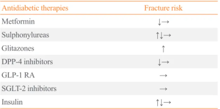
Multiple studies have investigated the effects of antidiabetic medication on fracture risk. Table 1 gives an overview of the ef- fects of antidiabetic therapies on fracture risk. However, most studies are registry based and concerning many recently mar- keted drugs, the follow-up period may not be long enough yet to allow for the detection of fractures. Glitazones increase the risk of fractures as BMD is decreased by switching of osteoblastic recruitment to the adipocytic lineage [107-109]. This has been shown in both randomized controlled trials and observational studies [110-112]. Insulin use have shown both a neutral out- come on fracture risk [100,113], increased fracture risk [114- 116], and decreased fracture risk [93] in a number of different studies. Long-acting insulins may be less prone to cause hypo- glycemia and have been associated with a decreased fracture
risk compared to other insulins [117]. Animal studies have indi- cated a beneficial effect of sulphonylureas on bone by increas- ing bone formation, but so far no effect has been shown in hu- mans [118]. Sulphonylureas are reported to have a neutral out- come on fracture risk [100,113,119], to increase fracture risk [120-122], and to decrease fracture risk [93]. Sulphonylureas increase the risk of hypoglycemia [123] and current use of sul- phonylureas [110] and recent initiation [114] have been associ- ated with an increased fracture risk in T2D, whereas ever use or cumulative dose have not been associated with increased frac- ture risk [110,120]. Compared with metformin monotherapy in- sulin monotherapy and metformin and sulphonylurea in combi- nation were associated with increased major osteoporotic frac- ture risk of 1.6- and 1.3-fold, respectively [114]. Metformin is first line therapy in the treatment of T2D. Cell and animal stud- ies suggest metformin to be osteogenic by promoting Runx2 [124]; however, metformin is associated with lower circulating levels of PINP [125]. Metformin has generally been associated with a decreased or neutral effect on the risk of fractures in T2D [93,100,110,112]. The severity of T2D determines whether sec- ond line therapy is added to the treatment and the effect of met- formin found in observational studies may be due to confound- ing. More recent drugs as the dipeptidylpeptidase 4 (DPP-4) in- hibitors, GLP-1 receptor agonists (GLP-1 RAs), and sodium- glucose co-transporter 2 (SGLT-2) inhibitors have also been in- vestigated in terms of fracture risk. The DPP-4 inhibitors may have beneficial effects on fracture risk as DPP-4 inhibitor-use could potentiate a favorable effect of GLP-1 RA. Sitagliptin, a DPP-4 inhibitor, is in vitro shown to decrease osteoclastogene- sis, whereas it is not known whether DPP-4 inhibitors affect os- teoblasts [126]. A clinical study has shown that 1-year treatment
Table 1. The Effects of Antidiabetic Therapies on Fracture Risk
Antidiabetic therapies Fracture risk
Metformin ↓→
Sulphonylureas ↑↓→
Glitazones ↑
DPP-4 inhibitors ↓→
GLP-1 RA →
SGLT-2 inhibitors →
Insulin ↑↓→
Increased fracture risk (↑), decreased fracture risk (↓), neutral effects on fracture risk (→).
DPP-4, dipeptidylpeptidase 4; GLP-1 RA, glucagon-like peptide 1 re- ceptor agonist; SGLT-2, sodium-glucose co-transporter 2.
with DPP-4 did not change postprandial levels of the bone re- sorption marker CTX nor calcium, phosphate, or serum alkaline phosphatase [127]. A number of retrospective studies have been conducted concerning DPP-4 inhibitors and fracture risk. One study showed that DPP-4 inhibitors were not associated with an increased fracture risk compared to sulphonylureas or insulin [113]. The DPP-4 inhibitors tended to reduce fracture risk com- pared to sulphonylureas with an hazard ratio of 0.8 (95% confi- dence interval, 0.51 to 1.24) and significantly reduced fracture risk compared to glitazones [113]. These effects may, however, represent harmful effects of sulphonylureas and glitazones. Ever use compared to no use of DPP-4 inhibitors have been associat- ed with a decreased risk of fractures [128] and in a study with 5 years follow-up, use of DPP-4 inhibitors have been associated with a 14% decreased risk of fractures in patients with T2D compared to no use of DPP-4 inhibitors [129]. However in other cohort studies, DPP-4 inhibitors showed neutral effect with short treatment duration (less than an year) and longer treatment duration (4 years) [130,131]. Concerning the different types of DPP-4 inhibitors, alogliptin was shown in a meta-analysis of randomized controlled trials to be associated with a lower risk of fracture compared to other DPP-4 inhibitors (saxagliptin and linagliptin) or placebo [132]. Although current evidence is am- biguous, studies with longer follow-up may reveal potential beneficial effects of DPP-4 inhibitors as most current studies are limited by a short treatment period. For GLP-1, as GLP-1 recep- tors are found on osteoblasts [58], GLP-1 is proposed to in- crease bone formation. In registry based studies and randomized controlled trials, GLP-1 RA were associated with neutral frac- ture risk. However, the GLP-1 RA treatment follow-up was too short to adequately evaluate fracture risk [133-135]. As stated previously, a large weight loss is associated with an increase in fracture risk [82]. GLP-1 RA may counteract this harmful effect of weight loss on fracture risk as GLP-1 RA-treatment has been shown to increase bone formation and reduce BMD-loss in obese non-diabetic women [136]. Further research and random- ized controlled trials with a longer follow-up period is needed to determine the role of GLP-1 RA in fracture prevention in T2D.
For the SGLT-2 inhibitors, concern has been raised as to wheth- er the increased glucosuria may lead to loss of calcium and bone loss. Current evidence does not confirm the hypothesis of any harmful effects of SGLT-2 on fracture risk in randomized con- trolled trials [137,138], although long-term effects are yet un- known. In conclusion, concerning the effect of antidiabetic ther- apies on diabetic bone disease caution should be applied to the use of glitazones in T2D in terms of fracture risk. Furthermore,
sulphonylureas may through hypoglycemic events increase fracture risk which should be taken into account when selecting second-line antidiabetic drugs. Further research, investigating the effects of longer durations of treatment is needed on the more recently marketed antidiabetic drugs (DPP-4 inhibitors, GLP-1 RA, and SGLT-2 inhibitors). However, data from obser- vational studies should be interpreted carefully as they are sub- ject to residual confounding.
ANTIOSTEOPOROTIC THERAPIES AND DIABETIC BONE DISEASE
Few studies have investigated the effects of antiosteoporotic treatment in patients with T2D. A post hoc analysis of the Frac- ture Intervention Trial revealed increased BMD and decreased circulating levels of CTX and bone specific alkaline phospha- tase in women with T2D randomized to antiresorptive treatment with the bisphosphonate alendronate [139]. Similar results are reported in a retrospective cohort [140]. A registry based study report similar effects of antiresorptive treatment in patients with T2D compared with controls [141]. Effects of antiresorptive treatment with denosumab on bone have not been investigated in patients with T2D. Studies on bone anabolic therapy with teriparatide report a reduction of fracture risk and increasing BMD in both patients with T2D and patients without diabetes [142]. A subgroup analysis of the recent VERtebral fracture treatment comparison in Osteoporotic women (VERO) trial comparing the effects of teriparatide with risedronate in women with severe osteoporosis demonstrated that the more pronounced anti-fracture efficacy of teriparatide was independent of preva- lence of T2D [143].
Based on current evidence, antiosteoporotic therapy show similar effects in patients with and without T2D. However, con- cerning antiresorptive treatment, it can be speculated whether a further lowering of the already low bone turnover in T2D may be detrimental for bone quality and may actually increase frac- ture risk.
CONCLUSIONS
T2D is associated with an increased risk of fracture which may be due to detrimental effects of low bone turnover. Osteocytic dysfunction due to hyperglycemia, an impaired incretin re- sponse, factors related to hyperglycemia (e.g., insulin resis- tance) and low physical activity may all play a role in the devel- opment of diabetic bone disease. Furthermore, hyperglycemia
may also cause hypermineralization relative to bone turnover and thus increase BMD and mask the actual fracture risk. Cur- rently, BMD is the best predictor of fracture in T2D although flawed and biochemical markers such as sclerostin and IGF-1 should be evaluated further as potential markers for fracture risk in patients with T2D. In the clinical setting, awareness should be aimed at the avoiding the use of glitazones and specific anti- diabetics that increase the risk of hypoglycemia (e.g., sulpho- nylureas) in order to reduce fractures in T2D. Currently, the knowledge of antiosteoporotic therapy in patients with T2D is sparse. More research on detection and treatment of diabetic bone disease and the associated increased risk of fractures is needed, but at present, current osteoporosis guidelines should be followed.
CONFLICTS OF INTEREST
Bente Lomholt Langdahl has received research grants (institu- tion) from Amgen and Novo Nordisk, honoraria for advisory boards and lectures from Amgen, UCB, Eli Lilly, and TEVA.
ORCID
Bente Lomholt Langdahl http://orcid.org/0000-0002-8712-7199
REFERENCES
1. International Diabetes Federation. IDF diabetes atlas. 8th ed. Brussels: International Diabetes Federation; 2017.
2. Janghorbani M, Van Dam RM, Willett WC, Hu FB. Sys- tematic review of type 1 and type 2 diabetes mellitus and risk of fracture. Am J Epidemiol 2007;166:495-505.
3. Vestergaard P. Discrepancies in bone mineral density and fracture risk in patients with type 1 and type 2 diabetes: a meta-analysis. Osteoporos Int 2007;18:427-44.
4. Holm JP, Jensen T, Hyldstrup L, Jensen JB. Fracture risk in women with type II diabetes. Results from a historical co- hort with fracture follow-up. Endocrine 2018;60:151-8.
5. Leslie WD, Johansson H, McCloskey EV, Harvey NC, Kanis JA, Hans D. Comparison of methods for improving fracture risk assessment in diabetes: the Manitoba BMD registry. J Bone Miner Res 2018 Jun 28 [Epub]. https://doi.
org/10.1002/jbmr.3538.
6. Hothersall EJ, Livingstone SJ, Looker HC, Ahmed SF, Cle- land S, Leese GP, et al. Contemporary risk of hip fracture in type 1 and type 2 diabetes: a national registry study from
Scotland. J Bone Miner Res 2014;29:1054-60.
7. Dobnig H, Piswanger-Solkner JC, Roth M, Obermayer-Pi- etsch B, Tiran A, Strele A, et al. Type 2 diabetes mellitus in nursing home patients: effects on bone turnover, bone mass, and fracture risk. J Clin Endocrinol Metab 2006;91:
3355-63.
8. Oei L, Zillikens MC, Dehghan A, Buitendijk GH, Castano- Betancourt MC, Estrada K, et al. High bone mineral densi- ty and fracture risk in type 2 diabetes as skeletal complica- tions of inadequate glucose control: the Rotterdam Study.
Diabetes Care 2013;36:1619-28.
9. Ivers RQ, Cumming RG, Mitchell P, Peduto AJ; Blue Mountains Eye Study. Diabetes and risk of fracture: the blue mountains eye study. Diabetes Care 2001;24:1198-203.
10. Jia P, Bao L, Chen H, Yuan J, Liu W, Feng F, et al. Risk of low-energy fracture in type 2 diabetes patients: a meta- analysis of observational studies. Osteoporos Int 2017;28:
3113-21.
11. Napoli N, Schwartz AV, Schafer AL, Vittinghoff E, Cawthon PM, Parimi N, et al. Vertebral fracture risk in diabetic elder- ly men: the MrOS study. J Bone Miner Res 2018;33:63-9.
12. Ni Y, Fan D. Diabetes mellitus is a risk factor for low bone mass-related fractures: a meta-analysis of cohort studies.
Medicine (Baltimore) 2017;96:e8811.
13. Voko Z, Gaspar K, Inotai A, Horvath C, Bors K, Speer G, et al. Osteoporotic fractures may impair life as much as the complications of diabetes. J Eval Clin Pract 2017;23:1375- 80.
14. Panula J, Pihlajamaki H, Mattila VM, Jaatinen P, Vahlberg T, Aarnio P, et al. Mortality and cause of death in hip frac- ture patients aged 65 or older: a population-based study.
BMC Musculoskelet Disord 2011;12:105.
15. Hasserius R, Karlsson MK, Jonsson B, Redlund-Johnell I, Johnell O. Long-term morbidity and mortality after a clini- cally diagnosed vertebral fracture in the elderly: a 12- and 22-year follow-up of 257 patients. Calcif Tissue Int 2005;
76:235-42.
16. Hernlund E, Svedbom A, Ivergard M, Compston J, Cooper C, Stenmark J, et al. Osteoporosis in the European Union:
medical management, epidemiology and economic burden.
A report prepared in collaboration with the International Osteoporosis Foundation (IOF) and the European Federa- tion of Pharmaceutical Industry Associations (EFPIA).
Arch Osteoporos 2013;8:136.
17. Starup-Linde J, Frost M, Vestergaard P, Abrahamsen B.
Epidemiology of fractures in diabetes. Calcif Tissue Int
2017;100:109-21.
18. Ma L, Oei L, Jiang L, Estrada K, Chen H, Wang Z, et al.
Association between bone mineral density and type 2 dia- betes mellitus: a meta-analysis of observational studies. Eur J Epidemiol 2012;27:319-32.
19. Krakauer JC, McKenna MJ, Buderer NF, Rao DS, White- house FW, Parfitt AM. Bone loss and bone turnover in dia- betes. Diabetes 1995;44:775-82.
20. Manavalan JS, Cremers S, Dempster DW, Zhou H, Dwora- kowski E, Kode A, et al. Circulating osteogenic precursor cells in type 2 diabetes mellitus. J Clin Endocrinol Metab 2012;97:3240-50.
21. Acevedo C, Sylvia M, Schaible E, Graham JL, Stanhope KL, Metz LN, et al. Contributions of material properties and structure to increased bone fragility for a given bone mass in the UCD-T2DM rat model of type 2 diabetes. J Bone Miner Res 2018;33:1066-75.
22. Hunt HB, Pearl JC, Diaz DR, King KB, Donnelly E. Bone tissue collagen maturity and mineral content increase with sustained hyperglycemia in the KK-Ay murine model of type 2 diabetes. J Bone Miner Res 2018;33:921-9.
23. Saito M, Marumo K. Bone quality in diabetes. Front Endo- crinol (Lausanne) 2013;4:72.
24. Karim L, Moulton J, Van Vliet M, Velie K, Robbins A, Malekipour F, et al. Bone microarchitecture, biomechanical properties, and advanced glycation end-products in the proximal femur of adults with type 2 diabetes. Bone 2018;
114:32-9.
25. Hygum K, Starup-Linde J, Harslof T, Vestergaard P, Lang- dahl BL. Mechanisms in endocrinology: diabetes mellitus, a state of low bone turnover. A systematic review and meta- analysis. Eur J Endocrinol 2017;176:R137-57.
26. Starup-Linde J, Vestergaard P. Biochemical bone turnover markers in diabetes mellitus: a systematic review. Bone 2016;82:69-78.
27. Purnamasari D, Puspitasari MD, Setiyohadi B, Nugroho P, Isbagio H. Low bone turnover in premenopausal women with type 2 diabetes mellitus as an early process of diabe- tes-associated bone alterations: a cross-sectional study.
BMC Endocr Disord 2017;17:72.
28. Mitchell A, Fall T, Melhus H, Wolk A, Michaelsson K, By- berg L. Type 2 diabetes in relation to hip bone density, area, and bone turnover in Swedish men and women: a cross- sectional study. Calcif Tissue Int 2018 Jun 26 [Epub].
https://doi.org/10.1007/s00223-018-0446-9.
29. Starup-Linde J, Lykkeboe S, Gregersen S, Hauge EM,
Langdahl BL, Handberg A, et al. Differences in biochemi- cal bone markers by diabetes type and the impact of glu- cose. Bone 2016;83:149-55.
30. Wang N, Xue P, Wu X, Ma J, Wang Y, Li Y. Role of sclerostin and dkk1 in bone remodeling in type 2 diabetic patients. Endocr Res 2018;43:29-38.
31. Manolagas SC, Almeida M. Gone with the Wnts: beta- catenin, T-cell factor, forkhead box O, and oxidative stress in age-dependent diseases of bone, lipid, and glucose me- tabolism. Mol Endocrinol 2007;21:2605-14.
32. Bonewald LF. The amazing osteocyte. J Bone Miner Res 2011;26:229-38.
33. Raska I Jr, Raskova M, Zikan V, Skrha J. Prevalence and risk factors of osteoporosis in postmenopausal women with type 2 diabetes mellitus. Cent Eur J Public Health 2017;25:
3-10.
34. Kang J, Boonanantanasarn K, Baek K, Woo KM, Ryoo HM, Baek JH, et al. Hyperglycemia increases the expres- sion levels of sclerostin in a reactive oxygen species- and tumor necrosis factor-alpha-dependent manner. J Periodon- tal Implant Sci 2015;45:101-10.
35. Tanaka K, Yamaguchi T, Kanazawa I, Sugimoto T. Effects of high glucose and advanced glycation end products on the expressions of sclerostin and RANKL as well as apop- tosis in osteocyte-like MLO-Y4-A2 cells. Biochem Bio- phys Res Commun 2015;461:193-9.
36. Farr JN, Khosla S. Determinants of bone strength and qual- ity in diabetes mellitus in humans. Bone 2016;82:28-34.
37. Rubin MR. Skeletal fragility in diabetes. Ann N Y Acad Sci 2017;1402:18-30.
38. Farr JN, Drake MT, Amin S, Melton LJ 3rd, McCready LK, Khosla S. In vivo assessment of bone quality in post- menopausal women with type 2 diabetes. J Bone Miner Res 2014;29:787-95.
39. Nilsson AG, Sundh D, Johansson L, Nilsson M, Mellstrom D, Rudang R, et al. Type 2 diabetes mellitus is associated with better bone microarchitecture but lower bone material strength and poorer physical function in elderly women: a population-based study. J Bone Miner Res 2017;32:1062- 71.
40. Napoli N, Strollo R, Defeudis G, Leto G, Moretti C, Zam- petti S, et al. Serum sclerostin and bone turnover in latent autoimmune diabetes in adults. J Clin Endocrinol Metab 2018;103:1921-8.
41. Gennari L, Merlotti D, Valenti R, Ceccarelli E, Ruvio M, Pietrini MG, et al. Circulating sclerostin levels and bone
turnover in type 1 and type 2 diabetes. J Clin Endocrinol Metab 2012;97:1737-44.
42. Deng X, Xu M, Shen M, Cheng J. Effects of type 2 diabetic serum on proliferation and osteogenic differentiation of mes- enchymal stem cells. J Diabetes Res 2018;2018:5765478.
43. Bartolome A, Lopez-Herradon A, Portal-Nunez S, Garcia- Aguilar A, Esbrit P, Benito M, et al. Autophagy impairment aggravates the inhibitory effects of high glucose on osteo- blast viability and function. Biochem J 2013;455:329-37.
44. Botolin S, McCabe LR. Chronic hyperglycemia modulates osteoblast gene expression through osmotic and non-os- motic pathways. J Cell Biochem 2006;99:411-24.
45. Wu YY, Yu T, Zhang XH, Liu YS, Li F, Wang YY, et al.
1,25(OH)2D3 inhibits the deleterious effects induced by high glucose on osteoblasts through undercarboxylated os- teocalcin and insulin signaling. J Steroid Biochem Mol Biol 2012;132:112-9.
46. Zayzafoon M, Stell C, Irwin R, McCabe LR. Extracellular glucose influences osteoblast differentiation and c-Jun ex- pression. J Cell Biochem 2000;79:301-10.
47. Liu Z, Jiang H, Dong K, Liu S, Zhou W, Zhang J, et al. Dif- ferent concentrations of glucose regulate proliferation and osteogenic differentiation of osteoblasts via the PI3 kinase/
Akt pathway. Implant Dent 2015;24:83-91.
48. Zhen D, Chen Y, Tang X. Metformin reverses the deleteri- ous effects of high glucose on osteoblast function. J Diabe- tes Complications 2010;24:334-44.
49. Wittrant Y, Gorin Y, Woodruff K, Horn D, Abboud HE, Mohan S, et al. High d(+)glucose concentration inhibits RANKL-induced osteoclastogenesis. Bone 2008;42:1122- 30.
50. Xu J, Yue F, Wang J, Chen L, Qi W. High glucose inhibits receptor activator of nuclear factor-κB ligand-induced os- teoclast differentiation via downregulation of v-ATPase V0 subunit d2 and dendritic cell specific transmembrane pro- tein. Mol Med Rep 2015;11:865-70.
51. Xu F, Ye YP, Dong YH, Guo FJ, Chen AM, Huang SL. In- hibitory effects of high glucose/insulin environment on os- teoclast formation and resorption in vitro. J Huazhong Univ Sci Technolog Med Sci 2013;33:244-9.
52. Tonks KT, White CP, Center JR, Samocha-Bonet D, Green- field JR. Bone turnover is suppressed in insulin resistance, independent of adiposity. J Clin Endocrinol Metab 2017;
102:1112-21.
53. Laurent MR, Cook MJ, Gielen E, Ward KA, Antonio L, Adams JE, et al. Lower bone turnover and relative bone
deficits in men with metabolic syndrome: a matter of insu- lin sensitivity? The European male ageing study. Osteopo- ros Int 2016;27:3227-37.
54. Kalimeri M, Leek F, Wang NX, Koh HR, Roy NC, Camer- on-Smith D, et al. Association of insulin resistance with bone strength and bone turnover in menopausal Chinese- Singaporean women without diabetes. Int J Environ Res Public Health 2018;15:E889.
55. Yang J, Hong N, Shim JS, Rhee Y, Kim HC. Association of insulin resistance with lower bone volume and strength in- dex of the proximal femur in nondiabetic postmenopausal women. J Bone Metab 2018;25:123-32.
56. Clowes JA, Allen HC, Prentis DM, Eastell R, Blumsohn A.
Octreotide abolishes the acute decrease in bone turnover in response to oral glucose. J Clin Endocrinol Metab 2003;88:
4867-73.
57. Bollag RJ, Zhong Q, Phillips P, Min L, Zhong L, Cameron R, et al. Osteoblast-derived cells express functional glu- cose-dependent insulinotropic peptide receptors. Endocri- nology 2000;141:1228-35.
58. Pacheco-Pantoja EL, Ranganath LR, Gallagher JA, Wilson PJ, Fraser WD. Receptors and effects of gut hormones in three osteoblastic cell lines. BMC Physiol 2011;11:12.
59. Nuche-Berenguer B, Portal-Nunez S, Moreno P, Gonzalez N, Acitores A, Lopez-Herradon A, et al. Presence of a func- tional receptor for GLP-1 in osteoblastic cells, independent of the cAMP-linked GLP-1 receptor. J Cell Physiol 2010;
225:585-92.
60. Zhong Q, Itokawa T, Sridhar S, Ding KH, Xie D, Kang B, et al. Effects of glucose-dependent insulinotropic peptide on osteoclast function. Am J Physiol Endocrinol Metab 2007;292:E543-8.
61. Chailurkit LO, Chanprasertyothin S, Rajatanavin R, Ong- phiphadhanakul B. Reduced attenuation of bone resorption after oral glucose in type 2 diabetes. Clin Endocrinol (Oxf) 2008;68:858-62.
62. Faerch K, Torekov SS, Vistisen D, Johansen NB, Witte DR, Jonsson A, et al. GLP-1 response to oral glucose is reduced in prediabetes, screen-detected type 2 diabetes, and obesity and influenced by sex: the ADDITION-PRO study. Diabe- tes 2015;64:2513-25.
63. Leslie WD, Aubry-Rozier B, Lamy O, Hans D; Manitoba Bone Density Program. TBS (trabecular bone score) and diabetes-related fracture risk. J Clin Endocrinol Metab 2013;98:602-9.
64. Dhaliwal R, Cibula D, Ghosh C, Weinstock RS, Moses
AM. Bone quality assessment in type 2 diabetes mellitus.
Osteoporos Int 2014;25:1969-73.
65. Rianon N, Ambrose CG, Buni M, Watt G, Reyes-Ortiz C, Lee M, et al. Trabecular bone score is a valuable addition to bone mineral density for bone quality assessment in older Mexican American women with type 2 diabetes. J Clin Densitom 2018;21:355-9.
66. Iki M, Fujita Y, Kouda K, Yura A, Tachiki T, Tamaki J, et al. Hyperglycemia is associated with increased bone miner- al density and decreased trabecular bone score in elderly Japanese men: the Fujiwara-kyo osteoporosis risk in men (FORMEN) study. Bone 2017;105:18-25.
67. Kim JH, Choi HJ, Ku EJ, Kim KM, Kim SW, Cho NH, et al. Trabecular bone score as an indicator for skeletal deteri- oration in diabetes. J Clin Endocrinol Metab 2015;100:475- 82.
68. Choi YJ, Ock SY, Jin Y, Lee JS, Kim SH, Chung Y. Urinary pentosidine levels negatively associates with trabecular bone scores in patients with type 2 diabetes mellitus. Os- teoporos Int 2018;29:907-15.
69. Shanbhogue VV, Hansen S, Hermann P, Jorgensen N, Hen- riksen JE, Brixen K. Diabetic microvascular disease and bone structure in patients with type 2 diabetes mellitus. Di- abetes 2015;64(Suppl 1):A174.
70. Patsch JM, Burghardt AJ, Yap SP, Baum T, Schwartz AV, Joseph GB, et al. Increased cortical porosity in type 2 dia- betic postmenopausal women with fragility fractures. J Bone Miner Res 2013;28:313-24.
71. Heilmeier U, Cheng K, Pasco C, Parrish R, Nirody J, Patsch JM, et al. Cortical bone laminar analysis reveals in- creased midcortical and periosteal porosity in type 2 dia- betic postmenopausal women with history of fragility frac- tures compared to fracture-free diabetics. Osteoporos Int 2016;27:2791-802.
72. Osima M, Kral R, Borgen TT, Hogestol IK, Joakimsen RM, Eriksen EF, et al. Women with type 2 diabetes mellitus have lower cortical porosity of the proximal femoral shaft using low-resolution CT than nondiabetic women, and in- creasing glucose is associated with reduced cortical porosi- ty. Bone 2017;97:252-60.
73. Starup-Linde J, Lykkeboe S, Gregersen S, Hauge EM, Langdahl BL, Handberg A, et al. Bone structure and pre- dictors of fracture in type 1 and type 2 diabetes. J Clin En- docrinol Metab 2016;101:928-36.
74. Samelson EJ, Demissie S, Cupples LA, Zhang X, Xu H, Liu CT, et al. Diabetes and deficits in cortical bone density,
microarchitecture, and bone size: Framingham HR-pQCT study. J Bone Miner Res 2018;33:54-62.
75. Li N, Li XM, Xu L, Sun WJ, Cheng XG, Tian W. Compari- son of QCT and DXA: osteoporosis detection rates in post- menopausal women. Int J Endocrinol 2013;2013:895474.
76. Yang L, Udall WJ, McCloskey EV, Eastell R. Distribution of bone density and cortical thickness in the proximal fe- mur and their association with hip fracture in postmeno- pausal women: a quantitative computed tomography study.
Osteoporos Int 2014;25:251-63.
77. Heilmeier U, Carpenter DR, Patsch JM, Harnish R, Joseph GB, Burghardt AJ, et al. Volumetric femoral BMD, bone geometry, and serum sclerostin levels differ between type 2 diabetic postmenopausal women with and without fragility fractures. Osteoporos Int 2015;26:1283-93.
78. Kiyohara N, Yamamoto M, Sugimoto T. Discordance be- tween prevalent vertebral fracture and vertebral strength estimated by the finite element method based on quantita- tive computed tomography in patients with type 2 diabetes mellitus. PLoS One 2015;10:e0144496.
79. Leslie WD, Morin SN, Lix LM, Majumdar SR. Does dia- betes modify the effect of FRAX risk factors for predicting major osteoporotic and hip fracture? Osteoporos Int 2014;
25:2817-24.
80. Sogaard AJ, Holvik K, Omsland TK, Tell GS, Dahl C, Schei B, et al. Abdominal obesity increases the risk of hip fracture. A population-based study of 43,000 women and men aged 60-79 years followed for 8 years. Cohort of Nor- way. J Intern Med 2015;277:306-17.
81. Tang X, Liu G, Kang J, Hou Y, Jiang F, Yuan W, et al. Obe- sity and risk of hip fracture in adults: a meta-analysis of prospective cohort studies. PLoS One 2013;8:e55077.
82. Komorita Y, Iwase M, Fujii H, Ohkuma T, Ide H, Jodai- Kitamura T, et al. Impact of body weight loss from maxi- mum weight on fragility bone fractures in Japanese patients with type 2 diabetes: the Fukuoka diabetes registry. Diabe- tes Care 2018;41:1061-7.
83. Strollo R, Soare A, Manon Khazrai Y, Di Mauro A, Paler- mo A, Del Toro R, et al. Increased sclerostin and bone turn- over after diet-induced weight loss in type 2 diabetes: a post hoc analysis of the MADIAB trial. Endocrine 2017;56:
667-4.
84. Shanbhogue VV, Hansen S, Frost M, Brixen K, Hermann AP. Bone disease in diabetes: another manifestation of mi- crovascular disease? Lancet Diabetes Endocrinol 2017;5:
827-38.
85. Lee RH, Sloane R, Pieper C, Lyles KW, Adler RA, Van Houtven C, et al. Clinical fractures among older men with diabetes are mediated by diabetic complications. J Clin En- docrinol Metab 2018;103:281-7.
86. Kim JH, Jung MH, Lee JM, Son HS, Cha BY, Chang SA.
Diabetic peripheral neuropathy is highly associated with nontraumatic fractures in Korean patients with type 2 dia- betes mellitus. Clin Endocrinol (Oxf) 2012;77:51-5.
87. Viegas M, Costa C, Lopes A, Griz L, Medeiro MA, Ban- deira F. Prevalence of osteoporosis and vertebral fractures in postmenopausal women with type 2 diabetes mellitus and their relationship with duration of the disease and chronic complications. J Diabetes Complications 2011;25:
216-21.
88. Vestergaard P, Rejnmark L, Mosekilde L. Diabetes and its complications and their relationship with risk of fractures in type 1 and 2 diabetes. Calcif Tissue Int 2009;84:45-55.
89. Timar B, Timar R, Gaita L, Oancea C, Levai C, Lungeanu D. The impact of diabetic neuropathy on balance and on the risk of falls in patients with type 2 diabetes mellitus: a cross-sectional study. PLoS One 2016;11:e0154654.
90. Yokomoto-Umakoshi M, Kanazawa I, Kondo S, Sugimoto T. Association between the risk of falls and osteoporotic fractures in patients with type 2 diabetes mellitus. Endocr J 2017;64:727-34.
91. Bonds DE, Larson JC, Schwartz AV, Strotmeyer ES, Rob- bins J, Rodriguez BL, et al. Risk of fracture in women with type 2 diabetes: the Women’s Health Initiative Observa- tional Study. J Clin Endocrinol Metab 2006;91:3404-10.
92. Schwartz AV, Sellmeyer DE, Ensrud KE, Cauley JA, Tabor HK, Schreiner PJ, et al. Older women with diabetes have an increased risk of fracture: a prospective study. J Clin En- docrinol Metab 2001;86:32-8.
93. Vestergaard P, Rejnmark L, Mosekilde L. Relative fracture risk in patients with diabetes mellitus, and the impact of in- sulin and oral antidiabetic medication on relative fracture risk. Diabetologia 2005;48:1292-9.
94. Schwartz AV, Vittinghoff E, Bauer DC, Hillier TA, Strot- meyer ES, Ensrud KE, et al. Association of BMD and FRAX score with risk of fracture in older adults with type 2 diabetes. JAMA 2011;305:2184-92.
95. Giangregorio LM, Leslie WD, Lix LM, Johansson H, Oden A, McCloskey E, et al. FRAX underestimates fracture risk in patients with diabetes. J Bone Miner Res 2012;27:301-8.
96. Valentini A, Cianfarani MA, De Meo L, Morabito P, Ro- manello D, Tarantino U, et al. FRAX tool in type 2 diabetic
subjects: the use of HbA(1c) in estimating fracture risk.
Acta Diabetol 2018 Jul 6 [Epub]. https://doi.org/10.1007/
s00592-018-1187-y.
97. Conway BN, Long DM, Figaro MK, May ME. Glycemic control and fracture risk in elderly patients with diabetes.
Diabetes Res Clin Pract 2016;115:47-53.
98. Li CI, Liu CS, Lin WY, Meng NH, Chen CC, Yang SY, et al. Glycated hemoglobin level and risk of hip fracture in older people with type 2 diabetes: a competing risk analysis of Taiwan diabetes cohort study. J Bone Miner Res 2015;
30:1338-46.
99. Puar TH, Khoo JJ, Cho LW, Xu Y, Chen YT, Chuo AM, et al. Association between glycemic control and hip fracture.
J Am Geriatr Soc 2012;60:1493-7.
100. Starup-Linde J, Gregersen S, Vestergaard P. Associations with fracture in patients with diabetes: a nested case-control study. BMJ Open 2016;6:e009686.
101. Schwartz AV, Margolis KL, Sellmeyer DE, Vittinghoff E, Ambrosius WT, Bonds DE, et al. Intensive glycemic con- trol is not associated with fractures or falls in the ACCORD randomized trial. Diabetes Care 2012;35:1525-31.
102. Ardawi MS, Akhbar DH, Alshaikh A, Ahmed MM, Qari MH, Rouzi AA, et al. Increased serum sclerostin and de- creased serum IGF-1 are associated with vertebral fractures among postmenopausal women with type-2 diabetes. Bone 2013;56:355-62.
103. Yamamoto M, Yamauchi M, Sugimoto T. Elevated scleros- tin levels are associated with vertebral fractures in patients with type 2 diabetes mellitus. J Clin Endocrinol Metab 2013;98:4030-7.
104. Miyake H, Kanazawa I, Sugimoto T. Decreased serum in- sulin-like growth factor-I is a risk factor for non-vertebral fractures in diabetic postmenopausal women. Intern Med 2017;56:269-73.
105. Tanaka KI, Kanazawa I, Kaji H, Sugimoto T. Association of osteoglycin and FAM5C with bone turnover markers, bone mineral density, and vertebral fractures in postmeno- pausal women with type 2 diabetes mellitus. Bone 2017;
95:5-10.
106. Filardi T, Carnevale V, Massoud R, Russo C, Nieddu L, Tavaglione F, et al. High serum osteopontin levels are asso- ciated with prevalent fractures and worse lipid profile in post-menopausal women with type 2 diabetes. J Endocrinol Invest 2018 Jun 18 [Epub]. https://doi.org/10.1007/s40618- 018-0914-0.
107. Meier C, Schwartz AV, Egger A, Lecka-Czernik B. Effects
of diabetes drugs on the skeleton. Bone 2016;82:93-100.
108. Palermo A, D’Onofrio L, Eastell R, Schwartz AV, Pozzilli P, Napoli N. Oral anti-diabetic drugs and fracture risk, cut to the bone: safe or dangerous? A narrative review. Osteopo- ros Int 2015;26:2073-89.
109. Harslof T, Wamberg L, Moller L, Stodkilde-Jorgensen H, Ringgaard S, Pedersen SB, et al. Rosiglitazone decreases bone mass and bone marrow fat. J Clin Endocrinol Metab 2011;96:1541-8.
110. Starup-Linde J, Gregersen S, Frost M, Vestergaard P. Use of glucose-lowering drugs and risk of fracture in patients with type 2 diabetes. Bone 2017;95:136-42.
111. Bazelier MT, de Vries F, Vestergaard P, Herings RM, Galla- gher AM, Leufkens HG, et al. Risk of fracture with thia- zolidinediones: an individual patient data meta-analysis.
Front Endocrinol (Lausanne) 2013;4:11.
112. Kahn SE, Zinman B, Lachin JM, Haffner SM, Herman WH, Holman RR, et al. Rosiglitazone-associated fractures in type 2 diabetes: an Analysis from A Diabetes Outcome Progression Trial (ADOPT). Diabetes Care 2008;31:845- 51.
113. Gamble JM, Donnan JR, Chibrikov E, Twells LK, Midodzi WK, Majumdar SR. The risk of fragility fractures in new users of dipeptidyl peptidase-4 inhibitors compared to sul- fonylureas and other anti-diabetic drugs: a cohort study. Di- abetes Res Clin Pract 2018;136:159-67.
114. Losada E, Soldevila B, Ali MS, Martinez-Laguna D, Nogues X, Puig-Domingo M, et al. Real-world antidiabetic drug use and fracture risk in 12,277 patients with type 2 di- abetes mellitus: a nested case-control study. Osteoporos Int 2018;29:2079-86.
115. Losada-Grande E, Hawley S, Soldevila B, Martinez-Lagu- na D, Nogues X, Diez-Perez A, et al. Insulin use and excess fracture risk in patients with type 2 diabetes: a propensity- matched cohort analysis. Sci Rep 2017;7:3781.
116. Wallander M, Axelsson KF, Nilsson AG, Lundh D, Lorent- zon M. Type 2 diabetes and risk of hip fractures and non- skeletal fall injuries in the elderly: a study from the frac- tures and fall injuries in the elderly cohort (FRAILCO). J Bone Miner Res 2017;32:449-60.
117. Pscherer S, Kostev K, Dippel FW, Rathmann W. Fracture risk in patients with type 2 diabetes under different antidia- betic treatment regimens: a retrospective database analysis in primary care. Diabetes Metab Syndr Obes 2016;9:17-23.
118. Fronczek-Sokol J, Pytlik M. Effect of glimepiride on the skeletal system of ovariectomized and non-ovariectomized
rats. Pharmacol Rep 2014;66:412-7.
119. Lapane KL, Yang S, Brown MJ, Jawahar R, Pagliasotti C, Rajpathak S. Sulfonylureas and risk of falls and fractures: a systematic review. Drugs Aging 2013;30:527-47.
120. Colhoun HM, Livingstone SJ, Looker HC, Morris AD, Wild SH, Lindsay RS, et al. Hospitalised hip fracture risk with rosiglitazone and pioglitazone use compared with oth- er glucose-lowering drugs. Diabetologia 2012;55:2929-37.
121. Napoli N, Strotmeyer ES, Ensrud KE, Sellmeyer DE, Bau- er DC, Hoffman AR, et al. Fracture risk in diabetic elderly men: the MrOS study. Diabetologia 2014;57:2057-65.
122. Rajpathak SN, Fu C, Brodovicz KG, Engel SS, Lapane K.
Sulfonylurea use and risk of hip fractures among elderly men and women with type 2 diabetes. Drugs Aging 2015;
32:321-7.
123. Schopman JE, Simon AC, Hoefnagel SJ, Hoekstra JB, Scholten RJ, Holleman F. The incidence of mild and severe hypoglycaemia in patients with type 2 diabetes mellitus treated with sulfonylureas: a systematic review and meta- analysis. Diabetes Metab Res Rev 2014;30:11-22.
124. Molinuevo MS, Schurman L, McCarthy AD, Cortizo AM, Tolosa MJ, Gangoiti MV, et al. Effect of metformin on bone marrow progenitor cell differentiation: in vivo and in vitro studies. J Bone Miner Res 2010;25:211-21.
125. Stage TB, Christensen MH, Jorgensen NR, Beck-Nielsen H, Brosen K, Gram J, et al. Effects of metformin, rosiglitazone and insulin on bone metabolism in patients with type 2 dia- betes. Bone 2018;112:35-41.
126. Wang C, Xiao F, Qu X, Zhai Z, Hu G, Chen X, et al. Sita- gliptin, an anti-diabetic drug, suppresses estrogen deficien- cy-induced osteoporosisin vivo and inhibits RANKL-in- duced osteoclast formation and bone resorption in vitro.
Front Pharmacol 2017;8:407.
127. Bunck MC, Poelma M, Eekhoff EM, Schweizer A, Heine RJ, Nijpels G, et al. Effects of vildagliptin on postprandial markers of bone resorption and calcium homeostasis in re- cently diagnosed, well-controlled type 2 diabetes patients. J Diabetes 2012;4:181-5.
128. Dombrowski S, Kostev K, Jacob L. Use of dipeptidyl pep- tidase-4 inhibitors and risk of bone fracture in patients with type 2 diabetes in Germany: a retrospective analysis of real- world data. Osteoporos Int 2017;28:2421-8.
129. Hou WH, Chang KC, Li CY, Ou HT. Dipeptidyl pepti- dase-4 inhibitor use is associated with decreased risk of fracture in patients with type 2 diabetes: a population-based cohort study. Br J Clin Pharmacol 2018;84:2029-39.
130. Driessen JH, van den Bergh JP, van Onzenoort HA, Henry RM, Leufkens HG, de Vries F. Long-term use of dipeptidyl peptidase-4 inhibitors and risk of fracture: a retrospective population-based cohort study. Diabetes Obes Metab 2017;
19:421-8.
131. Driessen JH, van Onzenoort HA, Starup-Linde J, Henry R, Neef C, van den Bergh J, et al. Use of dipeptidyl peptidase 4 inhibitors and fracture risk compared to use of other anti- hyperglycemic drugs. Pharmacoepidemiol Drug Saf 2015;
24:1017-25.
132. Yang J, Huang C, Wu S, Xu Y, Cai T, Chai S, et al. The ef- fects of dipeptidyl peptidase-4 inhibitors on bone fracture among patients with type 2 diabetes mellitus: a network meta-analysis of randomized controlled trials. PLoS One 2017;12:e0187537.
133. Driessen JH, van Onzenoort HA, Starup-Linde J, Henry R, Burden AM, Neef C, et al. Use of glucagon-like-peptide 1 receptor agonists and risk of fracture as compared to use of other anti-hyperglycemic drugs. Calcif Tissue Int 2015;97:
506-15.
134. Driessen JH, de Vries F, van Onzenoort H, Harvey NC, Neef C, van den Bergh JP, et al. The use of incretins and fractures: a meta-analysis on population-based real life data. Br J Clin Pharmacol 2017;83:923-6.
135. Mabilleau G, Mieczkowska A, Chappard D. Use of gluca- gon-like peptide-1 receptor agonists and bone fractures: a meta-analysis of randomized clinical trials. J Diabetes 2014;6:260-6.
136. Iepsen EW, Lundgren JR, Hartmann B, Pedersen O, Han- sen T, Jorgensen NR, et al. GLP-1 receptor agonist treat- ment increases bone formation and prevents bone loss in weight-reduced obese women. J Clin Endocrinol Metab
2015;100:2909-17.
137. Tang HL, Li DD, Zhang JJ, Hsu YH, Wang TS, Zhai SD, et al. Lack of evidence for a harmful effect of sodium-glucose co-transporter 2 (SGLT2) inhibitors on fracture risk among type 2 diabetes patients: a network and cumulative meta- analysis of randomized controlled trials. Diabetes Obes Metab 2016;18:1199-206.
138. Kohler S, Kaspers S, Salsali A, Zeller C, Woerle HJ. Analy- sis of fractures in patients with type 2 diabetes treated with empagliflozin in pooled data from placebo-controlled trials and a head-to-head study versus glimepiride. Diabetes Care 2018;41:1809-16.
139. Keegan TH, Schwartz AV, Bauer DC, Sellmeyer DE, Kelsey JL; fracture intervention trial. Effect of alendronate on bone mineral density and biochemical markers of bone turnover in type 2 diabetic women: the fracture intervention trial. Diabetes Care 2004;27:1547-53.
140. Iwamoto J, Sato Y, Uzawa M, Takeda T, Matsumoto H.
Three-year experience with alendronate treatment in post- menopausal osteoporotic Japanese women with or without type 2 diabetes. Diabetes Res Clin Pract 2011;93:166-73.
141. Vestergaard P, Rejnmark L, Mosekilde L. Are antiresorp- tive drugs effective against fractures in patients with diabe- tes? Calcif Tissue Int 2011;88:209-14.
142. Schwartz AV, Pavo I, Alam J, Disch DP, Schuster D, Harris JM, et al. Teriparatide in patients with osteoporosis and type 2 diabetes. Bone 2016;91:152-8.
143. Geusens P, Marin F, Kendler DL, Russo LA, Zerbini CA, Minisola S, et al. Effects of teriparatide compared with risedronate on the risk of fractures in subgroups of post- menopausal women with severe osteoporosis: the VERO trial. J Bone Miner Res 2018;33:783-94.