Jae Hoon Lee, M.D., Bi O Jeong, M.D.,
Jung Chul Hwang, M.D., Duke Whan Chung, M.D.
Department of Orthopedic Surgery, School of Medicine, Kyung Hee University, Seoul, Korea
Injuries to the distal ulnar epiphysis are uncommon.
Irreducible epiphyseal plate fracture of the distal ulna is a rare, having been reported in the English literature less than 10 times. We report two cases of irreducible frac- ture of the distal ulnar epiphysis. First case is Salter- Harris type II fracture of distal radius and ulna. Second case is Galeazzi-equivalent fracture. The distal ulnar epi- physis was irreducible due to interposition of extensor carpi ulnaris tendon and periosteum between the frag- ments, respectively.
Key Words: Distal ulna, Epiphyseal injury, Irreducible fracture, Galeazzi-equivalent fracture, Open reduction
도수 정복이 불가능한 척골 원위부 성장판 골절은 지금까지 문헌에서도 10편 미만으로 보고될 정도로 매 우 드물다1-3. 저자들은 원위 척골 성장판 골절 환자에 서 척골의 도수 정복이 불가능하였던 2예를 문헌 고찰 과 함께 보고하고자 한다.
증례 1
14세 남아로 자전거 타다 넘어져 발생한 좌측 손목 의 통증과 종창을 주소로 내원하였다. 진찰상 손목 관 절의 운동 제한과 압통이 있었으며 원위 척골이 반대 측에 비해 전방으로 전위되어 있었다. 감각 이상이나 수지 운동의 장애는 없었으며 근위 전완부 및 주관절 부 동통이나 압통은 없었다. 단순 방사선 사진상 요골 은 Salter-Harris 2형의 성장판 골절 소견을 보였고 원위 척골은 완전 전위된 Salter-Harris 2형의 성장 판 골절 소견을 보였다(Fig. 1A,B). Chinese fin- ger trap을 장착한 후 도수 정복을 시행하였다. 원위 요골은 쉽게 정복이 가능하였으나, 원위 척골은 정복 을 얻을 수 없어 다음날 전신 마취 하에 관혈적 정복 을 시행하였다. 원위 척골의 척측에 종적 피부 절개를 한 후 척골 신경의 배측 분지를 조심하며 골절부에 접 근하였다. 성장판은 척측 및 수장측으로 전위되어 있 었으며 척 수근 신건이 삽입되어 정복을 방해하고 있 었다(Fig. 2A). 골절부에 삽입된 척 수근 신건을 배 측으로 직접 당긴 후에는 쉽게 정복이 가능하였다 (Fig. 2B). 정복된 상태로 원위 척골에 2개의 K-강 선을 사용하여 고정하였고 원위 요골에는 1개의 K-강 선으로 고정하였다(Fig. 3). 술 후 U자형 부목으로 고정 하였다. 술 후 10일 째 수술 부위에 감염이 발생 하여 원위 척골의 2개의 K-강선 중 피부와 연부 조직 절개 부위에 있는 1개를 제거하고 상처를 약 1 cm 개 방하여 세척(irrigation)을 하였으며 수술 3주째 감염 이 조절되어 피부 봉합을 시행하였다. K-강선 및 부 목은 수술 5주째 완전히 제거하고 능동적 수동적 운동 을 시작하였다. 수술 3년 이후 최종 추시에서 단순 방 사선 사진에서는 원위 척골은 성장 장애가 발생하여 정상적인 성장이 이루어지지 못하였으나, 관절 운동은 정상적인 운동 범위를 보였다(Fig. 4).
통신저자: 정정 덕덕 환환
서울특별시 동대문구 회기동 1 경희대학교 의과대학 정형외과학 교실 TEL: 02-958-8368, FAX: 02-964-3865 E-mail: dukech@khmc.or.kr
증례 2
14세 남아로 자전거 타고 가다 차량과 충돌하여 넘 어져 발생한 좌측 손목의 통증, 종창으로 내원하였다.
이학적 소견상 부종이 심하였으며, 요골 원위 1/3 부
위와 원위 척골 부위에 국소적 압통을 보였다. 단순 방사선 사진에서 원위 요골의 1/3 부위에 골절 소견이 보였다. 원위 요척골 관절은 잘 유지되어 있었으며, 원위 척골은 골절된 근위 골편이 후방으로 심하게 전 위된 Salter-Harris 2형의 성장판 골절을 보였다 (Fig. 5). 도수 정복을 시도하였으나 원위 척골의 정 복이 불가능하여 관혈적 정복을 시행하였다. 원위 척 골의 후방에 피부 절개를 가하고 연부 조직을 박리하 자, 후방의 골막 조직이 근위 및 원위 골편 사이에 끼 어 있어서 정복을 방해 하고 있는 것을 확인할 수 있 었다(Fig. 6A). 이를 제거하여 쉽게 정복 할 수 있었 다. 1개의 K-강선을 이용하여 원위 척골 성장판을 고 정하였으며, 이후 전완부를 회내 상태로 하여 4주간 장상지 석고 고정을 하였다(Fig. 6B,C). 추후 2주간 단상지 석고 고정을 하였고, K-강선과 석고 고정은 술 후 6주에 완전히 제거하였다. 수술 후 1년 사진상 성장 장애와 골 변형은 관찰 되지 않았다(Fig. 7).
Fig. 2. (A) Intraoperative photographs show interposition of extensor carpi ulnaris tendon (arrow) between the frag- ments. (B) The tendon was retracted and ulnar epiphysis was easily reduced.
Fig. 3. Post-operative radiographs. (A) Posteroanterior radi- ograph. (B) Lateral radiograph. The epiphysis of the distal radius and ulna was fixed with small K-wires.
Fig. 1. Radiographs at initial visit. (A) Posteroanterior
radiograph of the left wrist. (B) Lateral radi-
ograph. Both radiographs show a severe dis-
placed Salter-Harris type II fracture of the distal
ulna and minimal displaced Salter-Harris type II
fracture of the distal radius.
고 찰
소아에 있어 전완부의 골절은 매우 흔하며 성장판 손상은 10~15%에서 동반된다4. 대부분이 원위 요골 의 성장판 손상이며, 원위 척골의 성장판 손상은 매우 드물게 발생한다5. 원위 척골 부위의 성장판 손상은 크 게 3가지 형태로 나타날 수 있다. 요골 성장판 동반 골절, 성장판을 침범하지 않은 요골 동반 골절이 있으 며, 가장 드문 형태로 원위 척골 단독의 성장판 골절 이 있다. 성장판을 침범하지 않은 요골의 동반 골절의 대표적인 경우는 증례 2와 같이 요골의 원위 1/3 부위 의 골절과 함께 원위 요척골 관절면은 보존하며 척골 의 성장판 손상이 일어나는 Galeazzi-equivalent 골
절이 있다2.
원위 척골 성장판 손상에서 골절은 대부분 도수 정 복이 가능하나, 관혈적 정복이 필요했던 주된 적응증 을 보면 척 수근 신건, 골막 등의 삽입된 구조물에 의 해 적절한 도수 정복이 되지 않는 경우, 척골 신경 결 손이 있거나 개방성 골절인 경우 등이 있다6.
관혈적 정복은 도수 정복이 실패할 경우 더 이상의 삽입된 구조물의 손상을 막기 위해 지체 없이 시행 해 야 한다7,8. 저자들의 경우에 있어서는 도수 정복이 실 패 후 바로 다음날 전신마취 하에 관혈적 정복을 시행 하였다. 수술 소견 상 척 수근 신건, 골막이 골절 사 이에 삽입 되어 있는 것을 확인 할 수 있었으며, 추가 손상 없이 쉽게 정복이 가능하였다.
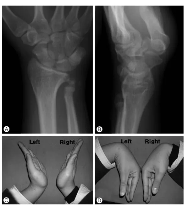
Fig. 4. Radiographs three years after the operation. (A) Posteroanterior radiograph. (B) Lateral radiograph. Both radiographs show a growth disturbance of the distal ulnar epiphyseal plate and ulnar-minus variance. (C, D) Active ranges of motion of the wrist were full, and the patient had no complaints.
Fig. 5. Radiographs at initial visit. (A) Posteroanterior radi- ograph of the left wrist. (B) Lateral radiograph. Both radiographs show a fracture of the distal radius at junc- tion of the middle and distal thirds with Salter -Harris Type II fracture of the distal ulna.
Fig. 7. Radiographs one year after the operation. ( A) Posteroanterior radiograph. (B) Lateral radiograph. The epiphyseal line of the distal ulna looks closed as well as in the epiphyseal line of the distal radius. The distal radio-ulnar joint was well preserved.
Fig. 6. (A) Intraoperative photograph. Show interposition of periosteum (arrow) between the fragments. (B, C) Post-operative radi- ographs. The epiphysis of the distal ulna was fixed with small K-wires.
참고문헌
01) Hanel DP, Scheid DK. Irreducible fracture-dislocation of the distal radioulnar joint secondary to entrapment of the extensor carpi ulnaris tendon. Clin Orthop Relat Res.
1988;234:56-60.
02) Imatani J, Hashizume H, Nishida K, Morito Y, Inoue H.
The Galeazzi-equivalent lesion in children revisited. J
07) Karlsson J, Appelqvist R. Irreducible fracture of the wrist in a child. A case report. Act Orthop. Scand. 1987;58:280-1.
08) Evans DL, Stauber M, Frykman GK. Irreducible epiphy- seal plate fracture of the distal ulna due to interposition of the extensor carpi ulnais tendon. A case report. Clin Orthop Relat Res 1990;251:162-5.
09) Nelson OA, Buchanan JR, Harrison CS. Distal ulnar growth arrest. J Hand Surg Am. 1984;9(2):164-70