Journal of Bacteriology and Virology 2006. Vol. 36, No. 2 p.51 – 57
Stabilizing Microtubular Network Facilitates the Intracellular Growth of Orientia tsutsugamushi
Mee-Kyung Kim2, Mi-Jeong Kim, Byung-Uk Lim and Jae-Seung Kang1* Department of Microbiology, Inha University College of Medicine,
2Inha Research Institute for Medical Sciences, 1Incheon 400-712, Republic of Korea
Received : March 20, 2006 Accepted : June 13, 2006
Microtubule network provides many intracellular microbes with an efficient way to move within host cells. Orientia tsutsugamushi move from the cell periphery to the microtubule organizing center (MTOC) by dynein-dependent mechanism. In this study, we investigated the role of microtubule on the growth of O. tsutsugamushi. The treatment of infected cells with taxol as well as daunomycin enhanced the bacterial growth in contrast to colchicine. Immunofluorescent (IF) staining of taxol-treated cells exhibited that O. tsutsugamushi clustered tightly near the nucleus with thick bundles of microtubules, whereas dispersed in the cytoplasm in colchicine-treated cells. These results suggest that microtubule network facilitate the growth of O. tsutsugamushi.
Key Words: Orientia tsutsugamushi, Microtubule, Taxol, Daunomycin, Colchicine
INTRODUCTION
Orientia tsutsugamushi, a causative agent of scrub typhus, belongs to a separate genus in the family Rickettsiaceae (28), and is an endemic pathogen of many Asian-Pacific countries, including Korea (2,21). This bacterium exhibits several unique biological features, such as lack of lipopoly- saccharide and peptidoglycan (1), tendency to stick to cell components (33), and the distinct ultrastructure of cell wall that differs significantly with that of its closest relatives, typhus and spotted fever group rickettsiae (24). Moreover, it leaves host cells by budding, which resembles virus (32).
Endothelial cells are one of the major target cells of O.
tsutsugamushi infection (17). Although the cellular receptor and its bacterial ligand still remained unknown (9,21), O.
tsutsugamushi induces phagocytosis for the invasion into cells (27). After entering host cells, it escapes the primary
phagosome into cytosol of host cells, and then moves along microtubules to the MTOC (14), where it replicates forming an aggregate near the nucleus. This movement is mediated dynein-dependent mechanism (14).
Microtubule network provides some intracellular bacteria as well as viruses with an efficient way to move inside host cells. Thus, intracellular pathogens could reach their site of replication or escape host cells to reach their new host. In addition, there are several reports that some cytotoxic che- micals with microtubule-modifying action affect the growth of intracellular microorganisms (15,20,26). Therefore, we investigated the role of microtubules on the growth of O.
tsutsugamushi. To address this issue, we treated infected cells with microtubule-modifying agents and examined the effect of these drugs on the growth of O. tsutsugamushi.
Our results suggest that microtubule network facilitate the growth of O. tsutsugamushi.
Abbreviations: Microtubule organizing center, MTOC;
Immunofluorescent, IF; fetal bovine serum, FBS; mono- clonal antibody, MAb; fluorescein isothiocyanate, FITC.
51
*Corresponding author: Jae-Seung Kang. Department of Microbiology, Inha University College of Medicine, Jungsuk B/D, 3rd street, Shinheung- Dong, Choong-Gu, Inchon 400-712, Republic of Korea.
Phone: 82-32-860-0952, Fax: 82-32-881-8559 e-mail: jaeskang@inha.ac.kr
MATERIALS AND METHODS 1. O. tsutsugamushi
The Boryong serotype of O. tsutsugamushi (2) was culti- vated in ECV304 cells, as described previously (12). The infected cells were cultivated until heavily infected cells detached from culture substrates. The bacterial inoculum was prepared by harvesting attached cells and subsequently disrupted with glass beads (diameter 1.0 mm). After centri- fugation at 1,500 rpm for 5 min, the supernatant containing O. tsutsugamushi was used for the infection of ECV304 cells.
2. Cell culture and infection of O. tsutsugamushi A subconfluent monolayer of ECV304 cells were grown on 13 mm coverslips placed into 24-well culture plates (Nunc, Denmark) at an initial cell concentration of 1~2 × 10⁵ cells per well. The cells were cultivated in M199 me- dium (Gibco BRL, Gaithersburg, USA) supplemented with 10% fetal bovine serum (FBS, Gibco BRL) and incubated at 37℃ in 5% CO2. This subconfluent monolayer of cells was infected with O. tsutsugamushi and incubated at 37℃
in 5% CO2-enriched environment for 3 h, allowing sufficient time for the pathogen to establish attachment and entry into the host cells. At the end of the initial incubation period, infected cells were washed with phosphate buffered saline (PBS) and then fresh medium was added to cells.
3. Treatment of cells with chemicals
Subconfluent cultures of ECV304 cells were prepared before 1 day in M199 containing 10% FBS. After O. tsutsu- gamushi infection, culture medium were switched to M199 containing 2% FBS with different concentration of taxol or colchicines, and re-incubated at 37℃ in 5% CO2 for 3 days before the fixation for IF staining. The working concentra- tion of each these chemicals were selected based on their effect on the cellular proliferation.
4. Antibodies
The methods for the production of monoclonal antibodies (MAbs) were as described previously (11). MAb FS15 is reactive against 56-kDa of O. tsutsugamushi (22) and MAb M686-8 is reactive against 47-kDa of O. tsutsugamushi (13).
For IF staining, FS15 was conjugated with fluorescein iso- thiocyanate (FITC) by using NHS-fluorescein (Pierce, Rock- ford) according to the guidelines specified by the manu- facturer. MAbs against α-tubulin (Clone No. DM1a, Sigma Chemical Co., St. Louis, USA) and acetylated tubulin (Sigma Chemical Co.) were used for IF staining and immu- noblot experiments. Monoclonal anti-β-actin (Sigma Che- mical Co.) was used for Western blotting. For IF staining, goat anti-mouse immunoglobulin G (Jackson Immuno- Research Lab., West Grove, USA) conjugated to rhodamine was used with a dilution of 1:200 as a secondary antibody.
5. IF staining and confocal laser scanning microscopy For the simultaneous observation of O. tsutsugamushi and microtubule, IF staining was performed using a MAb (FS15), which had been conjugated with FITC, and anti-α- tubulin. The secondary antibody was goat anti-mouse immu- noglobulin G (Jackson ImmunoResearch Laboratories) con- jugated to Rhodamine. The images were taken using a con- focal laser scanning system equipped with an Argon, green He-Ne, and blue laser diode mixed gas laser (Bio-Rad, Hercules, Ca.). The corresponding images were processed digitally using LaserSharp 2000 version 4.1 software (Bio- Rad).
6. Quantification of O. tsutsugamushi by counting bacterial particles
The growth of O. tsutsugamushi was determined as pre- viously described with minor modifications (29). For coun- ting particles of O. tsutsugamushi, ECV304 cells were in- fected and cultivated on a 24-well culture plate. After 3 days, infected cells were harvested and disrupted with glass beads, and then the resulting cell lysate was added to new ECV- 304 cells. After 3 h incubation, to allow the attachment of O. tsutsugamushi on the cell surface, the bacteria were stained with IF staining and photographs were taken. Quan- tification of the number of O. tsutsugamushi and cells was performed by the examination on photographs. The growth index was calculated by dividing total number of bacterial particles with total number of cells.
7. Western blot analysis
Cells were solubilized in lysis buffer (1% Triton X-100, 0.5% sodium-deoxycholate, 500 mM NaCl supplemented
with 1 mM Na3VO4, 1 mM NaF, 1 mM iodoacetamide, 1 mM phenylmethylsulfonyl fluoride, 1 mM aprotinin, 1 mM leupeptin). From each experiment, 25 µg of extracted pro- tein were separated on an 11% SDS-polyacrylamide gel electrophoresis. The resolved proteins were transferred elec- trophoretically to Immobilon-P polyvinylidene difluoride (PVDF)-membrane. The membranes were further incubated with primary antibodies, MAb M686-8. The membrane was incubated again with horseradish peroxidase-conjugated rabbit anti-mouse immunoglobulin G (Jackson Immuno- Research Lab. U.S.A.), and the signals were visualized using enhance chemiluminescence by Amersham ECL kit (Amer- sham, UK) according to the manufacturer's instructions.
RESULTS
1. Effect of microtubule-modifying agents on the gro- wth of O. tsutsugamushi
To address whether the microtubule structure could affect the growth of O. tsutsugamushi, we examined the effects of two kinds of microtubule-modifying agents: taxol and daunomycin are microtubule-stabilizing agents due to pre- vention of MT disassembly, and colchicine is a microtubule- destabilizing agent due to enhance of MT depolymerization.
For the evaluation of the effects of these agents on bacterial growth, we first infected ECV304 cells with O. tsutsuga- mushi and then cultured for 3 days in presence of each chemicals. As the total cell number was decrease conside- rably by the cytotoxic action of each chemical, total growth of bacteria could not be determined. Therefore, we counted the total the bacterial particles in the surviving cells, which were affected by microtubule-modifying chemicals. The bacterial growth in the surviving taxol-treated cells was much higher than in colchicine-treated cells (Fig. 1). Dau- nomycin, which is known to enhance the growth of O.
tsutsugamushi (8), also stimulated bacterial growth.
To confirm the growth-stimulating effect of taxol, we performed immunoblot analysis of 47-kDa protein of O.
tsutsugamushi to estimate the bacterial growth. Taxol treat- ment resulted in the increase of 47-kDa protein in immuno- blot (Fig. 2A). In contrast, colchicine treatment resulted in the decrease of 47-kDa protein in the cells treated with 5 ng/ml or higher concentrations of colchicine. Consistent with this result, the IF staining of infected cells, in which
tubulin and bacteria were stained simultaneously, demon- strated that large number of O. tsutsugamushi clustered tightly near a nucleus along with the stiffened and thick bundles of microtubules. Furthermore this effect was seemed to be dependent on the dose of taxol (Fig. 2B). In contrast, in the colchicine-treated cells, O. tsutsugamushi showed a dispersed pattern in the cytoplasm and they were elongated and enlarged as compared with that in taxol-treated cells.
This effect became evident in cells treated with 1 ng/ml or higher concentration of colchicine, which disintegrated mi- crotubular network.
2. Analysis of acetylated tubulin
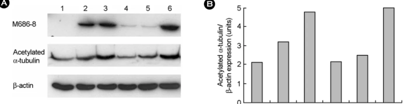
Next, we analyzed the amount of acetylated tubulin, which is known to increase the stabilized microtubules (19), and actin in cells treated with microtubule-active drugs. The data on immunoblot analysis demonstrated that treatment of cells with taxol and daunomycin increased levels of acetylated α-tubulin, while the β-actin levels remained the same as those of the control cells (Fig. 3).
DISCUSSION
We have observed opposite effects of taxol and colchicine on bacterial growth. Although these chemicals are cytotoxic, Figure 1. The effect of microtubule-modifying agents on the growth of O. tsutsugamushi. ECV304 cells were grown to subcon- fluence on 24-well culture plates. After infection, cells were incu- bated further for 3 days in the presence of 50 nM taxol (Tax), 10 ng/ml colchicine (Col), 40 ng/ml daunomycin (Dau) and without agent (C). The number of viable cells was counted using trypan blue exclusion assay. Growth index was calculated as described in Materials and Methods. Representative result of three independent experiments is shown.
which is evident in our experiment. However, there were changes in the microtubules as shown in IF staining of cells and the growth of O. tsutsugamushi was still observed in the surviving cells. Therefore, we could measure the effect
of these chemicals on the growth of O. tsutsugamushi. The differential effect was evident in immunoblot analysis as well as O. tsutsugamushi counts using IF staining. As it is well known that taxol binds to and stabilizes microtubules
1 2 3 4 5 6 7
A
0.25 2.5 25 250 (nM)
Taxol
Colchicine
0.1 1 10 100 (ng/ml)
B
Figure 2. The stimulating effect of taxol on the growth of O. tsutsugamushi. (A) The infected cells treated with taxol or colchicine for 3 days were solubilized in lysis buffer. The lysates (25 µg) was separated on an 11% SDS-PAGE and was further analyzed with the immunoblot analysis using M686-8 for the estimation of bacterial growth. Lane 1, untreated control; lane 2, 2.5 nM taxol; lane 3, 25 nM taxol; lane 4, 100 nM taxol; lane 5, 5 ng/ml colchicine; lane 6, 10 ng/ml colchicine; lane 7, 100 ng/ml colchicine. (B) The cells treated with the indicated concentration of taxol or colchicine were stained using an anti-α-tubulin MAb and FS15 to stain tubulin and O. tsutsugamushi simultaneously. Magnification, ×200.
A B
Figure 3. Immunoblot analysis of acetylated tubulin after treatment of cells with chemicals. The infected cells treated with indicated chemicals were solubilized in lysis buffer. (A) 25 µg of lysate was separated on 11% SDS-PAGE and was analyzed with M686-8 for bac- terial protein, anti-acetylated α-tubulin, and anti-β-actin. Lane 1, uninfected ECV304 cells; lane 2, infected cells without treatment; lane 3, 50 nM taxol; lane 4, 10 ng/ml colchicine; lane 5, 5 µM nocodazole; lane 6, 40 ng/ml daunomycin. (B) The density of each band was quantified using Quantaty One software and amount of acetylated tubulin was normalized with the density of β-actin.
by inhibiting microtubule dynamics and colchicine acts in opposite way, we hypothesized that the stabilization or de- stabilization of microtubules may play a specific role in the growth of O. tsutsugamushi. Consistent with this result, the IF staining of taxol-treated infected cells showed the large cluster of O. tsutsugamushi with the stiffened and thick bundles of microtubules, in contrast to colchicine-treated cells (Fig. 2B).
To confirm that microtubule stabilization enhances the growth of O. tsutsugamushi, we examined the acetylation of tubulin, which is known to occur on microtubules stabili- zed by normal cellular mechanisms (19). The data on wes- tern blot analysis has clearly demonstrated that the treat- ment of cells with taxol and daunomycin increased levels of acetylated α-tubulin, while the levels of β-actin remained the same as those of control cells. Therefore, it seems evi- dent that the treatment of taxol and daunomycin resulted in microtubule stabilization. Considering the enhanced growth of O. tsutsugamushi in cells treated with these chemicals, our results implicate that the growth enhancement of O.
tsutsugamushi may be correlated with microtubule stabili- zation.
Daunomycin has been reported earlier to stimulate the growth of O. tsutsugamushi (8). However, the mechanism of action remains unknown. One possible explanation is that the growth-stimulating effect of daunomycin is due to its microtubule-stabilizing action. Daunomycin has complex mechanisms for its action. It can intercalate between base pairs of DNA, produce single- and double-stranded DNA scission, and impair DNA repair. Moreover, it has been reported to influence on the microtubule and induce its stabilization (16,18). Consistent with our explanation, dau- nomycin increased the levels of acetylated α-tubulin (Fig.
3). Therefore, we suggest that daunomycin stimulate the growth of O. tsutsugamushi by its microtubule-stabilizing action.
Microtubules are the major component of cytoskeletal systems that are responsible for the regulation of distribution of cytoplasmic organelles. Microtubule-dependent clustering of mitochondria around the nucleus has been observed in various cells under experimental conditions (25). Further- more, microtubules regulate the distribution of various org- anelles, namely, endoplasmic reticulum (30), Golgi appar- atus (31), and lysosomes (4). They also provide tracks for
the movement of various vesicles and proteins. Conside- ring the previous report that O. tsutsugamushi moves to MTOC by microtubule- and dynein-mediated movement (14), our results suggest that the proper location in cyto- plasm by intact microtubules is required for the growth of O. tsutsugamushi.
Although host cells usually are considered to provide a suitable shelter and supply important nutrients for intracell- ular bacteria, cell cytoplasm may be in fact a hostile envi- ronment for intracellular parasites or bacteria (10). When, for example, extracellular bacteria are injected directly into the cytoplasm of cells, they are unable to grow. On the other hand, intracellular bacteria use specific mechanisms that enable their growth in the cytoplasm of cells (5). Thus, it seems likely that even the obligate intracellular bacterium, O. tsutsugamushi, requires the proper location in the cyto- plasm for its survival.
It is also notable that host cell microfilaments and micro- tubules are known to play a critical role in the life cycles of several pathogenic intracellular microbes by providing successful invasion and promoting movement of the patho- gen. In the case of Chlamydia trachomatis serovar L2, it was demonstrated earlier that the disruption of microfila- ments as well as microtubule with colchicine or nocodazole results in the formation of multiple small inclusions instead of one large inclusion (23). In this system, microtubules are required for apposition of Golgi vesicles to chlamydial in- clusions for supply of nutrients and membranes; disruption of microtubules inhibits this pathway, leading to a decrease in fusion of inclusion bodies (23). In other studies on C.
trachomatis, bacteria require a perinuclear location for effec- tive growth for disrupting the normal Golgi trafficking and intercepting endogenously synthesized shingomyelin, nor- mally destined for the plasma membrane (3,6,7). Therefore, one hypothesis explaining our results is that the main pur- pose of location of O. tsutsugamushi near MTOC may be an uptake of nutrient.
In summary, we have shown that taxol and daunomycin stimulated the growth of O. tsutsugamushi, suggesting mi- crotubular network stability is necessary for its intracellular life cycle.
Acknowledgements
This study was supported by a grant from Inha University.
We also thank Sun-Ho Kee of the Korea University for critical review of the manuscript.
REFERENCES
1) Amano K, Tamura A, Ohashi N, Urakami H, Kaya S, Fukushi K: Deficiency of peptidoglycan and lipo- polysaccharide components in Rickettsia tsutsugamushi.
Infect Immun 55: 2290-2292, 1987.
2) Chang WH, Kang JS, Lee WK, Choi MS, Lee JH:
Serological classification by monoclonal antibodies of Rickettsia tsutsugamushi isolated in Korea. J Clin Microbiol 28: 685-688, 1990.
3) Clausen JD, Christiansen G, Holst HU, Birkelund S: Chlamydia trachomatis utilizes the host cell micro- tubule network during early events of infection. Mol Microbiol 25: 441-449, 1997.
4) Collot M, Louvard D, Singer SJ: Lysosomes are asso- ciated with microtubules and not with intermediate filaments in cultured fibroblasts. Proc Natl Acad Sci USA 81: 788-792, 1984.
5) Goetz M, Bubert A, Wang G, Chico-Calero I, Vazquez-Boland JA, Beck M, Slaghuis J, Szalay AA, Goebel W: Microinjection and growth of bacteria in the cytosol of mammalian host cells. Proc Natl Acad Sci USA 98: 12221-12226, 2001.
6) Hackstadt T, Rockey DD, Heinzen RA, Scidmore MA: Chlamydia trachomatis interrupts an exocytic pathway to acquire endogenously synthesized sphin- gomyelin in transit from the Golgi apparatus to the plasma membrane. EMBO J 15: 964-977, 1996.
7) Hackstadt T, Scidmore MA, Rockey DD: Lipid meta- bolism in Chlamydia trachomatis-infected cells: direc- ted trafficking of Golgi-derived sphingolipids to the chlamydial inclusion. Proc Natl Acad Sci USA 92:
4877-4881, 1995.
8) Hanson B: Factors influencing Rickettsia tsutsugamushi infection of cultured cells. Am J Trop Med Hyg 36:
621-630, 1987.
9) Inn KS, Han SH, Kim HR, Huh MS, Seong SY, Kang JS, Han TH, Kim IS, Choi MS: Cellular in- vasion of Orientia tsutsugamushi requires initial inter- action with cell surface heparan sulfate. Microb Pathog 28: 227-233, 2000.
10) Jeon KW: The large, free-living amoebae: wonderful cells for biological studies. J Eukaryot Microbiol 42:
1-7, 1995.
11) Kang JS Chang WH: Antigenic relationship among the eight prototype and new serotype strains of Ori- entia tsutsugamushi revealed by monoclonal antibo- dies. Microbiol Immunol 43: 229-234, 1999.
12) Kim MK, Kee SH, Cho KA, Chung MH, Lim BU, Chang WH, Kang JS: Apoptosis of endothelial cell line ECV304 persistently infected with Orientia tsutsu- gamushi. Microbiol Immunol 43: 751-757, 1999.
13) Kim MK, Odgerel Z, Chung MH, Lim BU, Kang JS: Characterization of monoclonal antibody reacting exclusively against intracellular Orientia tsutsugamushi.
Microbiol Immunol 46: 733-740, 2002.
14) Kim SW, Ihn KS, Han SH, Seong SY, Kim IS, Choi MS: Microtubule and dynein-mediated movement of Orientia tsutsugamushi to the microtubule organizing center. Infect Immun 69: 494-500, 2001.
15) Meyer DH, Rose JE, Lippmann JE, Fives-Taylor PM: Microtubules are associated with intracellular movement and spread of the periodontopathogen Acti- nobacillus actinomycetemcomitans. Infect Immun 67:
6518-6525, 1999.
16) Molinari A, Calcabrini A, Crateri P, Arancia G:
Effects of daunomycin on the microtubular network: a cytochemical study on a human melanoma cell line.
Eur J Cell Biol 54: 291-298, 1991.
17) Moron CG, Popov VL, Feng HM, Wear D, Walker DH: Identification of the target cell of Orientia tsutsu- gamushi in human cases of scrub typhus. Mod Pathol 14: 752-759, 2001.
18) Na C, Timasheff SN: Physical-chemical study of daunomycin-tubulin interactions. Arch Biochem Bio- phys 182: 147-154, 1977.
19) Piperno G, LeDize M, Chang X: Microtubules con- taining acetylated α-tubulin in mammalian cells in culture. J Cell Biol 104: 289-302, 1987.
20) Rikihisa Y, Zhang Y, Park J: Inhibition of infection of macrophages with Ehrlichia risticii by cytochalasins, monodansylcadaverine, and taxol. Infect Immun 62:
5126-5132, 1994.
21) Seong SY, Choi MS, Kim IS: Orientia tsutsugamushi infection: overview and immune responses. Microbes
Infect 3: 11-21, 2001.
22) Seong SY, Kim MK, Lee SM, Odgerel Z, Choi MS, Han TH, Kim IS, Kang JS, Lim BU: Neutralization epitopes on the antigenic domain II of the Orientia tsutsugamushi 56-kDa protein revealed by monoclonal antibodies. Vaccine 19: 2-9, 2000.
23) Schramm N, Wyrick PB: Cytoskeletal requirements in Chlamydia trachomatis infection of host cells. Infect Immun 63: 324-332, 1995.
24) Silverman DJ, Wisseman Jr CL: Comparative ultra- structural study on the cell envelopes of Rickettsia prowazekii, Rickettsia rickettsii, and Rickettsia tsutsu- gamushi. Infect Immun 21: 1020-1023, 1978.
25) Smirnova E, Shurland DL, Ryazantsev SN, van der Bliek AM: A human dynamin-related protein controls the distribution of mitochondria. J Cell Biol 143: 351 -358, 1998.
26) Suikkanen S, Aaltonen T, Nevalainen M, Valilehto O, Lindholm L, Vuento M, Vihinen-Ranta M: Explo- itation of microtubule cytoskeleton and dynein during parvoviral traffic toward the nucleus. J Virol 77: 10270 -10279, 2003.
27) Tamura A: Invasion and intracellular growth of Ricke- ttsia tsutsugamushi. Microbiol Sci 5: 228-232, 1988.
28) Tamura A, Ohashi N, Urakami H, Miyamura S:
Classification of Rickettsia tsutsugamushi in a new genus, Orientia gen. nov., as Orientia tsutsugamushi comb. nov. Int J Syst Bacteriol 45: 589-591, 1995.
29) Tamura A, Urakami H: Easy method for infectivity titration of Rickettsia tsutsugamushi by infected cell counting. Jpn J Bacteriol 36: 783-785, 1981.
30) Terasaki M, Reese TS: Interactions among endopla smic reticulum, microtubules, and retrograde move- ments of the cell surface. Cell Motil Cytoskeleton 29:
291-300, 1994.
31) Thyberg J, Moskalewski S: Microtubules and the organization of the Golgi complex. Exp Cell Res 159:
1-16, 1985.
32) Urakami H, Tsuruhara T, Tamura A: Electron micro- scopic studies on intracellular multiplication of Ricke- ttsia tsutsugamushi in L cells. Microbiol Immunol 28:
1191-1201, 1984.
33) Winkler HH: Rickettsia species (as organisms). Annu Rev Microbiol 44: 131-153, 1990.