Background and Purpose Cutaneous nerve biopsies based on two-dimensional analysis have been regarded as a creditable assessment tool for diagnosing peripheral neuropathies.
However, advancements in methodological imaging are required for the analysis of intact struc- tures of peripheral nerve fibers. A tissue-clearing and labeling technique facilitates three-di- mensional imaging of internal structures in unsectioned, whole biological tissues without ex- cessive time or labor costs. We sought to establish whether a tissue-clearing and labeling technique could be used for the diagnostic evaluation of peripheral neuropathies.
Methods Five healthy individuals and four patients with small-fiber neuropathy (SFN) and postherpetic neuralgia (PHN) were prospectively enrolled. The conventional methods of indi- rect immunofluorescence (IF) and bright-field immunohistochemistry (IHC) were adopted in addition to the tissue-clearing and labeling method called active clarity technique-pressure re- lated efficient and stable transfer of macromolecules into organs (ACT-PRESTO) to quantify the intraepidermal nerve-fiber density (IENFD).
Results The mean IENFD values obtained by IF, bright-field IHC, and ACT-PRESTO in the healthy control group were 6.54, 6.44, and 90.19 fibers/mm2, respectively; the corresponding values in the patients with SFN were 1.99, 2.32, and 48.12 fibers/mm2, respectively, and 3.06, 2.87, and 47.21 fibers/mm2, respectively, in the patients with PHN.
Conclusions This study has shown that a tissue-clearing method provided not only rapid and highly reproducible three-dimensional images of cutaneous nerve fibers but also yielded reliable quantitative IENFD data. Quantification of the IENFD using a tissue-clearing and labeling tech- nique is a promising way to improve conventional cutaneous nerve biopsies.
Key Words cutaneous nerve biopsy, peripheral neuropathy, tissue-clearing and labeling technique, ACT-PRESTO, intraepidermal nerve-fiber density.
Tissue-Clearing Technique and Cutaneous Nerve Biopsies:
Quantification of the Intraepidermal Nerve-Fiber Density Using Active Clarity Technique-Pressure Related Efficient and Stable Transfer of Macromolecules Into Organs
INTRODUCTION
Cutaneous nerve biopsies have come to be considered one of the few reliable tools for eval- uating peripheral neuropathies, including small-fiber neuropathy (SFN).1-4 These conven- tional methods are confined to two-dimensional analyses, based on the XY plane of thin tis- sue sections of skin,1,2,5 which may fail to accurately quantify three-dimensionally arborized nerve fibers. Thus, alternative methods that take into account the entire, unsectioned nerve morphology would allow more-accurate quantifications and diagnoses of the relevant pa- thologies.
The recently reported tissue-clearing and labeling technique called active clarity tech- Dai Hyun Kima
Se Jeong Leea Eunsoo Leea Ji Hyuck Hongb Soo Hong Seob Hyo Hyun Ahnb Byung-Jo Kimc Woong Suna,d Im Joo Rhyua,d
a Departments of Anatomy,
b Dermatology, and cNeurology, Korea University College of Medicine, Seoul, Korea
d Division of Brain Korea 21 Plus Program for Biomedical Science, Korea University College of Medicine, Seoul, Korea
pISSN 1738-6586 / eISSN 2005-5013 / J Clin Neurol 2019;15(4):537-544 / https://doi.org/10.3988/jcn.2019.15.4.537
Received May 13, 2019 Revised July 4, 2019 Accepted July 4, 2019 Correspondence Im Joo Rhyu, MD, PhD Department of Anatomy,
Korea University College of Medicine, 73 Goryeodae-ro, Seongbuk-gu, Seoul 02841, Korea
Tel +82-2-2286-1150 Fax +82-2-2286-1387 E-mail irhyu@korea.ac.kr
cc This is an Open Access article distributed under the terms of the Creative Commons Attribution Non-Com- mercial License (https://creativecommons.org/licenses/by-nc/4.0) which permits unrestricted non-commercial use, distribution, and reproduction in any medium, provided the original work is properly cited.
JCN
Open Access ORIGINAL ARTICLEVolume Imaging of Intact Cutaneous Nerve Fibers
JCN
nique-pressure related efficient and stable transfer of macro- molecules into organs (ACT-PRESTO) can clear biological tissues as a whole without damaging the original architec- ture, and can also accelerate antibody penetration into thick specimens by applying pressure.6,7 These characteristics mean that the method could reduce the processing time relative to other tissue-clearing protocols7 and three-dimensional image reconstruction techniques,8,9 which is advantageous for clini- cal implementations as a diagnostic tool.
In the present study we sought to establish the usefulness of ACT-PRESTO in the three-dimensional quantification of intraepidermal nerve fibers density (IENFD) and in diagnos- ing peripheral neuropathies.
METHODS
The present data were obtained from a prospective clinical study evaluating the diagnostic value of the ACT-PRESTO skin-clearing and labeling technique for assessing peripheral neuropathies. The institutional review board of Korea Univer- sity Medical Center (KUMC), Anam Hospital approved the clinical trial (approval no. 2016AN2056).
Participants
Healthy participants and neuropathy patients who were ex- pected to show a reduced IENFD including SFN or posther- petic neuralgia (PHN) were recruited for this study. SFN was defined according to its indicative clinical symptoms, includ- ing distal dominant paresthesia despite normal results in a neurological examination, nerve conduction study (NCS), and electromyography (EMG). PHN was determined according to a medical history of herpes zoster infection and subsequent paresthesia appearing in the compromised area. All partici- pants were fully informed about the possible risks before the study. A neurologist and dermatologists working at KUMC performed the clinical and/or neurological examinations, as well as the NCS and EMG.
Cutaneous nerve biopsies
Cutaneous nerve biopsies were performed with a commonly used 3-mm punch device under local anesthesia with lido- caine, as reported previously.10,11 Two separate skin samples were collected from the distal leg of healthy participants and patients with SFN. For patients with PHN, cutaneous biopsy samples were obtained in the area where the neuropathy symp- toms were most prominent. One pair of the biopsied tissues was cryosectioned and immunostained for protein gene prod- uct 9.5 (PGP 9.5; CEDARLANE, Burlington, NC, USA) and/
or collagen type IV antibodies using two conventional meth- ods: bright-field immunohistochemistry (IHC) and indirect immunofluorescence (IF). The other pair of biopsied tissues was processed using ACT-PRESTO followed by PGP 9.5 im- munolabeling.
Conventional two-dimensional assessments
Biopsy specimens were fixed in Zamboni’s solution (2% para- formaldehyde and picric acid) for two-dimensional assess- ments. The fixed samples were cryosectioned at thicknesses of 50 and 80 µm for IHC and IF, respectively. The first few and last few sections were discarded owing to potential arti- facts.1,2 Three randomly selected sections of each thickness were used for subsequent immunostaining. The 50-µm-thick sections were immunolabeled with PGP 9.5 (1:2,000) using 3,3'-diaminobenzidine staining for IHC. The immunolabeled sections were imaged using bright-field microscopy (BX51, Olympus, Tokyo, Japan). The 80-µm-thick sections were im- munostained with PGP 9.5 (1:800) and type IV collagen (1:800;
Sigma-Aldrich, St. Louis, MO, USA) using indirect IF. We used confocal microscopy (LSM 700, Carl Zeiss, Jena, Ger- many) for image acquisition, and serial optical sections taken at 1-μm intervals were reconstructed using the related soft- ware (ZEN, Carl Zeiss). The maximum projection image de- rived from the stacked results was used to quantify cutaneous nerve fibers. The guidelines of the European Federation of Neurological Societies (EFNS) were adopted for quantifica-
Day 0 Skin biopsy
3-mm punch biopsy
1.0 M
NaCl Zamboni’s
solution Immersion in
RIMS Confocal
microscopy c-PRESTO
1) Hydrogel monomer embedding 2) Polymerization 3) ETC
Tissue
clearing RI
matching Immunolabeling
SSST Fixation Imaging
Day 1 Days 1–2 Days 2–3
Cutaneous ACT-PRESTO
Days 3–4 Days 3–4 Days 4–5
Fig. 1. Summary of the processing steps of the cutaneous ACT-PRESTO protocol, from separating the epidermis from biopsied skin samples to im- aging with confocal microscopy. ACT-PRESTO: active clarity technique-pressure related efficient and stable transfer of macromolecules into organs, c-PRESTO: centrifugal-PRESTO, ETC: electrophoretic tissue clearing, RI: reflective index, RIMS: reflective-index matching solution, SSST: salt split skin test.
Kim DH et al.
JCN
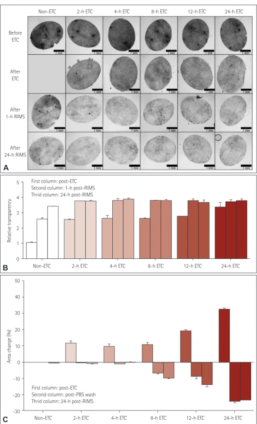
Fig. 2. Serial changes in transparency and area during the cutaneous ACT-PRESTO process, focusing on ETC and immersion in RIMS. A: Morphologi- cal changes in transparency according to the duration of ETC. B: Quantitative alterations of the relative transparency according to the duration of ETC. The improvement in relative transparency was proportional to the duration of ETC, but the differences were standardized after immersion in RIMS. C: Extent of area change related to the duration of ETC. The skin specimens consistently expanded after ETC in proportion to the duration of ETC.
The enlarged tissues returned to their original size after washing with PBS. However, ETC durations exceeding 4 h resulted in irreversible shrinkage of the original tissues to an extent proportional to the ETC duration. ACT-PRESTO: active clarity technique-pressure related efficient and stable trans- fer of macromolecules into organs, ETC: electrophoretic tissue clearing, PBS: phosphate-buffered saline, RIMS: reflective-index matching solution.
Non-ETC
Before ETC
2-h ETC 4-h ETC 8-h ETC 12-h ETC 24-h ETC
After ETC
After 1-h RIMS
After 24-h RIMS
A
5 4 3 2 1
0
Non-ETC 2-h ETC 4-h ETC 8-h ETC 12-h ETC 24-h ETC
First column: post-ETC Second column: 1-h post-RIMS Thrid column: 24-h post-RIMS
Relative transparency
B
50 40 30 20 10 0 -10 -20 -30
Area change (%)
Non-ETC 2-h ETC 4-h ETC 8-h ETC 12-h ETC 24-h ETC
First column: post-ETC Second column: post-PBS wash Thrid column: 24-h post-RIMS
C
Volume Imaging of Intact Cutaneous Nerve Fibers
JCN
tion of the IENFD and the unit of results were represented as
‘IENFs/mm’.1,2
Three-dimensional evaluations using ACT-PRESTO The remaining biopsied skin samples were processed accord- ing to the cutaneous ACT-PRESTO protocol (Fig. 1), which was registered as a patent in South Korea in August 2018 (patent number: 10-1888782). The epidermis was separated from the dermis using the salt split skin test12 and was fixed in Zamboni’s solution. The ACT tissue-clearing method es- sentially comprised three stages: hydrogel monomer embed- ding, polymerization, and electrophoretic tissue clearing (ETC). More specifically, the epidermis was immersed in a 4%
acylamide solution mixed with the thermal initiator 2,2'-azobis [2-(2-imidazolin-2-yl) propane] dihydrochloride (Wako Pure Chemical, Osaka, Japan) overnight at 4°C. The hydrogel monomer infused into the tissue was polymerized at -70 kPa and 37°C in a vacuum chamber. The polymerized sample was then transported to an ETC chamber filled with 4% sodium dodecyl sulfate in a solution of 200 mM boric acid in H2O (pH 8.5). ETC proceeded for 4 h under static conditions, in- cluding 1.5 A and 37°C; precise conditions are required for ETC to ensure effective tissue-clearing and prevent signifi- cant morphological changes that could interfere with IENFD quantification (Fig. 2). The cleared epidermis was immuno- labeled with PGP 9.5 (1:400) aided by centrifugal force; this process is thus called c-PRESTO (Fig. 3). The cleared skin was immersed in a solution with PGP 9.5 or Cy3 (1:500;
Jackson Immuno Research, West Grove, PA, USA) and cen- trifuged at 800×g for 3 and 2 h to accelerate the penetration of primary and secondary antibodies, respectively. The im- munostained tissue was subsequently immersed in a reflec-
tive-index matching solution (RIMS) to increase the trans- parency.
The immunostained cutaneous nerve fibers were imaged by confocal microscopy (LSM 700) with z-stacking at inter- vals of 1 μm. Twenty images with an overall area of 0.58 mm2 in the XY plane were consecutively imaged and merged by a tile scan, and the reconstructed three-dimensional cutaneous nerve fibers were quantified and unit of the results were repre- sented as ‘IENF/mm’. All of the PGP 9.5-stained nerve fibers were manually counted, and the nerve fibers that clearly arose from the basement of the epidermis were regarded as signifi- cant data. The basal side of the epidermis could be readily verified by its more-compact cellular composition compared with the upper side in the nuclear staining.
Statistical analysis
Statistical differences between the results obtained from con- ventional two-dimensional assessments and the three-dimen- sional evaluations were analyzed using Wilcoxon’s signed- rank test. All analyses were performed with IBM SPSS software (version 20.0 for Windows, IBM Corp., Armonk, NY, USA).
RESULTS
Nine subjects comprising five healthy individuals and four patients with peripheral neuropathies voluntarily participated in the study (Table 1). The sex ratio was 1:0.8, and they were aged 52.56±18.93 years (mean±SD). The patient group com- prised one patient with SFN and three with PHN. The com- promised regions of patients with PHN were confined to the trunk (T4 or T6 dermatome).
The ETC process transformed each opaque skin specimen
Centrifugal force
Target antigen Specific antibody Penetration of antibodies
Centrifugal force
Whole tissue Whole tissue
Sectioned tissue
Free diffusion c-PRESTO
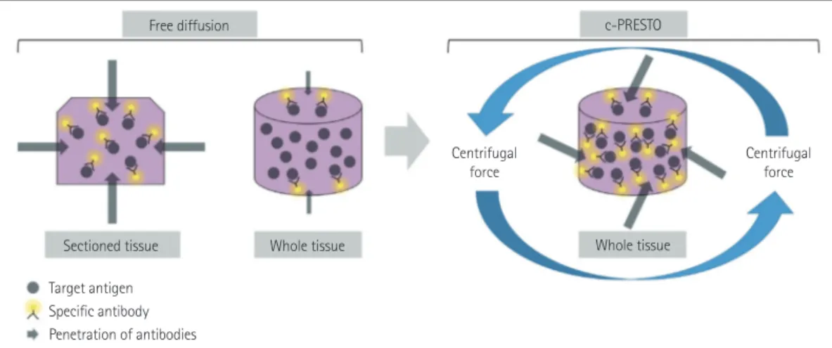
Fig. 3. Schematic diagram of immunohistochemistry for whole-tissue processing after tissue-clearing. The effectiveness of combinations of target antigens and specific antibodies was substantially impeded by the thicker whole tissue compared with conventional sectioned tissue. Tissues for c- PRESTO were centrifuged to obtain sufficient penetration depths of the primary and secondary antibodies, and so the technique could increase the probability of binding between target antigens and specific antibodies. c-PRESTO: centrifugal-pressure related efficient and stable transfer of mac- romolecules into organs.
Kim DH et al.
JCN
into an optically translucent sample, and adjustment of the reflective index with RIMS additionally increased the trans- parency (Fig. 2A, B) without substantially changing any ar- eal dimensions from the original state (Fig. 2C). This pro- cess also made the skin specimen more penetrable by macro- molecules, such as antibodies, through effective lipid removal, without deterioration of the original architecture. Within a few days the ACT-PRESTO process yielded distinctive three- dimensional images of IENFs (Figs. 4E, F, and 5) that differed from conventional two-dimensional images (Figs. 1 and 4A- D) and also provided reliable quantitative data on the IENFD (Table 1).
The IENFD values obtained by IF and IHC in the healthy participant group were 6.54±1.21 and 6.44±1.41 fibers/mm, respectively; the corresponding values for patients with SFN were 1.99±0.76 and 2.32±0.26 fibers/mm, respectively, and those for the patients with PHN were 3.06±2.57 and 2.87±
1.72 fibers/mm, respectively. The three-dimensional evaluation using ACT-PRESTO showed that the IENFD in the healthy groups was 90.19±30.18 fibers/mm2. The mean IENFD for the patients with SFN was 48.12 fibers/mm2, and the IENFD in the patients with PHN was 47.21±28.00 fibers/mm2. There was a significant correlation between the IENFD val- ues quantified using conventional two-dimensional methods and the new three-dimensional approach, with a correlation coefficient of 0.65 for IF (p<0.01) and 0.56 for IHC (p<0.05).
DISCUSSION
Cutaneous nerve biopsies are a credible method for evaluating the loss and regeneration of sensory nerve fibers in patients with peripheral neuropathies.1-5 A cutaneous nerve biopsy with quantification of the IENFD could be used to diagnose SFN or groups of conditions in which small nerve fibers are compromised.4 The presence of PHN after herpes zoster infec-
tion is also reportedly related to a significant loss of IENFs.13,14 In addition, nerve regeneration after multiple types of treat- ment is reportedly associated with neuropathies including di- abetes mellitus and HIV.15,16
The EFNS guidelines for performing cutaneous nerve biop- sies describe a series of procedures, including 3-mm punch biopsies at the distal leg; examination of the linear epider- mal density in at least three sections per biopsy, fixation in Zamboni’s solution, and staining with anti-PGP 9.5 antibod- ies; and imaging by bright-field IHC or IF using confocal mi- croscopy as the diagnostic standard for SFN.1,2
However, this traditional method has two unavoidable lim- itations. First, the original skin biopsy sample must be sec- tioned into thin specimens to ensure the effective penetra- tion of macromolecules and valid microscopic visualization without unwanted light scattering,17-19 and this cutting pro- cess could disturb the intrinsic structural organization. Sec- ond, previous assessments have been restricted to images of tissue sections in the XY plane.1,2,7 Consequently, intact com- plex arborizing patterns of cutaneous nerve fibers have never been evaluated in peripheral neuropathies, and extremely limited aspects of whole structures have been used in conven- tional quantification procedures.
The ability to perform volume imaging of intact cutaneous nerve fibers at high speed and in a consistent manner by us- ing cutaneous ACT-PRESTO could offer a new perspective in the field of diagnostic assessment for sensory nerve dysfunc- tions. Previously reported tissue-clearing methods for vol- ume imaging not only require prolonged times for clearing (typically more than 1–2 weeks), these techniques also have a possible risk of tissue damage owing to unstable tempera- tures, uncontrolled alteration of pH, and impeding of circula- tory currents by air bubbles.7,19 In comparison, the cutaneous ACT-PRESTO technique considerably shortens the duration of the overall procedure to 4 days, including only 4 h of tissue- Table 1. Demographics and quantitative IENFD values of Healthy participants and patients with neuropathy
Participant number Category Age (years) IENFD
IF (fibers/mm) IHC (fibers/mm) Skin clearing (fibers/mm2)
1 Healthy 40–45 4.45±1.99 4.40±0.50 78.74
2 Healthy 36–40 7.13±0.99 6.93±0.48 113.66
3 Healthy 76–80 6.62±2.16 6.71±0.18 63.34
4 Healthy 46–50 6.93±0.67 6.97±1.71 54.99
5 Healthy 20–25 7.55±0.93 7.21±3.64 130.24
6 SFN 36–40 1.99±0.76 2.32±0.26 48.12
7 PHN 66–70 0.87±0.32 1.02±0.18 17.32
8 PHN 70–75 2.42±0.54 4.42±0.67 51.50
9 PHN 60–65 5.89±1.67 3.16±0.46 72.82
Healthy: healthy individual, IENFD: intraepidermal nerve-fiber density, IF: indirect immunofluorescence, IHC: immunohistochemistry, PHN: posther- petic neuralgia, SFN: small-fiber neuropathy.
Volume Imaging of Intact Cutaneous Nerve Fibers
JCN
Fig. 4. Characteristics of the IENFs of a healthy participant (A, C, and E) and a PHN patient (B, D, and F). IENFs were quantified using the conven- tional methods of indirect IF (A and B) and IHC (C and D), as well as the ACT-PRESTO tissue-clearing technique (E and F). Cross-sectional views (A- D) and bird’s-eye views (E and F) of IENFs. IF images (A and B) were obtained using confocal microscopy with a Plan-Apochromat 20×/0.8 M27 lens (maximum projection; stack size, 80 μm; stack step, 1 μm). IHC images (C and D) were produced using bright-field microscopy with a UPlanA- po 40×/0.85 lens. Cutaneous nerve fibers (arrows) and the basal layer of the epidermis (arrowheads) (A-D). Images associated with skin-clearing (E and F) were also obtained using confocal microscopy with a Plan-Apochromat 20×/0.8 M27 lens (stack size, 200 μm; stack step, 1 μm). ACT-PRESTO:
active clarity technique-pressure related efficient and stable transfer of macromolecules into organs, Der: dermis, Ep: epidermis, IENFs: intraepider- mal nerve fibers, IF: immunofluorescence, IHC: immunohistochemistry, PGP 9.5: protein gene product 9.5, PHN: postherpetic neuralgia.
Healthy individual
IFIHCSkin-clearing
PHN
A B
D
F C
E
Ep Ep
Der Der
Der Der
Ep
Ep
PGP 9.5 PGP 9.5
PGP 9.5 / collagen type IV PGP 9.5 / collagen type IV
Kim DH et al.
JCN
clearing, with little damage to the cleared tissue owing to con- trolling the electric current, pH, temperature, and air bubbles.6,7 These improvements in the required time and reliability could facilitate the acquisition of three-dimensional infor- mation on cutaneous nerve fibers in a clinical setting. When using ACT-PRESTO, patients with sensory nerve dysfunc- tion can expect to receive examination results based on vol- ume imaging within 4 days from the time of performing the cutaneous nerve biopsy to obtaining images of intact periph- eral nerve fibers and related quantitative data.
The present study was originally designed to determine whether three-dimensional imaging and related volume in- formation could be utilized to diagnose sensory nerve dys- functions. The obtained results indicated that the quantified IENFD values obtained from volume imaging were also sig- nificantly correlated with the results obtained using the con- ventional gold-standard approach. However, this study was subject to the limitations of relatively small numbers of sub-
jects and the heterogeneous constitution of the included neu- ropathy patients, although both the SFN and PHN patients showed reduced IENFD values.
In our future work we will assess intact three-dimensional structures of cutaneous nerve fibers in more-detailed mor- phological analyses, including of the total length, complexi- ty, and various angles among distinct segments of nerve fi- bers. Such a study involving larger numbers of subjects and detailed analyses of volume information could contribute to elucidating the pathophysiological mechanisms of certain peripheral neuropathies at the subclinical stage, by broaden- ing the understanding of important morphological changes in sensory nerve endings.
Author Contributions
Conceptualization: Byung-Jo Kim, Dai Hyun Kim, Im Joo Rhyu, Woong Sun. Data curation: Dai Hyun Kim, Eunsoo Lee, Se Jeong Lee. Formal analysis: Dai Hyun Kim. Funding acquisition: Hyo Hyun Ahn, Im Joo Rhyu, Woong Sun. Investigation: Byung-Jo Kim, Dai Hyun Kim, Hyo
Healthy individualPHN 80 μm80 μm80 μm80 μmy (μm)y (μm) z (μm)z (μm)z (μm)z (μm)
x (μm)
x (μm) 50 μm
50 μm 50 μm 50 μm
50 μm 50 μm
PGP9.5 / DAPI
x (μm)
x (μm)
x (μm)
A
D
B
E
Fig. 5. Images of IENFs produced by the skin-clearing technique. IENFs of a healthy subject (A-C) and a PHN patient (D-F). Selected areas of three- dimensional images (A and D) in Fig. 4E and F, respectively, were optically sectioned at a thickness of 80 μm (B-F). All images were obtained and processed using confocal microscopy with a UPlanApo 40×/0.85 lens and the related software. Ep: epidermis, IENFs: intraepidermal nerve fibers, PGP 9.5: protein gene product 9.5, PHN: postherpetic neuralgia.
C
F
x (μm)
Volume Imaging of Intact Cutaneous Nerve Fibers
JCN
Hyun Ahn, Ji Hyuck Hong, Soo Hong Seo. Methodology: Byung-Jo Kim, Dai Hyun Kim, Im Joo Rhyu, Woong Sun. Project administration: Byung- Jo Kim, Hyo Hyun Ahn, Im Joo Rhyu, Soo Hong Seo, Woong Sun. Re- sources: Eunsoo Lee, Woong Sun. Supervision: Byung-Jo Kim, Hyo Hyun Ahn, Im Joo Rhyu, Woong Sun. Validation: Byung-Jo Kim, Dai Hyun Kim, Im Joo Rhyu. Visualization: Dai Hyun Kim, Se Jeong Lee. Writing—
original draft: Dai Hyun Kim. Writing—review & editing: Byung-Jo Kim, Dai Hyun Kim, Hyo Hyun Ahn, Im Joo Rhyu, Soo Hong Seo, Woong Sun.
ORCID iDs
Dai Hyun Kim https://orcid.org/0000-0002-3546-2366 Se Jeong Lee https://orcid.org/0000-0002-4437-8270 Eunsoo Lee https://orcid.org/0000-0002-3157-5267 Ji Hyuck Hong https://orcid.org/0000-0003-1301-6995 Soo Hong Seo https://orcid.org/0000-0002-3836-0445 Hyo Hyun Ahn https://orcid.org/0000-0002-1129-5305 Byung-Jo Kim https://orcid.org/0000-0002-0445-7185 Woong Sun https://orcid.org/0000-0003-1792-4894 Im Joo Rhyu https://orcid.org/0000-0002-5558-6278 Conflicts of Interest
The authors have no potential conflicts of interest to disclose.
Acknowledgements
This study was supported by the Brain Research Program through the National Research Foundation (NRF) funded by the Korean Ministry of Science, ICT, & Future Planning (NRF-2015M3C7A1028790) and Korea University Grant K1710481.
REFERENCES
1. Lauria G, Cornblath DR, Johansson O, McArthur JC, Mellgren SI, Nolano M, et al. EFNS guidelines on the use of skin biopsy in the di- agnosis of peripheral neuropathy. Eur J Neurol 2005;12:747-758.
2. Joint Task Force of the EFNS and the PNS. European Federation of Neurological Societies/Peripheral Nerve Society Guideline on the use of skin biopsy in the diagnosis of small fiber neuropathy. Report of a joint task force of the European Federation of Neurological Societies and the Peripheral Nerve Society. J Peripher Nerv Syst 2010;15:79-92.
3. Devigili G, Tugnoli V, Penza P, Camozzi F, Lombardi R, Melli G, et al. The diagnostic criteria for small fibre neuropathy: from symptoms to neuropathology. Brain 2008;131:1912-1925.
4. Mellgren SI, Nolano M, Sommer C. The cutaneous nerve biopsy:
technical aspects, indications, and contribution. Handb Clin Neurol 2013;115:171-188.
5. Lauria G. Small fiber neuropathies. Curr Opin Neurol 2005;18:591- 6. Chung K, Wallace J, Kim SY, Kalyanasundaram S, Andalman AS, Da-597.
vidson TJ, et al. Structural and molecular interrogation of intact bio- logical systems. Nature 2013;497:332-337.
7. Lee E, Choi J, Jo Y, Kim JY, Jang YJ, Lee HM, et al. ACT-PRESTO:
rapid and consistent tissue clearing and labeling method for 3-dimen- sional (3D) imaging. Sci Rep 2016;11:18631.
8. Gong H, Zeng S, Yan C, Lv X, Yang Z, Xu T, et al. Continuously tracing brain-wide long-distance axonal projections in mice at a one-micron voxel resolution. Neuroimage 2013;74:87-98.
9. Li A, Gong H, Zhang B, Wang Q, Yan C, Wu J, et al. Micro-optical sectioning tomography to obtain a high-resolution atlas of the mouse brain. Science 2010;330:1404-1408.
10. Kennedy WR, Wendelschafer-Crabb G. The innervation of human epidermis. J Neurol Sci 1993;115:184-190.
11. McCarthy BG, Hsieh ST, Stocks A, Hauer P, Macko C, Cornblath DR, et al. Cutaneous innervation in sensory neuropathies: evaluation by skin biopsy. Neurology 1995;45:1848-1855.
12. Gammon WR, Briggaman RA, Inman AO 3rd, Queen LL, Wheeler CE. Differentiating anti-lamina lucida and anti-sublamina densa anti- BMZ antibodies by indirect immunofluorescence on 1.0 M sodium chloride-separated skin. J Invest Dermatol 1984;82:139-144.
13. Rowbotham MC, Yosipovitch G, Connolly MK, Finlay D, Forde G, Fields HL. Cutaneous innervation density in the allodynic form of postherpetic neuralgia. Neurobiol Dis 1996;3:205-214.
14. Oaklander AL, Romans K, Horasek S, Stocks A, Hauer P, Meyer RA.
Unilateral postherpetic neuralgia is associated with bilateral sensory neuron damage. Ann Neurol 1998;44:789-795.
15. Boucek P, Havrdova T, Voska L, Lodererova A, He L, Saudek F, et al.
Epidermal innervation in type 1 diabetic patients: a 2.5-year prospec- tive study after simultaneous pancreas/kidney transplantation. Dia- betes Care 2008;31:1611-1612.
16. Schifitto G, Yiannoutsos C, Simpson DM, Adornato BT, Singer EJ, Hol- lander H, et al. Long-term treatment with recombinant nerve growth factor for HIV-associated sensory neuropathy. Neurology 2001;57:1313- 1316.
17. Helmchen F, Denk W. Deep tissue two-photon microscopy. Nat Meth- ods 2005;12:932-940.
18. Tang J, Germain RN, Cui M. Superpenetration optical microscopy by iterative multiphoton adaptive compensation technique. Proc Natl Acad Sci U S A 2012;109:8434-8439.
19. Tomer R, Ye L, Hsueh B, Deisseroth K. Advanced CLARITY for rapid and high-resolution imaging of intact tissues. Nat Protoc 2014;9:1682- 1697.