© 2016 Korean Breast Cancer Society. All rights reserved. http://ejbc.kr | pISSN 1738-6756 eISSN 2092-9900
This is an Open Access article distributed under the terms of the Creative Commons Attribution Non-Commercial License (http://creativecommons.org/
licenses/by-nc/3.0) which permits unrestricted non-commercial use, distribution, and reproduction in any medium, provided the original work is properly cited.
INTRODUCTION
Lymphedema is tissue swelling caused by localized fluid retention. Its natural course is chronic, progressive, and often destructive. It can either be congenital or acquired following trauma or surgery for cancer treatment that includes removal of the lymph nodes and radiotherapy [1].
The International Society of Lymphology classifies the se- verity of lymphedema into three stages. Karri et al. [2] report- ed a modified staging system, classifying the severity of lymphedema into four stages. According to their system, ad- vanced lymphedema is characterized by irreversible skin fi- brosis (stage IIIb) and nonpitting edema, with leather-like skin, skin crypts, and ulcers with or without involvement of the toes (stage IVa and IVb, respectively) [3].
Microvascular techniques such as lymphaticovenous anas- tomosis and vascularized lymph node flap transfer (VLNT) are effective for early stage lymphedema. Interestingly, lipo- suction, first used to treat lymphedema in 1989, has been shown to be an effective method of reducing limb volume and
has good long-term results both cosmetically and functionally, In addition, liposuction has a low rate of complications and does not further disrupt the lymphatic system [4].
In this study, we performed a combined two-stage opera- tion in an advanced lymphedema patient to achieve better re- sults. First, a debulking procedure was performed using lipo- suction. VLNT was conducted 10 weeks after the first opera- tion because there were no changes in the circumferences of various parts of the upper extremity after 2 months.
CASE REPORT
A 52-year-old woman had lymphedema in the right upper extremity caused by partial mastectomy with axillary lymph node dissection; she had under gone surgery and radiation therapy 10 years earlier for breast cancer. There was no im- provement in symptoms following conservative treatment with rehabilitation and medication therapies. There was sig- nificant fibrosis in the upper extremity and pitting edema was not observed, indicating a reversible state of lymphedema. We defined this patient’s lymphedema stage as IIIa (Figure 1).
Presurgical evaluation was done with lymphoscintigraphy, which indicated that there was no uptake in the right axillary lymph node. In addition, marked dermal back flow was noted, which is specific to lymphedema (Figure 2).
We planned a two-stage operation. First, a debulking oper-
Vascularized Free Lymph Node Flap Transfer in Advanced Lymphedema Patient after Axillary Lymph Node Dissection
Kyung Hoon Cook, Myong Chul Park, Il Jae Lee, Seong Yoon Lim, Yong Sik Jung1
Departments of Plastic and Reconstructive Surgery and 1Surgery, Ajou University Hospital, Ajou University School of Medicine, Suwon, Korea CASE REPORT
J Breast Cancer 2016 March; 19(1): 92-95 http://dx.doi.org/10.4048/jbc.2016.19.1.92
Lymphedema is a condition characterized by tissue swelling caused by localized fluid retention. Advanced lymphedema is characterized by irreversible skin fibrosis (stage IIIb) and non- pitting edema, with leather-like skin, skin crypts, and ulcers with or without involvement of the toes (stage IVa and IVb, respectively).
Recently, surgical treatment of advanced lymphedema has been a challenging reconstructive modality. Microvascular techniques such as lymphaticovenous anastomosis and vascularized lymph node flap transfer are effective for early stage lymphedema. In this study, we performed a two-stage operation in an advanced
lymphedema patient. First, a debulking procedure was per- formed using liposuction. A vascularized free lymph node flap transfer was then conducted 10 weeks after the first operation.
In this case, good results were obtained, with reduced circum- ferences in various parts of the upper extremity noted immedi- ately postoperation.
Key Words: Breast neoplasms, Lipectomy, Lymphedema, Mastectomy, Vascularized composite allotransplantation
Correspondence to: Il Jae Lee
Department of Plastic and Reconstructive Surgery, Ajou University Hospital, 164 Worldcup-ro, Yeongtong-gu, Suwon 16499, Korea
Tel: +82-31-219-5614, Fax: +82-31-219-5610 E-mail: [email protected]
Received: April 9, 2015 Accepted: July 17, 2015
Journal of
Breast
Cancer
Vascularized Free Lymph Node Flap Transfer in Advanced Lymphedema Patient 93
http://dx.doi.org/10.4048/jbc.2016.19.1.92 http://ejbc.kr
ation using liposuction was arranged. Then, VLNT was planned for physiologic realignment of lymphatics.
Liposuction was performed with a cannulated catheter.
During liposuction, fibrous septation was disrupted and 800 mL of fluid was drained. Immediately after liposuction, a gar- ment was applied to prevent swelling. After 10 weeks, we per- formed a lymph node transfer using vascularized free flaps from the cervical region. We elevated the flap based around
the transverse cervical artery as sigmoid shape in 1.5 cm above the clavicle. We then elevated the subplatysmal flap and identified the external jugular vein and omohyoid muscle be- tween the sternocleidomastoid muscle and trapezius muscle.
Figure 3. Flap elevation and anastomosis. (A) Preoperative surgical design on donor site of right cervical area. (B) Recipient design on right forearm area. (C) Anastomosis procedure of transverse cervical artery based lymph node flap. (D) Immediate postoperative state.
A
C
Figure 2. Lymphoscintigraphy evaluation. (A) Preoperative evaluation which seen in dermal back flow of right forearm. (B) Postoperative 1-year image shows increased lymphatic uptake in wrist area and re- duced dermal back flow.
A
B
B
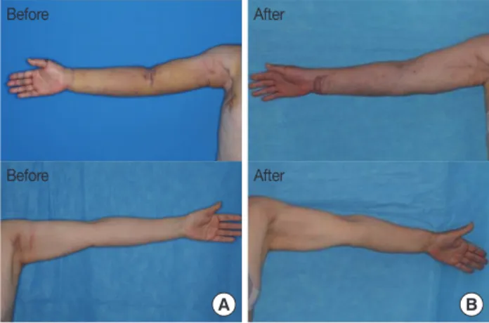
D Figure 1. Preoperative evaluation and postoperative 1-year evaluation.
(A) Preoperative status before vascularized lymph node transfer. (B) Postoperative 1-year evaluation.
A B
Before After
After Before
94 Kyung Hoon Cook, et al.
http://ejbc.kr http://dx.doi.org/10.4048/jbc.2016.19.1.92 Table 1. Clinical data of upper extremity circumferences during staged operations
Location Unaffected side (left) (cm) Affected side (right) (cm)
Preop Postliposuction Post-VLNT Post 1-year
Wrist 16.5 17 16 20 19
10 cm distal from the cubital fossa 22 30 26 (-50%*) 21 26 (-50%)
Cubital fossa 24.5 31.5 29 (-35.7%) 24.5 28 (-50%)
10 cm proximal from cubital fossa 27 35 30 (-62.5%) 26 30 (-62.5%)
Axillary area 32 33 32 (-100%) 31 31 (-200%)
VLNT=vascularized lymph node transfer.
*%=[Preoperative circumferences-postoperative (liposuction or VLNT) circumferences/Preoperative circumferences-unaffected side circumferences]×100.
Next, we identified the transverse cervical artery, which is be- neath the omohyoid muscle toward the anterior scalene mus- cle, and raised from the thyrocervical trunk. After flap eleva- tion, we anastomosed the radial artery with the end-to-side method (Figure 3).
The severity of the lymphedema was evaluated by measur- ing the circumferences of various parts of the upper extremity.
There were five points of measurement, including the wrist area, 10 cm distal from the cubital fossa, the cubital fossa, 10 cm proximal from the cubital fossa, and the axillary area.
There was improvement in the lymphedema condition fol- lowing the operations. Ten weeks after the first liposuction operation, decreased circumferences ranging from 35.7% to 100% compared to those of the contralateral side were noted.
Of note, we defined “%” as the ratio between the decreased circumferences and the difference between the normal side and affected side at their preoperative circumferences. One year after the second-stage lymph node transfer operation, all measurement points on the right upper limb except the wrist point had a smaller circumference than their preoperative measurement (Table 1, Figure 1).
DISCUSSION
The main mechanism of VLNT is re-establishment of lym- phatic circulation in the transferred flap. In the postoperative period, healing of transplanted lymphatics to native lymphat- ics at the recipient site and removal of lymphatic fluid by a di- rect pumping mechanism may provide further fluid drainage [5]. In some patients, improvement in lymphedema swelling can be observed immediately in the hospital after VLNT, be- fore the healing of the donor and native lymphatics. While the mechanism of improvement is unclear, the release of scar tis- sue in the previously operated and/or irradiated lymphatic bed has been postulated to account for this observation [6]. In addition, our patient showed immediate dramatic reduction of circumference length; 10 days after VLNT, the affected side arm had a smaller circumference than the contralateral nor-
mal side arm. This may have been caused by lymphatic ab- sorption due to earlier scar tissue release caused by liposuc- tion combined with strict arm elevation. We encouraged the patient to maintain a position with passive elevation of the arm 24 hours a day, using an elastic bandage sling attached to a standing pole.
Liposuction has been shown to remove circumferential subcutaneous fatty tissue in affected limbs. In addition, suc- tion-assisted lipectomy has been demonstrated to maximally reduce excess solid volume remaining in lymphedema after the fluid component has been removed with conventional therapy. While effective in removing excess volume, debulk- ing does not address the pathophysiology causing the lymph- edema. Therefore, patients must continue postoperative com- pression to prevent reaccumulation of excess fluid, and addi- tional postoperative therapy is required. Therefore, custom-fit compression garments are measured by the therapist and must be placed on the patient immediately postsurgery in the operating room.
It should be noted, however, that procedures that remove the fluid component of lymphedema, such as VLNT, are less likely to achieve the large reduction in excess volumes ob- served after liposuction. Rather, the procedures significantly decrease the postoperative need for compression garments and lymphedema therapy. Conversely, liposuction results in large volume reductions because it removes large amounts of solid fat and lymphatic material, but it does not address the ongoing lymphatic stasis and obstruction. Therefore, we have combined the liposuction and VLNT procedures in a two- staged approach to manage advanced staged lymphedema.
First, liposuction was performed to remove the solid com- ponents and reduce excess volume. After postoperative swell- ing stabilized, VLNT was used to improve lymphatic drainage and address subsequent fluid retention. Previously, this com- bined approach has resulted in volume reductions of over 83%, with compression garment use required only in the eve- nings and at night [7].
Furthermore, in this case report, we had to perform VLNT
Vascularized Free Lymph Node Flap Transfer in Advanced Lymphedema Patient 95
http://dx.doi.org/10.4048/jbc.2016.19.1.92 http://ejbc.kr
after liposuction to maintain superficial venous circulation. If we had performed VLNT before the liposuction, there would have been circulatory impairment caused by the liposuction procedure, which could have progressed to venous congestion.
The compression garment applied immediately after liposuc- tion also contributed to the prevention of venous retention.
VLNT requires microanastomosis, and there are three re- cipient sites available. The axillary area is usually operated on and irradiated, resulting in fibrotic changes, which make the dissection of recipient vessels more tedious. The anterior re- current ulnar artery is sometimes very small. Therefore, the ulnar artery may be used with an end-to-side technique. For recipient vessels, the radial artery’s dorsal branch and the ce- phalic vein are superficial and therefore easily dissected [8].
There is a hypothesis that VLNT may act by means of an in- ternal pump and suction mechanism using pathways for lym- phatic clearance of the lymphedematous limb. The “pump”
mechanism is driven by the high-pressure inflow of the arteri- al anastomosis from the radial artery, which provides a strong hydrostatic force into the vascularized groin lymph node flap.
The “suction” is continued by the large-caliber, superficially located, low-pressure venous drainage provided by the ce- phalic vein [9].
In this case, good results were obtained; immediately post- operation, the patient with advanced staged lymphedema had reduced circumferences in various parts of the upper extrem- ity . However, there were limitations to this study. One limita- tion is that an increase in diameter was seen at each point be- tween the immediate postoperative evaluation and the evalua- tion 1-year postoperation. We hypothesize that the increased circumferences after 1 year were the results of the stabilization of the grafted flap, rather than recurrence of lymphedema.
Long-term follow-up observations would be needed to con- firm this hypothesis. In addition, we did not evaluate the pa-
tient’s satisfaction in psychological aspects of the study and physical activity; assessments of object findings are necessary in a later study.
CONFLICT OF INTEREST
The authors declare that they have no competing interests.
REFERENCES
1. Murdaca G, Cagnati P, Gulli R, Spanò F, Puppo F, Campisi C, et al. Cur- rent views on diagnostic approach and treatment of lymphedema. Am J Med 2012;125:134-40.
2. Karri V, Yang MC, Lee IJ, Chen SH, Hong JP, Xu ES, et al. Optimizing outcome of Charles procedure for chronic lower extremity lymphoede- ma. Ann Plast Surg 2011;66:393-402.
3. Sapountzis S, Ciudad P, Lim SY, Chilgar RM, Kiranantawat K, Nicoli F, et al. Modified Charles procedure and lymph node flap transfer for advanced lower extremity lymphedema. Microsurgery 2014;34:439-47.
4. O’Brien BM, Khazanchi RK, Kumar PA, Dvir E, Pederson WC. Lipo- suction in the treatment of lymphoedema: a preliminary report. Br J Plast Surg 1989;42:530-3.
5. Granzow JW, Soderberg JM, Kaji AH, Dauphine C. Review of current surgical treatments for lymphedema. Ann Surg Oncol 2014;21:1195- 201.
6. Chang DW, Kim S. Breast reconstruction and lymphedema. Plast Reconstr Surg 2010;125:19-23.
7. Granzow JW, Soderberg JM, Dauphine C. A novel two-stage surgical approach to treat chronic lymphedema. Breast J 2014;20:420-2.
8. Cheng MH, Chen SC, Henry SL, Tan BK, Lin MC, Huang JJ. Vascu- larized groin lymph node flap transfer for postmastectomy upper limb lymphedema: flap anatomy, recipient sites, and outcomes. Plast Reconstr Surg 2013;131:1286-98.
9. Lin CH, Ali R, Chen SC, Wallace C, Chang YC, Chen HC, et al. Vascu- larized groin lymph node transfer using the wrist as a recipient site for management of postmastectomy upper extremity lymphedema. Plast Reconstr Surg 2009;123:1265-75.