Clinical Study on the Efficacy of the Autogenous Tooth Bone Graft Material (AutoBT)
전체 글
수치
관련 문서
From the results of this study, we concluded that two different sized graft materials have positive effects on new bone formation.. Additionally, smaller
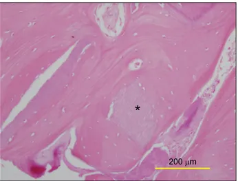
The grafted particulated tooth driven allografts were partially surrounded with new bone matrix and fairly supported graft associated new bone formation with some
Long-term outcomes of short dental implants supporting single crowns in posterior region: a clinical retrospective study of 5-10 years.. Osteotome sinus
Clinical and histomorph ometric evaluation of extraction sockets treated with an autologous bone marro w graft... Barone A, Aldini NN, Fini M, Giardino R, Calvo Guirado
The OSFE (osteotome sinus floor elevation) technique has been used for maxillary sinus augmentation.. The implants were clinically and radiographically followed
5) After GBR, the membrane was removed i n i ni ti al ti me, the usage of nonabsorbabl e membrane and autogenous bone resul ted i n the mostfavorabl e bone formati
success rates of dental implants placed at the time of or after alveolar ridge augmentation with an autogenous mandibular bone graft and titanium mesh: a 3-to
In 4-week group, the group filled with bone graft with decortication revealed larger new bone formation area than shown in the group that had a defect area
