조지호∙김수관∙문성용∙오지수
조선대학교 치의학전문대학원 구강악안면외과학교실
탈단백우골과 비탈회 동종골을 사용한 상악동 거상술의 임상적 연구
Clinical Comparative Study for Maxillary Sinus Augmentation Using Deproteinized Bovine Bone Mineral and Mineralized Allograft
Ji-Ho Jo, Su-Gwan Kim, Seong-Yong Moon, Ji-Su Oh
Department of Oral and Maxillofacial Surgery, School of Dentistry, Chosun University
Purpose: The purpose of this study was to compare the clinical efficacy of popular bone graft materials mineralized allograft and deproteinized bovine bone mineral.
Materials and Methods: One hundred seven implants of 78 patients, accompanied by sinus lift using the lateral window technique and simultaneous implantation, were sampled. In addition, some patients with severe systemic conditions were excluded. The initial bone heights of all patients ranged from 3-6 mm. All of the sample cases were treated at our hospital from January 2005 to January 2008. Techniques other than the lateral window technique were excluded, and only one graft material (Tutoplast
�or Bio-Oss
�) was accepted for inclusion. Tutoplast
�was used in 63 implants of 41 patients, whereas Bio-Oss
�was chosen for the remaining 44 implants of 37 patients. The diameters of the particles used ranged from 0.25-1.0 mm, and the volume was 0.5-2 cc (mean, 1.5 cc).
Results: The survival rate of the implant fixtures was 99.07% when the lateral window technique was used. Among all of the cases, cases in which Tutoplast
�was used demonstrated a survival rate of 98.4%, whereas Bio-Oss
�resulted in 100% survival. With respect to the alveolar bone height, no significant differ- ences were detected between the two graft materials that failed.
Conclusion: According to the result reported above, the two common materials for sinus augmentation do not have clinically significant difference. Rather, host factors, such as the height of residual bone, which could be disclosed during questioning patients' systemic conditions, might have greater effects on the prognosis.
Key words: Allograft, Maxillary sinus graft, Xenograft Abstract
서 론
무치악 부위에 대한 보철적 치료로서 이미 임플란트는 널 리 받아들여지고 있는 술식이며, 이는 인공 매식체와 유기 체인 골 사이의 상호 유착을 그 기본으로 하고 있다. 잔존 골량 및 골질, 환자의 전신적인 상태, 식립부위의 국소적인 환경 및 해부학적 요소, 흡연 여부, 환자의 교육수준 및 건 강에 대한 의지 등 다양한 숙주요소가 임플란트의 성공과
실패에 적지 않은 영향을 미칠 수 있다. 이러한 면에서 해부 학적인 제한이 큰 상악 구치부는 치조골 흡수와 함께 상악 동 함기화가 함께 진행되기 때문에 그 골량이 현저히 적고 골질 또한 불량한 경우가 많아 임플란트 식립에 있어서 불 리한 조건을 갖는다.
상악동 거상술이 일반화 되지 않았던 과거에는 골량이 적
은 상악 구치부는 임플란트 식립의 비적응증 이었다. 때때
로 적은 골량을 극복하기위해 전방의 긴 임플란트와 함께
상악구치부에 대해서는 길이가 짧은 임플란트가 식립하여 보철적 치료가 시행되기도 하였다. 하지만, 상악동 이식술 의 일반화된 요즘 상악동 이식술은 상악구치부의 부족한 골 량을 증가시키기 위한 술식으로 널리 사용되고 또 그 안정 성이 입증된 방법이다.
1,2)골이식에 있어 골 형성의 잠재적 능력을 갖고 있는 자가골이 가장 훌륭한 이식재라는 것은 이미 모두가 동의 하는 사실이다.
3-6)하지만, 구강 내 또는 구강 외의 골 공여부에 대한 추가적인 술식이 필요하며 이 에 따른 환자의 불편감 및 부작용의 가능성 때문에 자가골 이 최상의 이식재라고는 할 수 없다.
7,8)최근 다양한 이식재가 개발되어 자가골의 대체골로 사용 되고 있으며, 그 중 임상에서 널리 사용되는 이식재로 Bio- Oss
�(Geistlich-Pharma, Wolhusen, Switzerland)와 Tutoplast
�(Tutogen Medical GmbH, Germany) 가 있 다. Bio-Oss
�(Anorganic bovine bone material)는 소뼈 에서 채취되어 가공 멸균된 물질로 무기물로만 구성되어져 있으며, Tutoplast
�(Mineralized cancellous bone allo- graft)는 사체로부터 얻어진 사람 뼈를 가공 멸균한 것이다.
본 연구에서는 임상에서 널리 사용되는 이 두 가지 이식재 를 상악동 거상술에 적용한 증례에 대해 2년 후 잔존골량의 비교 및 임플란트 생존률을 평가함으로써 두 이식재의 임상 적 효용성을 평가하고자 한다.
연구 대상 및 방법
본 연구는 2005년 1월부터 2008년 1월까지 3년 동안 조 선대학교 치과병원에서 상악동 거상술을 시행한 환자 중 측 방개창법(lateral window technique)을 통해 상악동 거상 술 및 임플란트 동시 식립술을 시행한 78명의 환자, 107개 의 임플란트를 대상으로 하였다. Tutoplast
�를 사용한 군 은 41명에서 63개의 임플란트가 식립되었으며, Bio-Oss
�를 사용한 군은 37명 44개의 임플란트가 식립되었다. 또한, 본 연구에서는 상악동 거상술에 사용한 두 재료의 임상적 효용성을 임플란트의 생존율과 잔존 골 높이를 통해 비교 하는데 목적을 두고 있기 때문에 임플란트의 성공에 영향을 줄 수 있는 다양한 인자들을 제한할 수밖에 없었고, 연구 대 상은 다음의 조건에 따라 제한되었다.
1. 결과에 영향을 미칠 수 있는 전신질환을 가진 환자는 대상에서 제외 되었다. 당뇨, 고혈합, 골다공증 그 외
내과적 질환을 가진 환자는 제외하였고, 건강한 무치악 환자를 대상으로 하였다.
2. 잔존골의 높이는 3-6 mm로 제한하였으며, lateral window technique 외 다른 방법을 통한 상악동 거상 술은 본 연구에서 제외하였고, 임플란트를 동시에 식립 한 경우에 한하였다.
3. Bio-Oss
�혹은 Tutoplast
�를 단독으로 사용한 경우로 대상을 제한하였으며 이식재를 혼용한 경우는 제외하 였다.
임플란트 식립 후 주기적인 내원을 통해 임상검사를 시행 하였으며, 필요시 예방치과에 의뢰하여 구강위생교육 및 관 리를 시행하였다. 진료기록지, 방사선 사진 및 임상적 검사 를 토대로 하여 총 107개의 임플란트를 대상으로 수술 전, 술후 즉시, 술후 6개월, 술후 2년 추적 검사시 치조골의 높 이 변화를 비교하였으며, 추적기간 중 임플란트 탈락의 경 우 실패로 간주하였다.
두 재료 간 임플란트의 생존률은 SPSS v12.0 프로그램을 사용한 Cross tab의“카이제곱 검정” 을 통해 이루어졌다.
9,10)연구 결과
Tutoplast
�를 이식재로 사용한 군은 41명, 63개 임플란 트였다. 환자의 나이 분포는 28세에서 71세로 평균 나이는 53세였으며, 50대와 40대가 각각 20명, 9명으로 가장 많 았고, 남자가 30명, 여자는 11명이었다(Table 1). Bio- Oss
�를 이식재로 사용한 군은 37명, 44개 임플란트였다.
환자의 나이 분포는 23세에서 69세로 평균 나이는 51세였 으며, 40대와 50대가 각각 13명, 12명으로 가장 많았고, 남자가 25명, 여자는 12명이었다(Table 1).
임플란트의 식립 부위는 모두 상악 대구치부로 CT상 잔 존골 높이는 3-6 mm로 Tutoplast
�군은 평균 3.8 mm, Bio-Oss
�군은 평균 3.7 mm였으며, 두 군 모두에서 3.5-4 mm의 잔존 골높이를 갖는 환자가 가장 많았다(Table 2).
사용된 이식재의 입자크기는 모두 0.25-1 mm였으며, 이식 된 양은 0.5-2 cc (mean 1.5 cc)였다. 식립된 임플란트는 Astra, 3i, ITI, Neobiotech, Dentis, Osstem, Dentium, Biohorizon였고, 직경은 제조사에 따라 약간의 차이는 있 지만 4.5 mm와 5 mm가 사용되었으며, 길이는 10-13 mm로 평균 11.5 mm였다.
Table 1. Age distribution in Tutoplast
�group and Bio-Oss
�group
Age of patient (years)
20-29 30-39 40-49 50-59 60-69 70-79
Tutoplast Men 1 1 7 15 3 3
Women 1 1 2 5 1 1
Bio-oss Men 2 1 10 8 4 0
Women 0 0 3 4 5 0
상악동 거상술 및 임플란트 즉시식립이 이루어진 후 2차 수술까지 걸린 기간은 4-10개월로 두 군 모두 평균 6.6개 월이 지난 후 2차 수술이 시행되었다(Table 3). 수술 후 임 플란트 생존률 평가는 주기적인 외래 내원을 통해 이루어졌 으며, 추적기간은 최소 20개월이었다.
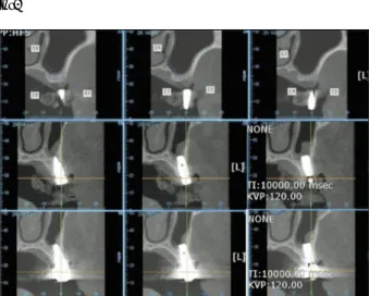
잔존골량은 술전, 수술 직후, 술후 6개월, 술후 2년의 치 근단 방사선 사진, Dental panorama 및 CT를 통하여 평 가하였다. CT에 나타난 술 전∙후 골 높이 변화의 평가는 fixture 중앙부위에서 측정하였다(Fig. 1). 임플란트 식립 2년후의 fixture 상방 골높이는 이식재의 종류 및 임플란트 길이와 상관없이 평균 0.5 mm였다(Table 4). CT를 통한 골 높이 평가에서 Tutoplast
�를 사용한 군과 Bio-Oss
�를 사용한 군 모두에서 비슷한 양상이 관찰되었는데, 두 이식 재 모두 임플란트 fixture 근단 상방에서 불투과성이 시간 에 따라 감소되었고, 임플란트 fixture 근단 측방과 상악동 외벽쪽의 불투과성은 2년 후에도 잔존된 것을 관찰할 수 있 었다(Fig. 2).
총 107개 임플란트 중 임플란트가 탈락한 경우는 단지 1 개의 임플란트로 Tutoplast
�를 사용한 60세 남자였다. 실 패한 임플란트는 상악 좌측 제 2대구치였으며, 술 전 잔존
골의 높이는 3 mm였고, 2 cc의 Tutoplast
�가 사용된 경우 로 보철 치료 전 골유착이 실패하여 재수술이 시행되었다.
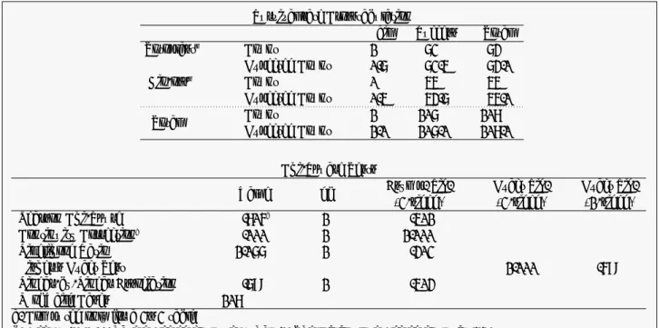
본 연구에서 임플란트 생존률은 99.07%였으며, Tutoplast
�를 사용한 군의 생존률은 98.4%, Bio-Oss
�를 사용한 군에 서는 100%였다(Table 5). 두 군의 생존률은 SPSS분석 결 과 유의할 만한 차이를 나타내지 못하였다(P = 0.401, Fig. 3).
Table 2. Residual bone height preoperatively Residual bone height (mm) 3-3.5 3.5-4 4-4.5 4.5-5 5-5.5 5.5-6
Tutoplast
�19 27 10 5 1 1
Bio-Oss
�15 19 6 3 1 0
Table 3. The mean period to 2nd surgery
Mean period to 2nd surgery (months) 4-5 5-6 6-7 7-8 8-9 9-10
Tutoplast
�6 11 25 15 5 1
Bio-Oss
�7 4 17 12 3 1
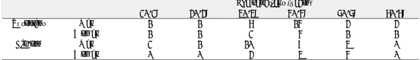
Table 4. Comparison of bone height : pre-OP and post-OP
MRBH MLI Mean total gain Mean apex gain
OP date 2 yrs after OP date 2 yrs after
Tutoplast
�3.8 11.5 9.7 8.2 2 0.5
Bio-Oss
�3.7 11.5 10.2 8.3 2.4 0.5
MRBH: Mean residual bone height, MLI: Mean length of implant.
Fig. 1. Diagram of sinus augmentation. AG: apex gain, LI:
length of implant, TG: total gain, RBH: residual bone
height. Fig. 2. The first molar of right maxilla. CT image preopera-
tively, immediately after, 2 years after surgery. The amount of residual bone is 3 mm before surgery, 15.3 mm right after surgery, and 14 mm 2 years after surgery. The resorption appeared to begin from the center of the site.
Table 5. The survival rate of implant
Implant Failed implant Survival rate
(n) (n) (%)
Tutoplast
�63 1 98.4
Bio-Oss
�44 0 100
According to the result reported above, the two common
materials for sinus augmentation do not have clinically
significant difference (P = 0.401).
고 찰
골이식에 있어 자가골은“Golden standard”로 이미 알 려져 있다.
3-5)하지만, 골 채취 과정으로 인한 환자의 부담과 예기치 못한 골흡수에 대한 문제점으로 상악동 거상술에 있 어 다양한 골이식재들이 사용되고 있으며, 이 이식재들은 자가골 채취에 따른 위험을 피할 수 있게 해 주었다.
11)본 연구에서 사용된 Bio-Oss
�와 Tutoplast
�는 임플란트 및 치주 수술에서 널리 사용되는 골 이식재이다. Bio-Oss
�(Anorganic bovine bone material)는 소뼈에서 추출된 다 공성의 생체 수산화인회석(hydroxyapatite)로 인체 골과 유사한 다공성을 가지고 있어 다른 골이식재에 비해 혈관의 신생이 용이하고 구조적으로 안정적이며, 이미 많은 연구에 서 그 골전도 능력이 보고된 바 있다.
12-15)Valentini 등과 Hising 등의 연구에 의하면 100% 이종골을 이용한 상악동 거상술에서 자가골 단독 사용이나 자가골과 이종골을 혼합 사용한 경우보다 임플란트의 생존률이 오히려 더 높다고 보 고되기도 했다.
13,15)Klinge 등은 다른 이식재에 비해 Bio- Oss
�에서 빠른 골형성 보고 하기도 하였으며,
16)다른 연구 에서도 Bio-Oss
�가 새로이 형성된 골과의 긴밀한 접촉을 통해 골형성에 유리한 점을 갖고 있다고 보고되었다.
17-20)Tutoplast
�(Mineralized cancellous bone allograft)는 사체로부터 채취한 골을 Tutoplast processing technique 을 통해 용매 탈수(solvent-dehydrated)시킨 것으로 골유 도에 필요한 골형성단백질(BMP)과 무기물을 모두 갖고 있
다는 유리한 점이 있다.
21)Bio-Oss
�와 비교했을 때 Tutoplast
�를 이식한 군에서 조직학적 평가를 통해 더 많은 vital bone을 관찰할 수 있었다는 보고가 있으며,
22,23)이는 Tutoplast
�안에 존재하고 있는 교원질이 잠재적인 골전도 능력에 영향을 미친 것으로 보인다고 하였다.
24)Groeneveld 등은 이런 다양한 골 이식재들이 성장인자와 함께 사용되었 을 때 더 큰 골 유도능을 갖는다고 보고하였다.
25,26)여러 문헌에서 조직학적 평가시 vital bone의 생성 정도 에 대한 의견이 다양하지만, 본 연구에서 Bio-Oss
�와 Tutoplast
�를 이용한 상악동 거상술을 시행 후 2년간의 추 적기간 동안 임플란트 생존률은 두 군 모두에서 높은 생존 률을 보였으며, 두 군 간에 유의할 만한 차이는 없었다. 본 연구를 토대로 상악동 거상술 시 골이식재의 단독 사용뿐만 아니라 여러 골이식재의 혼합 사용에 대한 임상적인 평가가 필요하리라 생각된다.
결 론
본 연구에서는 Bio-Oss
�와 Tutoplast
�를 사용한 상악동 거상술에서의 임플란트 생존률을 비교하고자 하였다.
1. 상악동 거상술 및 임플란트 동시 식립술을 시행한 78 명의 환자, 107개의 임플란트를 대상으로 하였다.
2. Tutoplast
�를 사용한 군은 41명에서 63개의 임플란트 가 식립되었으며, Bio-Oss
�를 사용한 군은 37명에서 44개의 임플란트가 식립되었다.
Fig. 3. The statistic analysis of implant survival rate.
Chi-Square Tests
Value df Asymp. Sig. Exact Sig. Exact Sig.
(2-sided) (2-sided) (1-sided)
Pearson Chi-Squre .705
b1 .401
Continuity Correction
a.000 1 1.000
Likelihood Ratio 1.066 1 .302
Fisher's Exact Test 1.000 .589
Linear-by-Linear Association .689 1 .403
N of Vaild Cases 107
a. Computed only for a 2×2 table
b. 2 cells (50.0%) have expected count less than 5. The minimum expected count is .41.
Survival rate Crosstabulation
Fail Success Total
Totoplast
�Count 1 62 63
Expected Count 0.6 62.4 63.0
Bio-oss
�Count 0 44 44
Expected Count 0.4 43.6 44.0
Total Count 1 106 107
Expected Count 1.0 106.0 107.0
3. 잔존 골높이는 3-6 mm로 Tutoplast
�군은 평균 3.8 mm, Bio-Oss
�군은 평균 3.7 mm였다.
4. 임플란트 생존률은 99.07%였으며, Tutoplast
�를 사 용한 군의 생존률은 98.4%, Bio-Oss
�를 사용한 군에 서는 100%였다.
본 연구의 결과에 따른면 상악동 거상술 및 동시 임플란트 식립술에 있어서 Tutoplast
�를 사용한 군과 Bio-Oss
�를 사용한 군 사이의 유의할 만한 임상적 차이를 나타내지 못 하였으며(P = 0.401), 두 이식재 모두 상악동 거상술에서 의 골이식재로서 충분한 가치가 있다고 생각된다.
References