교신저자: 권건영, 700-712 대구광역시 중구 달성로 56, 계명대학교 의과대학 병리학교실
Kun Young Kwon, M.D., Department of Pathology, Keimyung University School of Medicine 56 Dalseong-ro, Jung-gu, Daegu 700-712, Korea
Tel: 82-53-580-3812 E-mail: [email protected]
Department of Pathology, Keimyung University School of Medicine, Daegu, Korea
Ilseon Hwang, M.D., Kun Young Kwon, M.D.
황일선·권건영 계명대학교 의과대학 병리학교실
2011 IASLC/ATS/ERS 분류에 근거한 비소세포폐암종의 병리 진단
Pathologic Diagnosis of Nonsmall Cell Lung Carcinoma According to 2011 IASLC/ATS/ERS Classification
Abstract
1999 and 2004 WHO classifications of lung cancer has used widely with few major advances for about 10 years. Recently the lung cancer classification has changed to a markedly evolving field with rapid progression of molecular discovery in the lung adenocarcinoma. The exact pathologic diagnosis and subclassification of the non-small cell lung carcinoma in small biopsies and cytologic specimens are key roles in the clinical management of lung cancer patients. We need special stains or immunohistochemical stains using several useful markers as an aid to diagnosis, particularly in the setting of poorly differentiated tumors that do not show clear differentiation by light microscopy. Also every institution needs to develop a multidisciplinary cooperation system to make an exact diagnosis and plan an appropriate targeted treatment in the patients with lung cancer. In this article, we describe the concept and diagnostic criteria of the 2011 IASLC/ATS/ERS classification of nonsmall cell lung carcinoma in the small biopsy and resected specimens.
Key Words :
2011 IASLC/ATS/ERS classification, Adenocarcinoma, Immunohistochemistry, Non-small cell lung carcinoma서 론
International Association for the Study of Lung Cancer (IASLC), American Thoracic Society (ATS) 및 European Respiratory Society (ERS)의 국제적인 호흡기 분야의 학술단체 지원으로 병리학, 분자생물학, 영상의학, 호흡기 내과학, 종양학 및 흉부외과 의사 등 각 영역별 전문가들이 다수 참여하여 다학제적 검토와 많은 토론 과정을 거쳐서 2011 IASLC/ATS/ERS 폐암 분류가 개정되어 폐암의 진단 기준과 분류에서 2004년 WHO 폐암 분류와 비교하여 큰 변화를 보여주고 있다[1-4]. 2004년 WHO 폐암 분류에서는 주로 현미경 소견에 기준하여 비소세포폐암종의 진단이 임상에서 오랜 기간 사용되어 왔으며, 편평세포암종과 선암종의 감별이나 선암종에서 자세한 세분류가 요구되지 않았다[1].
최근 다수의 분자 바이오물질의 발굴과 함께 EGFR 유전자 돌연변이를 가진 환자에서 tyrosine kinase inhibitor (TKI) 치료에 잘 반응함이 알려져 진단과 치료에 일치하는 세분류의 필요성이 제기되어 왔다[5- 7]. 특히 2011년 IASLC/ATS /ERS 폐암 분류의 새로운 기준에 의해 초기 및 진행성 폐선암종 환자에서 다학제적 접근의 진단과 치료에 큰 변화를 보이고 있으며, 적출한 폐조직과 폐생검 및 세포진 검체에서 최신 치료 경향에 부합하는 폐선암종의 분류와 진단이 이루어지고 있다[8,9].
여기서는 2011 IASLC/ATS/ERS 최신 폐암분류에 의한 폐선암종의 특성을 주로 기술하고, 절제된 폐조직과 생검 및 세포진 검체에서 폐선암종의 진단 기준을 알아본다. 그리고 면역조직화학적 바이오마커 적용에 따른 비소세포폐암종의 감별진단과 폐선암종의 아형에 대해 기술하고자 한다.
1. 2011 IASLC/ATS/ERS 폐암 분류의 특성
폐암의 약 70% 환자는 진행된 병기를 가지며, 수술적 치료를 하지 않기 때문에 생검 또는 세포진 검사를 통해 진단을 얻고 있다[3]. 2004년 WHO 폐암 분류는 수술로 절제된 폐조직에서 진단하는 기준만을 제시하였지만 2011 IASLC/ATS/ERS 폐암 분류에서는
수술로 절제된 폐조직 뿐만 아니라 생검 및 세포진 검체에서 폐암종을 진단하는 기준과 요령을 제시하고 있다[3](Table 1,2). 1999년 및 2004년 WHO 폐암 분류에서는 생 검 조 직 에 서 비 소 세 포 폐 암 종 에 서 선 암 종 과 편평세포암종을 감별하여 분류해야할 필요성이 크지 않았으나, 그 동안 분자생물학의 획기적인 발전으로 임상에서 이들 조직 유형에 따른 표적치료제가 달리 사용되고 있어서 최근에는 작은 생검에서도 가능한 조직 아형을 세분류하여 진단하고 있다[2,3,10].
2. 수술로 절제된 폐조직에서 폐암의 병리 진단
2011년 IASLC/ATS/ERS 폐암 분류는 2004년 WHO 분류와 비교하여 큰 변화를 보인다[3,4,7] (Table 1).
특히 폐선암종에서 분류와 진단 기준이 새롭게 정리되었다. 종양의 직경이 3 cm 이하인 폐선암종에서 간 질 , 혈 관 및 림 프 관 침 윤 이 없 는 종 양 을
“상피내선암종 (adenocarcinoma in situ, AIS)”로 진단하며, 간질 내로 종양세포 침윤 범위의 직경이 0. 5 cm 이하인 경우 “최소침 윤성 선암종(minimally invasive adenocarcinoma, MIA)”으로 진단하고 간질 내 침윤이 0.5 cm를 넘거나 혈관 및 림프관 침윤이 있는 경우 “침윤성 선암종(invasive adenocarcinoma)”으로 진단 한다[3]. AIS와 MIA인 환자는 완전히 종양 절제술을 하는 경우 5년간 재발하지 않을 가능성이 각각 100%와 거의 100%를 보이므로 특히 침윤성 선암종과 감별이 필요하다[11].
세기관지폐포암종(bronchioloalveolar carcinoma, BAC)의 진단 용어는 2011 새분류법에서는 더 이상 사용하지 않는다. 2004 WHO 분류에서 BAC는 AIS로 정의하였으나, BAC로 진단되었던 증례에서 MIA 및 침 윤 성 선 암 종 의 소 견 을 보 이 는 증 례 가 많 이 보고되어서 2011 새 분류법에서는 혼돈을 피하기 위해서 BAC란 용어를 더 이상 사용하지 않게 되었다[2,3,6].
2004 WHO 분류에서 점액성 세기관지폐포암종 (mucinous BAC)은 점액성 AIS, MIA 또는 침윤성 점액선암종 (invasive mucinous adenocarcinoma)로 분류하며, 이 암종은 K-Ras 돌연변이를 갖는 경우가 많고, thyroid transcription factor-1 (TTF-1)에 음성을 보인다[5]. CT 영상에서 흔히 결절성 종양 병변을
보이며, 다결절을 갖거나 다엽성 분포를 한다[2,3].
2004년 WHO 분류에서 침윤성 선암종에서 “혼합형 선암종(adenocarcinoma, mixed subtype)”이란 용어를 사용하였으나, 2011년 IASLC/ATS/ERS 분류법에서는
“혼합형”을 사용하지 않으며, 현미경상에서 관찰되는 다양한 조직 아형을 %로 각각 기술하고 있다[2,12].
“Lepidic predominant adenocarcinoma” 는 과거 비 점 액 성 B A C 의 형 태 를 주 로 보 이 는 침 윤 성 선암종이며, 미세유두 (micropapillary) 양상을 주로 보이는 침윤성 선암종은 “micropapillary predominant adenocarcinoma” 로 진단한다.
정상 폐조직이나 침윤이 없는 lepidic pattern 선암종의 폐포벽(간질)에서는 면역조직화학염색에서 CD34에 양성이며, Smooth muscle actin (SMA)에는 음성인 섬유모세포를 관찰할 수 있다. 그러나 간질에 침윤이 있는 MIA의 종양 병변과 침윤성 선암종의 간질에서는 CD34에 음성을 보이고, SMA에 양성을 보이는 섬유모세포를 관찰할 수 있어서 폐암에서 간질의 침윤 여부를 결정하는데 있어서 이들 면역 조직화학염색을 사용하면 진단적 가치가 큰 것으로 보고되어 있다[13]. 그리고 2011 폐암의 새분류법에 따라 폐선암종의 조직학적 특성과 아형을 정확히 판독함으로서 다발성 폐결절을 가지는 폐암 증례에서 원발성 폐암종과 폐내 전이성 암종과의 감별을 하는데도 도움을 받을 수 있다[14,15].
3. 생검 또는 세포진 검체에서 폐암의 병리진단 및 판독요령
2011 IASLC/ATS/ERS 분류 및 진단 기준에 의해 작은 생검에서 폐암의 진단 요령은 수술로 절제된 조직에서 진단 기준과 다소 상이하며 다음과 같은 병리 판독 요령이 필요하다(Table 2).
(1) 작은 폐생검에서 분화가 좋지 않은 비소세포암종 (non-small cell lung carcinoma, NSCLC)에서도 가능하면 선암종과 편평세포암종을 감별하여 진단한다.
(2) 비소세포폐암종 진단에서 “비편평세포암종 (non-squamous cell carcinoma)” 이란 진단 용어를
가능하면 사용하지 않도록 하고 선암종, 편평세포암종 및 비소세포폐암종-NOS로 분류한다.
(3) 광학현미경상 선암종 또는 편평세포암종의 뚜렷한 분화를 보이지 않으면서 비소세포암종-NOS의 양상을 보이는 증례는 면역조직화학염색을 추가로 실시하여 “비소세포폐암종, 선암종 유망(Non-small cell lung carcinoma, favor adenocarcinoma)” 또는
“비소세포폐암종, 편평세포암종 유망(Non-small cell lung carcinoma, favor squamous cell carcinoma)”으로 진단하며, 면역조직화학염색상에서도 감별이 되지 않을 경우에만 “비소세포폐암종-NOS”로 진단한다. 또한 비소세포폐암종-NOS 소견을 보이는 증례에서는 조직 아 형 을 위 한 면 역 조 직 화 학 염 색 을 최 소 화 하 고 분자병리검사를 위해 파라핀 블록의 남은 조직절편을 확보한다.
(4) 2004 WHO 분류에 사용된 혼합형 선암종은 새 분류법에서는 사용하지 않으며, 현미경상 관찰되는 형태학적 유형 즉 선형(acinar), 유두형(papillary), 고형(solid) 및 미세유두형(micropapillary)이 관찰되면 모두 기술한다. 만일 생검상에서 lepidic 형태만을 보이는 선암종은 “adenocarcinoma with lepidic pattern”으로 진단하고, “침윤의 가능성을 배제할 수 없음” 이라는 기술을 추가한다.
(5) 비점액성 세기관지폐포암종의 양상을 보이는 증례에서는 “adenocarcinoma with lepidic pattern”
으로 진단한다. 점액성 세기관지폐포암종의 형태를 보 이 는 증 례 에 서 는 “ 점 액 선 암 종 ( m u c i n o u s adenocarcinoma)” 으로 진단하며, 다른 조직 유형(선, 유두, 고형, 미세유두)이 관찰될 때 추가 기술한다.
( 6 ) 반 지 세 포 ( s i g n e t r i n g c e l l s ) 또 는 투명세포(clear cells)가 추가로 관찰되면 “반지세포 또는 투명세포 양상 선암종(adenocarcinoma with signet ring cells or clear cell feature)”으로 진단한다.
(7) 고형 종양(solid tumor) 병변을 보이나 점액
염 색 또 는 면 역 조 직 화 학 염 색 상 선 암 종 또 는 편평세포암종의 특성을 보이면 “비소세포폐암종, 선암종 유망(Non-small cell lung carcinoma, favor adenocarcinoma)” 또는 “비소세포폐암종, 편평세포암종 유망(Non-small cell lung carcinoma, favor squamous cell carcinoma)”으로 진단한다.
(8) 2004 WHO 분류상 대세포암종(large cell carcinoma)의 소견을 보이면 “Non-small cell lung carcinoma-NOS” 로 우선 판독하며, 점액 염색과 면 역 조 직 화 학 염 색 을 추 가 로 실 시 한 다 . 현 미 경 소 견 에 서 대 세 포 신 경 내 분 비 암 종 ( l a r g e c e l l neuroendocrine carcinoma, LCNEC)을 의심할 수 있는 양상을 보이는 증례에서는 면역조직화학염색을 실 시 하 여 신 경 내 분 비 마 커 ( c h r o m o g r a n i n , s y n a p t o p h y s i n, C D56 등)에 양성을 보이면
“ 신 경 내 분 비 형 태 를 가 진 비 소 세 포 폐 암 종 , 대세포신경내분비암종 가능(Non-small cell lung carcinoma with neuroendocrine morphology, possible large cell neuroendocrine carcinoma)” 으로 진 단 하 며 , 신 경 내 분 비 마 커 에 음 성 을 보 이 면
“신경내분비 형태를 가진 비소세포폐암종(Non-small cell lung carcinoma with neuroendocrine morphology)” 으로 진단하며, “이 증례는 대세포신경 내 분 비 암 종 이 의 심 되 는 비 소 세 포 폐 암 종 이 며 , 신경내분비 분화는 확인되지 않았음 ”을 추가 기술한다.
(9) 편평선암종(adenosquamous carcinoma)의 형태를 보이는 증례에서는 “편평세포암종 및 선암종 양상을 가진 비소세포폐암종(Non-small cell lung carcinoma with squamous cell carcinoma and adenocarcinoma patterns)” 으로 진단한다.
(10) 암육종(sarcomatoid carcinoma)의 형태를 보이면 “방추세포 및/또는 거대세포암종을 가진 저분화성 비소세포폐암종(poorly differentiated Non- small cell lung carcinoma with spindle and/or giant c e l l c a r c i n o m a)” 으로 진단하며, 선암종 또는 편 평 세 포 암 종 소 견 이 함 께 관 찰 되 면 추 가 로 기술한다[1,3].
4. 생검 또는 세포진 검체에서 폐암 진단을 위한 특수염색 및 면역조직화학염색 적용
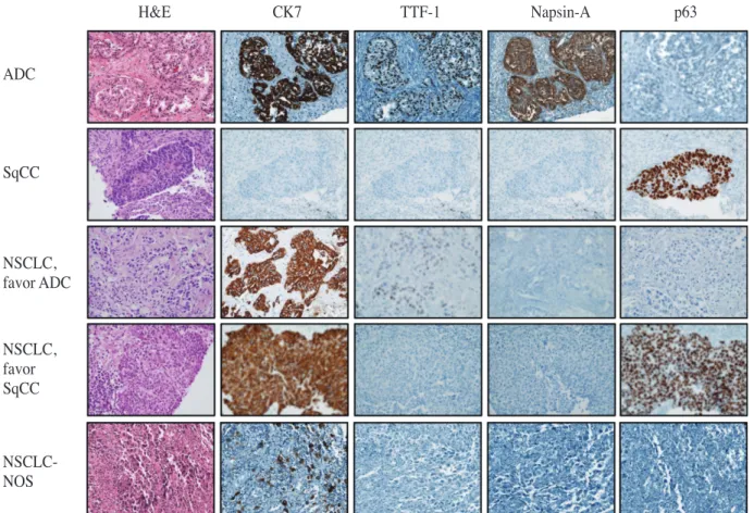
종양조직에서 뚜렷한 편평세포 또는 선(glandular) 분화를 보이는 증례는 “편평세포암종” 또는 “선암종” 으로 진단한다. 그러나 작은 생검 또는 세포진 검체에서 광학현미경상 뚜렷한 편평세포 또는 선분화를 보이지 않는 경우 우선 “비소세포폐암종” 으로 진단하고 파라핀블록을 세절하여 CK7, CK20, TTF-1, Napsin-A, p63, CK5/6, EGFR 등 진단에 유용한 면역마커를 패널로 사 용 하 여 면 역 조 직 화 학 염 색 을 하 거 나 점액염색(mucicarmine stain)을 실시한다[3] (Table 3).
고형 양상을 보이는 비소세포폐암종의 종양세포에서 점액 염색에 양성을 보이거나 면역조직화학염색 결과 선암종의 면역마커 (CK7, TTF-1, Napsin-A 등)에 양성을 보이면 “비소세포폐암종, 선암종 유망”으로 진단하며, 선암종의 마커에 음성이면서 편평세포암종 면 역 마 커 ( p 6 3 , C K 5 / 6 등 ) 에 양 성 을 보 이 면
“비소세포폐암종, 편평세포암종 유망”으로 진단한다.
만일 면역조직화학염색 및 점액염색 결과 감별진단에 도 움 될 만 한 뚜 렷 한 소 견 을 보 이 지 않 을 경우에는“비소세포 폐암종-NOS”로 진단한다[2,3](Fig. 1).
5. 폐생검에서 NSCLC 진단에 대한 임상적 이해
병리의사가 폐생검에서 “비소세포폐암종, 선암종 유망” 또는 “비소세포폐암종, 편평세포암종 유망” 으로 진단하면 광학현미경상 뚜렷한 조직 아형의 분화를 보이지 않았지만 면역조직화학염색 또는 점액 염색을 통해 선암종 또는 편평세포암종에 가까운 양상을 보였음을 시사하며, 환자 치료를 위해 EGFR, k-Ras, A L K 등 유전자에 대한 분자병리검사를 추가로 실시한다[5,16-18]. 2004년 WHO 폐암 분류에는 절제된 폐조직에서 대세포암종을 진단하는 경우가 있었으나 [1,19], 2011년 새 분류에는 “대세포암종” 진단 용어를 사용하지 않는다[3]. 특히 작은 생검 조직에서 이루어지는 “비소세포폐암종-NOS” 진단은 비세포폐암종 전체의 20~40% 정도이며, 임상시험 연구보고에서
“비소세포 폐암종-NOS” 진단 빈도가 증가하는 경향을 보이고 있다[20-22]. 그러나 최근 특이성이 높은
면역조직화학염색 마커가 많이 개발되어 있으며, 생검 또 는 세 포 진 검 체 에 서 비 소 세 포 폐 암 종 증 례 의 감별진단에 필요한 몇가지 면역표지 마커를 패널로 사용하면 대부분의 증례에서 “비소세포폐암종, 선암종 유망” 또는 “비소세포 폐암종, 편평세포암종 유망” 으로 진단할 수 있으며, 극히 일부 증례에서 “비소세포 폐암종-NOS” 란 진단을 하고 있다[3](Table 2).
6. 비소세포 폐암종의 유전자 돌연변이
비소세포 폐암종은 그 분류에 따라 매우 상이한 유전자 돌연변이를 보인다. 폐선암종의 체세포돌연변이(somatic mutation)를 분석한 결과를 보면 약 65-70%에서
TP53의 체세포돌연변이가 관찰되고, KRAS가 약 60%, EGFR과 STK11이 약 35% 내외에서 관찰된다[23].
특징적으로 EGFR의 돌연변이가 있을 경우 KRAS 돌연변이는 잘 관찰되지 않으며, 반대로 K R A S 돌연변이가 있을 경우 EGFR 돌연변이가 잘 관찰되지 않음이 잘 알려져 있고, 이로 인하여 KRAS 돌연변이가 있을 때에는 EGFR을 타겟으로 하는 표적치료에 반응하지 않는 것으로 잘 알려져 있다[23-25].
폐선암종의 경우에는 체세포돌연변이 이외에도 임상적으로 중요한 유전자 돌연변이가 있으며, 이는 EML4-ALK 융합유전자 전환이다[18]. EML4-ALK 융합유전자 전환은 유전자 전위에 의하여 EML4 유전자와 ALK 유전자의 융합이 일어나는 돌연변이로 Table 1. 2011 IASLC/ATS/ERS classification of lung adenocarcinoma in resection specimens [2]
Pre-invasive lesions
Atypical adenomatous hyperplasia
Adenocarcinoma in situ (=<3 cm, formerly solitary BAC) Nonmucinous
Mucinous
Mixed mucinous/nonmucinous
Minimally invasive adenocarcinoma (=<3 cm, lepidic predominant tumour with =<5 mm invasion) Nonmucinous
Mucinous
Mixed mucinous/nonmucinous Invasive adenocarcinoma
Lepidic predominant (formerly nonmucinous BAC pattern with >5 mm invasion) Acinar predominant
Papillary predominant Micropapillary predominant Solid predominant
Variants of invasive adenocarcinoma
Invasive mucinous adenocarcinoma (including formerly mucinous BAC) Colloid
Fetal (low and high grade) Enteric
IASLC: International Association for the Study of Lung Cancer, ATS: American Thoracic Society, ERS:
European Respiratory Society, BAC: bronchioloalveolar carcinoma.
전체 폐선암종의 약 5%에서 나타나며, EGFR이나 KRAS 돌연변이와는 함께 나타나지 않는 편이다[26]. EML4- ALK 융합유전자 전환이 발견되는 경우에는 그에 상응하는 표적치료에 잘 반응하는 것으로 알려져 있으므로, 치 료 제 의 선 택 에 매 우 중 요 하 다 . 그 에 반 하 여 편평상피세포암종에서는 특징적인 돌연변이가 아직 관찰되지 않고 있으며, 표적치료제의 개발도 활발히 이루어지지 못하고 있다.
7. Stage 1 선암종에서 조직학적 유형과 예후와의 상관성
조직학적 유형과 예후와의 상관관계에서 저등급
선암종에는 AIS와 MIA가 포함되며, 수술로 절제한 경우 5년 간 종양재발이 없을 가능성이 100%에 이른다[11]. 중등급 선암종에는 비점액성 lepidic 우세(predominant), 융모 우세(papillary predominant) 및 선우세(acinar predominant) 유형이 포함되며, 5년 간 종양재발이 없을 가능성이 90%, 83% 및 84%에 이른다. 그리고 고등급 선암종에는 침윤성 점액 선암종, 콜로이드 우세, 고형 우세 및 미세융모 우세 유형이 포함되며, 5년 간 종양 재발이 없을 가능성은 각각 75%, 71%, 70% 및 67%에 이른다(p=0.001)[4]. 이외에 통계학적으로 의미를 가지는 예후가 불량한 요소로 병기(stage 1B > 1A), 세포학적 고등급, 종양 침윤의 크기, 혈관침윤, 괴사 등을 들 수 있다[27,28].
Fig. 1. Diagnosis and subclassification of nonsmall cell lung carcinoma according to 2011 IASLC/ATS/
ERS classification in the small lung biopsy specimens. ADC: adenocarcinoma, SqCC: squamous cell carcinoma, NSCLC: non-small cell lung carcinoma, NSCLC-NOS: non-small cell lung carcinoma- not otherwise specified.
ADC
H&E CK7 TTF-1 Napsin-A p63
SqCC
NSCLC, favor ADC
NSCLC, favor SqCC NSCLC- NOS
요 약
2011년 IASLC/ATS/ERS 폐암 분류가 적용되면서 과거 병리 진단에서 흔히 이루어지던 비소세포폐암종 또 는 비 소 세 포 폐 암 종 - N O S 는 면 역 조 직 화 학 적
검사용으로 최근 개발된 유용한 진단적 마커들을 패널로 사용함으로서 작은 생검 조직에서도 선암종 또는 편평세포암종으로 세분류의 진단이 가능해지고 있다. 최근에는 폐암 환자에 대한 표적 항암치료가 활성화되고 있으며, 이를 위한 다양한 분자유전검사가 Table 2. Proposed diagnostic criteria of the lung carcinomas according to IASLC/ATS/ERS classification
compared to 2004 WHO classification [2,3]
Adenocarcinoma Adenocarcinoma with
Mixed subtype do not use mixed type Acinar describe histologic types
(acinar, papillary, solid, micropapillary) Papillary
Solid
BAC, nonmucinous Adenocarcinoma with lepidic pattern BAC, mucinous Mucinous adenocarcinoma
Fetal Adenocarcinoma with fetal pattern
Mucinous (colloid) Adenocarcinoma with colloid pattern Signet ring Adenocarcinoma with signet ring feature Clear cell Adenocarcinoma with clear cell feature
NSCLC, favour adenocarcinoma
Squamous cell carcinoma Squamous cell carcinoma
Papillary do not describe subtype
Clear cell Small cell Basaloid
NSCLC, favour SqCC
Small cell carcinoma Small cell carcinoma
Large cell carcinoma NSCLC, NOS
LCNEC LCNEC with NE morphology
(positive NE markers), possible LCNEC Large cell carcinoma with NE morphology (LCNEM) NSCLC with NE morphology
(negative NE markers)
Adenosquamous carcinoma NSCLC with squamous cell and
adenocarcinoma pattern
Sarcomatoid carcinoma Poorly differentiated NSCLC with
spindle and/or giant cell carcinoma 2004 WHO Classification* 2011 IASLC/ATS/ERS Classification**
*criteria applicating in the resected lung tumor specimens, **criteria applicating in the small biopsy/
cytology specimens, IASLC: International Association for the Study of Lung Cancer, ATS: American Thoracic Society, ERS: European Respiratory Society, BAC: bronchioloalveolar carcinoma, NSCLC:
non-small cell lung carcinoma, SqCC: squamous cell carcinoma, NOS: not otherwise specified, LCNEC:
large cell neuroendocrine carcinoma, NE: neuroendocrine.
비소세포폐암종, 특히 선암종에서 기본적으로 실시하는 경향이다. 따라서 작은 폐생검 조직에서도 정확한 폐암의 조직 유형을 진단하는 병리의사의 역할이 중요하며, 환자의 표적 치료를 위해 객관적이면서도 재현성이 높은 병리 진단이 요구되고 있다.
참 고 문 헌
1. Travis WD. Pathology and genetics of tumours of the lung, pleura, thymus and heart. Geneva: WHO; 2004.
2. Travis WD, Brambilla E, Noguchi M, Nicholson AG, Geisinger K, Yatabe Y, et al. International Association for the Study of Lung Cancer/American Thoracic Society/European Respiratory Society: International M u l t i d i s c i p l i n a r y C l a s s i f i c a t i o n o f L u n g Adenocarcinoma: Executive Summary. Am Thoracic Soc; 2011. p. 381.
3. Travis WD, Brambilla E, Van Schil P, Scagliotti GV, Huber RM, Sculier JP, et al. Paradigm shifts in lung cancer as defined in the new IASLC/ATS/ERS lung adenocarcinoma classification. Eur Respir J 2011;38:239-43.
4. Yoshizawa A, Motoi N, Riely GJ, Sima CS, Gerald WL, Kris MG, et al. Impact of proposed IASLC/ATS/
ERS classification of lung adenocarcinoma: prognostic subgroups and implications for further revision of staging based on analysis of 514 stage I cases. Mod Pathol 2011;24:653-64.
5. Sakuma Y, Matsukuma S, Yoshihara M, Nakamura Y, Noda K, Nakayama H, et al. Distinctive evaluation of n o n m u c i n o u s a n d m u c i n o u s s u b t y p e s o f bronchioloalveolar carcinomas in EGFR and K-ras gene-mutation analyses for Japanese lung adenocarcinomas: confirmation of the correlations with histologic subtypes and gene mutations. Am J Clin Pathol 2007;128:100-8.
6. Travis WD, Garg K, Franklin WA, Wistuba, II, Sabloff B, Noguchi M, et al. Bronchioloalveolar carcinoma and lung adenocarcinoma: the clinical importance and research relevance of the 2004 World Health Organization pathologic criteria. J Thorac Oncol 2006;1:S13-9.
7. Van Schil PE, Asamura H, Rusch VW, Mitsudomi T, Tsuboi M, Brambilla E, et al. Surgical implications of the new IASLC/ATS/ERS adenocarcinoma classification. Eur Respir J 2012;39:478-86.
8. Solis LM, Behrens C, Raso MG, Lin HY, Kadara H, Yuan P, et al. Histologic patterns and molecular characteristics of lung adenocarcinoma associated with Table 3. Useful immunohistochemical markers for the differential diagnosis between adenocarcinoma and
squamous cell carcinoma [3]
Expression Expression
CK7 p63
TTF-1* CK5/6
Napsin-A EGFR (membranous expression)
Less frequently expression Rarely expression
CK20** CK7
p63 TTF-1
Adenocarcinoma Squamous cell carcinoma
* mainly expressed in the nonmucinous adenocarcinoma, ** frequently expressed in the mucinous adenocarcinoma, TTF-1: thyroid transcription factor-1.
clinical outcome. Cancer 2011.
9. Sica G, Yoshizawa A, Sima CS, Azzoli CG, Downey RJ, Rusch VW, et al. A grading system of lung adenocarcinomas based on histologic pattern is predictive of disease recurrence in stage I tumors. Am J Surg Pathol 2010;34:1155-62.
10. Riquet M, Foucault C, Berna P, Assouad J, Dujon A, Danel C. Prognostic value of histology in resected lung cancer with emphasis on the relevance of the adenocarcinoma subtyping. Ann Thorac Surg 2006;81:1988-95.
11. Noguchi M, Morikawa A, Kawasaki M, Matsuno Y, Yamada T, Hirohashi S, et al. Small adenocarcinoma of the lung. Histologic characteristics and prognosis.
Cancer 1995;75:2844-52.
12. Motoi N, Szoke J, Riely GJ, Seshan VE, Kris MG, Rusch VW, et al. Lung adenocarcinoma: modification of the 2004 WHO mixed subtype to include the major histologic subtype suggests correlations between papillary and micropapillary adenocarcinoma subtypes, EGFR mutations and gene expression analysis. Am J Surg Pathol 2008;32:810-27.
13. Roh MS, Choi JW, Lee HW, Kwon HC, Park TH, Choi PJ, et al. Differential expression of CD34 and smooth muscle actin in the stroma of small lung adenocarcinoma with mixed bronchioloalveolar and invasive components. Korean J Pathol 2005;39:158- 63.
14. Finley DJ, Yoshizawa A, Travis W, Zhou Q, Seshan VE, Bains MS, et al. Predictors of outcomes after surgical treatment of synchronous primary lung cancers. J Thorac Oncol 2010;5:197-205.
15. Girard N, Deshpande C, Lau C, Finley D, Rusch V, Pao W, et al. Comprehensive histologic assessment helps to differentiate multiple lung primary nonsmall cell carcinomas from metastases. Am J Surg Pathol 2009;33:1752-64.
16. Fukui T, Yatabe Y, Kobayashi Y, Tomizawa K, Ito S, Hatooka S, et al. Clinicoradiologic characteristics of patients with lung adenocarcinoma harboring EML4-
ALK fusion oncogene. Lung Cancer 2012.
17. Mitsudomi T, Morita S, Yatabe Y, Negoro S, Okamoto I, Tsurutani J, et al. Gefitinib versus cisplatin plus docetaxel in patients with non-small-cell lung cancer harbouring mutations of the epidermal growth factor receptor (WJTOG3405): an open label, randomised phase 3 trial. Lancet oncol 2010;11:121- 8.
18. Soda M, Choi YL, Enomoto M, Takada S, Yamashita Y, Ishikawa S, et al. Identification of the transforming EML4-ALK fusion gene in non-small-cell lung cancer. Nature 2007;448:561-6.
19. Travis WD, Sobin L. Histological typing of lung and pleural tumours. Berlin: Springer; 1999.
20. Ou SH, Zell JA. Carcinoma NOS is a common histologic diagnosis and is increasing in proportion among non-small cell lung cancer histologies. J Thorac Oncol 2009;4:1202-11.
21. Santis G, Angell R, Nickless G, Quinn A, Herbert A, Cane P, et al. Screening for EGFR and KRAS mutations in endobronchial ultrasound derived transbronchial needle aspirates in non-small cell lung cancer using COLD-PCR. PLoS One 2011;6:e25191.
22. Syrigos KN, Vansteenkiste J, Parikh P, von Pawel J, Manegold C, Martins RG, et al. Prognostic and predictive factors in a randomized phase III trial comparing cisplatin-pemetrexed versus cisplatin- gemcitabine in advanced non-small-cell lung cancer.
Ann Oncol 2010;21:556-61.
23. Ding L, Getz G, Wheeler DA, Mardis ER, McLellan MD, Cibulskis K, et al. Somatic mutations affect key pathways in lung adenocarcinoma. Nature 2008;455:1069-75.
24. Linardou H, Dahabreh IJ, Kanaloupiti D, Siannis F, Bafaloukos D, Kosmidis P, et al. Assessment of somatic< i> k-RAS</i> mutations as a mechanism associated with resistance to EGFR-targeted agents: a systematic review and meta-analysis of studies in advanced non-small-cell lung cancer and metastatic colorectal cancer. Lancet oncol 2008;9:962-72.
25. Pao W, Wang TY, Riely GJ, Miller VA, Pan Q, Ladanyi M, et al. KRAS mutations and primary resistance of lung adenocarcinomas to gefitinib or erlotinib. PLoS Med 2005;2:e17.
26. Wong DWS, Leung ELH, So KKT, Tam IYS, Sihoe ADL, Cheng LC, et al. The EML4-ALK fusion gene is involved in various histologic types of lung cancers from nonsmokers with wild-type EGFR and KRAS.
Cancer 2009;115:1723-33.
27. Brunelli A, Pompili C, Berardi R, Mazzanti P, Onofri A, Salati M, et al. Performance at Preoperative Stair- Climbing Test Is Associated With Prognosis After Pulmonary Resection in Stage I Non-Small Cell Lung Cancer. Ann Thorac Surg 2012.
28. Wu Q, Qian YM, Zhao XL, Wang SM, Feng XJ, Chen XF, et al. Expression and prognostic significance of centromere protein A in human lung adenocarcinoma.
Lung Cancer 2012.