ISSN (print) 1226-8496 ISSN (online) 2288-3819
A Survey on the Prevalence and Risk Indicators of Dental Erosion among 13-15 Year Old Adolescents in Yangsan, Korea
Taehwan Noh, Guemlang Lee, Jiyeon Kim, Shin Kim
Department of Pediatric Dentistry, School of Dentistry, Pusan National University
It is a trend that carbonated drink intake among adolescents is increasing, which makes young people more vulnerable to dental erosion. However, in Korea, public knowledge about dental erosion is very insufficient. The aim of this study was to investigate the prevalence of dental erosion and to assess its risk indicators among 13- 15 years old students in Yangsan, Korea.
A total of 1,371 adolescents were examined by one calibrated clinician. Dental erosion was assessed by using the Visual Erosion Dental Examination system. Correlation between their dietary habit, oral hygiene and dental erosion was assessed.
The data showed that 676 (49.3%) adolescents had dental erosion. The prevalence of dental erosion was sig- nificantly higher in females than in males. The prevalence of tooth erosion in mandible is higher than in maxilla.
Dental erosion was generalized to develop mostly on anterior teeth, especially lateral incisor, however, the severity score was highest in canines. Following questionnaire analysis, dental erosion was significantly associ- ated with milk and flavored milk. No other associations were detected.
The prevalence of dental erosion in this study is higher than those of previous reports. On the contrary to pre- viously reported studies, the prevalence of dental erosion in females is higher than in males.
Key words :
Dental erosion, Adolescents, Prevalence, Risk indicators Abstract
Ⅰ. Introduction
The damage to the structure of the dental hard tissue is the result of complex reactions of physical and chemi- cal stimuli. One of the main causes of damage is the chemical stimulus by acid. Dental caries is the represen- tative oral disease. This is the demineralization of teeth by acid and the destruction of internal organics damag- ing dental tissues
1). Non-carious factors are: abrasion appeared by the friction of extrinsic substances, attrition caused by antagonist teeth, abfraction resulted by ten-
sile and compressive forces, and erosion, which is gradu- ally losing hard dental tissues and causing irreversible damage due to chemical reactions of acid or chelating materials
2). Erosion is the biggest risk factor to loss of dental tissues among these non-carious risk factors
3).
World Health Organization (WHO) set up a goal until the year 2,000 to decrease the average index of people with permanent teeth that had experienced caries
4). As a result, dental caries decreased steadily and still continue to decrease
5-7). However, the prevalence of dental erosion has consistently increased especially in young ages
8-12).
Corresponding author : Shin Kim
Department of Pediatric Dentistry, School of Dentistry, Pusan National University, 20, Geumo-ro, Mulgeum-eup, Yangsan, 50612, Korea Tel: +82-55-360-5180 / Fax: +82-55-360-5174 / E-mail: [email protected]
Received October 2, 2015 / Revised November 19, 2015 / Accepted November 11, 2015
※This work was supported by a 2-Year Research Grant of Pusan National University.
This has brought a change in perception about dental erosion and increased awareness worldwide. The preva- lence and severity of dental erosion clearly increased over the past few decades, and it is evidently worse es- pecially in adolescents
13).
Dental erosion is a multifactorial disease caused by in- teractions of oral hygiene habits, lifestyle, health status, and eating habits
14). Among these contributing factors, excessive consumption of acidic drinks and food is one of the significant factors causing dental erosion. There is an increasing tendency in the frequency and total amount of acidic drink and food intake due to changes in lifestyle, particularly in adolescence
15). According to the sixth Korea National Health and Nutrition Examination Survey (KNHNES)(2013), adolescents’daily intake of carbonated drink show a growing trend, which means adolescents could be more vulnerable to dental erosion
16). Dental erosion causes an irreversible loss of dental tis- sues, and requires extensive restorative treatment after reaching a certain stage. Detection as early as possible, is very critical to prevent such a progress
6). It is also im- portant to find the risk factors causing dental erosion.
Although dental erosion has come in to the worldwide spotlight, it has not received much attention domestical- ly. In fact, research and clinical interest of dentistry is not particularly high
17,18), especially in adolescents.
The purpose of this study is to research the prevalence of dental erosion, and to compare and analyze similari- ties of contributing factors related to this, such as eating habits, oral hygiene habits, food and beverages. This was done by surveying these factors appearing in middle school students (13-15 years old) who attend schools in Yangsan, Korea.
Ⅱ. Materials and Method 1. Subjects
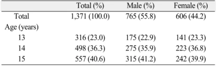
The status of dental erosion was researched on 1,380 students (13-15 years old) from four different middle schools in Yangsan and the risk factors were evaluated through this survey. The questionnaire was done prior to performing oral examination. 1,371 students (response rate = 99.3%) became final survey subjects, except nine students who did not participate on the survey or return the questionnaire. Age and gender distribution of the survey are as followed (Table 1).
2. Examination method
1) Preliminary investigation
The evaluation training was conducted prior to an epi- demiological survey by using clinical oral pictures of ado- lescents who are the same age range of the survey sub- jects. Visual Erosion Dental Examination (VEDE) sys- tem
19)was used as diagnostic criteria for dental erosion (Table 2). The oral pictures of 50 adolescents, which in- clude mandibular and maxillary labial, buccal, occlusal, and lingual surfaces, were evaluated by the diagnostic criteria of this research.
2) Oral examination
The oral examination was performed, and the status of dental erosion was investigated and recorded by one trained examiner using headlights and a dental mirror in each school’ s health unit. Food residue and plaque were removed by using gauze when necessary before the oral examination. Any retained primary teeth, missing per- manent teeth, and extensive restoration teeth were ex- cluded from the examination subject.
3. Survey method
1) Survey
The questionnaire including questions about eating habits, oral hygiene habits, and the obesity of adoles-
Table 2. VEDE system used in this study
Score Definition
0 No erosion
1 Initial loss of enamel and contour with no exposed dentin Smooth surface with silky-glazed appearance
2 Pronounced loss of enamel with no exposed dentin Possible absence of developmental ridges 3 Exposure of dentin involving < ⅓ of the surface 4 Exposure of dentin involving ⅓ - ⅔ of the surface 5 Exposure of dentin involving > ⅔ of the surface or the pulp Table 1. Distribution of gender and age of the examinees
Total (%) Male (%) Female (%) Total 1,371 (100.0) 765 (55.8) 606 (44.2) Age (years)
13 316 (23.0) 175 (22.9) 141 (23.3)
14 498 (36.3) 275 (35.9) 223 (36.8)
15 557 (40.6) 315 (41.2) 242 (39.9)
cents, was designed to analyze the correlation between dental erosion and these factors. The obesity was evalu- ated by Body Mass Index (BMI). BMI is a person’ s weight in kilograms divided by the square of height in meters. A high BMI can be an indicator of high body fat- ness
20). The eating habits include frequency, time, and methods of various drink consumption. The oral hygiene habits include frequency, time, and techniques of brush- ing teeth. The question about vomiting symptoms which is an intrinsic factor causing dental erosion, was also added.
2) Data analysis
The mean and standard deviation were used for gener- al characteristics of the subjects, and the difference be- tween each group was analyzed by an independent t- test. A chi-squared test was used to determine whether or not there is an association with the risk factors. The correlation between drink consumption habits and dental erosion was tested by binary logistic regression analysis.
SPSS 13.0 (SPSS Inc., U.S.A.) for Windows was used for statistical analysis. All statistics’significance level was set to be 0.05.
4. Ethical considerations
Deliberation exemption was approved to this study, which is in accordance with the examination of the bioethics committee of the Pusan National University Dental Hospital (PNUDH-2013-019).
Ⅲ. Results 1. The prevalence of dental erosion
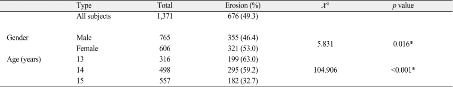
The prevalence of dental erosion about the survey sub- jects of the 1,371 adolescents is shown in Table 3. At least one tooth with dental erosion was observed in 676 out of 1,371 (49.3%) adolescents survey subject, 355 out of 765 males (46.4%) and 321 out of 606 females (53.0%), showing that it was found significantly more in females than in males. The prevalence of the age of the patient in the case of 13 year olds was 199 out of 316 people (63.0%). In the case of 14 year olds, it was 295 out of 498 people (59.2%), and in the case of 15 year olds, it was 182 out of 557 people (32.7%). Studies showed that the prevalence of dental erosion was statis- tically low as the age increased.
The comparison between maxilla and mandible about the prevalence distribution of dental erosion shows that it is significantly higher in the mandible, which was 590 people (43.0%), and 539 people (39.3%) in the case of the maxilla (p < 0.05, Table 4). When the left and right sides were compared by using the midline of maxilla and mandible as its standard, it was 675 out of 1,371 people (49.2%) in case of the left and 673 (49.1%) in case of the right, but it did not show a significant difference.
2. Distribution and severity of dental erosion The result of distribution and severity from dental ero-
Table 3. Prevalence of tooth erosion by gender and age
Type Total Erosion (%) X2 p value
All subjects 1,371 676 (49.3)
Gender Male 765 355 (46.4)
5.831 0.016*
Female 606 321 (53.0)
Age (years) 13 316 199 (63.0)
14 498 295 (59.2) 104.906 <0.001*
15 557 182 (32.7)
Independent t-test (* : p < 0.05)
Table 4. Prevalence of tooth erosion by jaw and side
Type Total Erosion (%) X2 p value
Upper 1371 539 (39.3)
3.916 0.048*
Lower 1371 590 (43.0)
Left 1371 675 (49.2)
0.006 0.939
Right 1371 673 (49.1)
Independent t-test (* : p < 0.05)
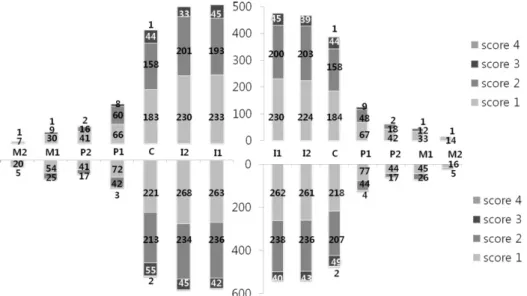
sion on each tooth showed that mandibular right lateral incisor had the highest erosion rates of 39.9%, and the lowest erosion rates on the maxillary right second molar (Table 5). The severity of dental erosion using VEDE score was indicated with the average value after mea- suring severities of each tooth. The severity was the highest on the mandibular right canine and the lowest on the maxillary left second molar (Table 6). The distri- bution and severity of dental erosional lesions of each tooth is shown in Fig. 1 and the most subjects to show dental erosion have the scores of 1 and 2.
3. The relationship between dental erosion and eat- ing habits of adolescents
1) Frequency of consuming carbonated drinks, fruit juices, milk, and fruit
Sugar free milk intake frequency was lower in the ero- sion group but it was higher for flavored milk (Table 7).
Flavored milk is a sweetened dairy drink made with milk, sugar, colorings and artificial or natural flavorings.
It was revealed that there is a significant relationship between the frequency of milk and flavored milk intake
Table 5. Distribution of prevalence value of dental erosion by tooth type
Right (%) Left (%)
Upper M2 M1 P2 P1 CI2 I1 I1 I2 CP1 P2 M1 M2
0.6 2.9 4.3 9.8 28.2 33.8 34.4 34.6 34.0 28.2 9.0 4.5 3.4 1.1
Lower 1.8 5.8 4.2 8.5 35.8 39.9 39.5 39.4 39.4 34.7 9.1 4.4 5.2 1.5
M2 M1 P2 P1 CI2 I1 I1 I2 CP1 P2 M1 M2
M2 : second molar, M1 : first molar, P2 : second premolar, P1 : first premolar, C : canine, I2 : lateral incisor, I1 : central incisor
Table 6. The VEDE score of each tooth, and the severity
Right (%) Left (%)
Upper M2 M1 P2 P1 CI2 I1 I1 I2 CP1 P2 M1 M2
1.13 1.28 1.34 1.57 1.65 1.58 1.60 1.61 1.60 1.64 1.53 1.35 1.30 1.07
Lower 1.20 1.32 1.29 1.41 1.67 1.59 1.60 1.59 1.60 1.65 1.42 1.28 1.37 1.24
M2 M1 P2 P1 CI2 I1 I1 I2 CP1 P2 M1 M2
VEDE : Visual Erosion Dental Examination, M2 : second molar, M1 : first molar, P2 : second premolar, P1 : first premolar, C : canine, I2 : lateral incisor, I1 : central incisor
Fig. 1. Distribution of prevalence value and the VEDE score of dental erosion by tooth type.
VEDE : Visual Erosion Dental Examination, M2 : second molar, M1 : first molar, P2 : second premolar, P1 : first premolar, C : canine, I2 : lateral incisor, I1 : central incisor
and dental erosion (p < 0.05). The association of dental erosion with other beverages was significantly lower (p >
0.05).
2) The methods of consuming carbonated drinks, fruit juice, milk, and fruits.
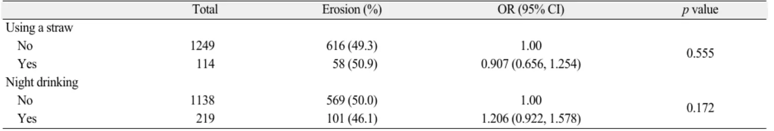
There was one investigation whether or not a straw was used as a way of consuming, and another investiga- tion whether or not drink or food was consumed before bedtime (Table 8). The group that used a straw showed the higher rate of dental erosion, but there was no sta- tistical correlation (p > 0.05).
4. The relationship between BMI of adolescents and dental erosion.
There was no statistically significant correlation be- tween dental erosion and BMI (Table 9). Commonly ac-
cepted BMI ranges are underweight : under 18.5, nor- mal weight : 18.5 to 25, overweight : 25 to 30, obese : over 3020).
5. The relationship between oral hygiene habits and dental erosion
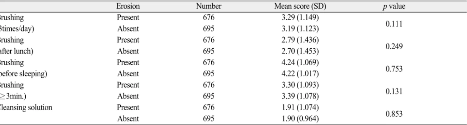
Regarding the relationship between oral hygiene habits and dental erosion, there was no statistical corre- lation (Table 10).
6. The relationship between the frequency of vomiting and dental erosion
The frequency of vomiting in adolescents and dental erosion showed no statistically significant results (Table 11).
Table 8. Association between dental erosion and intake habits
Total Erosion (%) OR (95% CI) p value
Using a straw
No 1249 616 (49.3) 1.00
Yes 114 58 (50.9) 0.907 (0.656, 1.254) 0.555
Night drinking
No 1138 569 (50.0) 1.00
Yes 219 101 (46.1) 1.206 (0.922, 1.578) 0.172
Binary logistic regression (* : p < 0.05) OR : odd ratio, CI : confidence interval
Table 7. Association between dental erosion and frequency of consumption of variable drinks and fruit
Erosion Number Mean score (SD) p value
Carbonated drinks Present 676 1.92 (0.789)
0.079
(sugar free) Absent 695 2.00 (0.859)
Carbonated drinks Present 676 1.97 (0.794)
0.067
(flavored) Absent 695 2.05 (0.884)
Fruit juice Present 676 2.01 (0.806)
0.114
(sugar free) Absent 695 2.09 (0.844)
Fruit juice Present 676 2.03 (0.768)
0.413
(flavored) Absent 695 2.07 (0.821)
Milk Present 676 2.57 (1.097)
0.003*
(sugar free) Absent 695 2.74 (1.031)
Milk Present 676 2.55 (0.769)
0.012*
(flavored) Absent 695 2.43 (0.888)
Fruit Present 676 2.94 (1.457)
0.141
Absent 695 3.05 (1.453)
Chi-squared test (* : p < 0.05)
Mean score : The frequency of consumption of variable drinks and fruit was converted into scores and the mean values are shown in the table above.
Frequency : §5: ≥three time/day, 4: twice/day, 3: once/day, 2: <once/day, 1:none SD : Standard deviation
Ⅳ. Discussion
Adolescence is a stage gets into the permanent denti- tion, and if exposed to oral disease caused by neglected oral hygiene at this period, there would have an adverse effect on oral health. Dugmore and Rock
15)conducted a survey of dental erosion for the age of 12 and two years later in the group of 1,753 adolescents. At the age of 12, 59.7% of the adolescents had an erosion lesion and 2.7%
of them had dentin exposure. Two years later, dentin exposure was increased by 8.9% in the dental erosion test, reporting that the severity of dental erosion was significantly higher after two years. Severity of dental
erosion was increased over time which showed a newly created pattern. This suggests that it could fail to mini- mize damage to the permanent dentition unless the pre- ventive treatment is performed by early detection of den- tal erosion. On the basis of these reasons, this study was conducted to investigate the prevalence and risk factors of dental erosion in the permanent dentition.
In the initial stage of dental erosion, it is hard to make a diagnosis of erosion because it appears without clinical symptoms of pain or sensitivity. Wang and Lussi
21)said that it is important to determine by using the clinical characteristics of dental erosion because there is no de- vice can accurately measure dental erosion and its pro-
Table 9. Association between dental erosion and BMITotal Erosion (%) OR (95% CI) p value
BMI
Under 403 214 (53.1) 1.00
Normal 662 326 (49.2) 1.073 (0.765, 1.505) 0.681
Over 100 45 (45.0) 0.912 (0.662, 1.256) 0.572
Obesity 124 63 (50.8) 0.746 (0.459, 1.212) 0.236
Binary logistic regression (* : p < 0.05)
BMI: Body mass index, OR : odd ratio, CI : confidence interval
Table 10. Association between dental erosion and oral hygiene habits
Erosion Number Mean score (SD) p value
Brushing Present 676 3.29 (1.149)
0.111
(3times/day) Absent 695 3.19 (1.123)
Brushing Present 676 2.79 (1.436)
0.249
(after lunch) Absent 695 2.70 (1.453)
Brushing Present 676 4.24 (1.069)
0.753
(before sleeping) Absent 695 4.22 (1.017)
Brushing Present 676 3.30 (1.093)
0.131
(≥3min.) Absent 695 3.39 (1.078)
Cleansing solution Present 676 1.91 (1.074)
0.853
Absent 695 1.90 (0.964)
Chi-squared test (* : p < 0.05)
Mean score : The frequency of oral hygiene habits was converted into scores and the mean values are shown in the table above.
Frequency : §1: not at all, 2: pretty negative, 3: normal, 4: pretty positive, 5: very positive SD : Standard deviation
Table 11. Association between dental erosion and vomiting
Erosion Number Mean score (SD) p value
Vomiting Present 676 4.49 (0.914)
0.371
Absent 695 4.54 (0.821)
Chi-squared test (* : p < 0.05)
Mean score : The frequency of vomiting was converted into scores and the mean values are shown in the table above.
Frequency : §5: not at all, 4: pretty negative, 3: normal, 2: pretty positive, 1: very positive SD : Standard deviation
gression. Larsen et al.
22)reported that a clinician is hard to distinguish between healthy enamel and enamel le- sions.
The clinical characteristics of dental erosion are as fol- lows
21): when affected in an enamel, tooth surface is smooth and glossy, and the lesion is often located in the crown side from the cemento-enamel junction. This is because health enamel margin is appeared in the gingiva where gingival cervical fluid and plaque exist. Plaque blocks the acid and gingival cervical fluid neutralize it.
When the occlusal erosion is progressed, the position of the restoration is higher than the height of the tooth surface of the adjacent tooth. In the severe case, the en- tire occlusal morphology is lost which can lead to dentin and pulp exposure
23).
The dental erosion should be distinguished from attri- tion
24). If an attrition is an exclusive cause, it should be appeared in the occlusal contact point and does not ap- pear from the labial and lingual side of teeth but can oc- cur from the lateral and anterior movement of the mandible. The morphological features of an attrition are shiny, flat, and sharp edge. Also, attrition appears con- sistently in the antagonist teeth with the teeth that the attrition appears. If attrition appears in the maxillary anterior teeth, attrition not appeared in the mandibular anterior teeth is not possible. In case of dental erosion, it can not be appeared in the occlusal contact part like labial and lingual surface of teeth.
The cupping phenomenon is shown rather than show- ing morphologically smooth, polished, and flat surface of teeth and the cusp and groove are round rather than sharp. It does not appear in the maxillary and mandibu- lar incisor together, but each appears. This appears in the person who has an anterior open bite, and when try- ing anterior transposition it can determine the type of abnormalities in the anterior teeth although having no contact on the anterior teeth, which can be thought of as tooth erosion caused by acid. It was difficult to distin- guish among attrition, abrasion, and dental erosion ac- curately. This is because there exists a possibility that attrition, abrasion, and dental erosion often occur at the same time. Because teeth become weak followed by demineralized enamel layer cased by dental erosion, it becomes more vulnerable to the attrition and abrasion.
In the past, most indices to clinically diagnose dental erosion were to modify the Eccles index
25)and Smith and knight index
26). Many indices were developed afterwards but dental erosion index universally agreed does not ex-
ist so far
27). The ideal characteristics of dental erosion proposed by Bardsley
28)are as follows. It should be sim- ply usable and understandable. Also, the criteria of scores should be clear and reproducible. When investi- gating the cause, it should be useful in the prevention and monitoring. Lastly, it should be able to be used as an epidemiologic and clinical tool. The two kinds of index most commonly used to assess dental erosion are Visual Erosion Dental Examination (VEDE) and Basic Erosive Wear Examination (BEWE)
29). VEDE and BEWE are widely used because of their high reproducibility and ap- propriateness, but they are not free from error. VEDE system and BEWE system are similar in many ways.
But VEDE system conducts a test without distinction of tooth surface and divides the affected part into a two - step and the range of affected dentin into 1/3 unit. Thus when using the VEDE system it tends to increase index score of dental erosion. In the early stage of enamel ero- sion, the VEDE system which departmentalizes affected enamel displays easier tendency to commit errors than BEWE system
19,30). In this study, the VEDE system was applied in the initial state mostly limited to the enamel in order to separate the dental erosion affected to the enamel in detail. It was reported that distinguishing the early enamel lesion from the health enamel exists the difficulty, but they tried to overcome it through pre- training
31).
For the subject of this investigation, 13-15 year-old
middle school student of 1,371 people, the prevalence of
dental erosion was about 676 people (49.3%). This was
relatively higher than other research papers which has
used the same dental erosion index targeting a similar
age. Arnadottir et al.
32)reported that the prevalence of
dental erosion for 1,507 people whose age is 12 and 15
year old living in Iceland was 349 people (23.1%) and
Mulic et al.
33)reported that the prevalence of dental ero-
sion for 1,456 people of 18 year-old living in Norway was
554 people (38.0%). There exist many studies that has
mentioned dental erosion is observed more significantly
in males than in females
33-35). This is because females
have a significantly thicker enamel than males, while
males have stronger muscular strength and masticatory
muscle, and more consumption of carbonated drinks
than females
36). However, this study appeared to be sig-
nificantly higher in females and males (p < 0.05) as a re-
sult of 355 out of 765 in case of men (46.4%) and 321
out of 606 in case of women (53.0%). Bere et al.
37)re-
ported that female has a higher tendency in the intake
of fruits and juices. Certain research paper has reported that in one’ s sleep, if the acidic product touches the tooth, dental erosion often occurs which is due to re- duced saliva production. Also, the fact that female has lower salivary secretion than male could be another rea- son. Wang and Lussi
21)suggested that the flow rate and buffering capacity of saliva is one of the etiological fac- tors, so it is important to determine them.
It was reported that the consumption of acidic bever- ages had a significant contribution to the presence and progression of dental erosion increases
38). However, this study has reported that the relationship between the dental erosion and the acidic beverage is not statistically significant, but male has higher intake number of the acidic beverage than female. Therefore, the link between milk intake and dental erosion was statistically signifi- cant. Amaechi et al.
39)and Gedalia et al.
12)have reported that intake of dairy products such as fresh milk reminer- alizes the softened tooth surfaces and this shows similar result with the study has reported that it would be used in situations such as erosion. According to Magalha ̃es et al.
40), the fluorine-containing milk will prevent dental erosion and this effects will be increased at higher con- centrations of fluoride. Also, in case of affected dentin by erosion, drinking the milk does not contain fluorine after the erosion will prevent erosion afterward.
The prevalence in relation to the subject’ s age is 199 out of 316 people (63.0%) in case of 13 year olds, 295 out of 489 people (59.2%) in case of 14 year olds, and 182 out of 557 people (32.7%) in case of 15 year olds, which shows as age increases, the prevalence of dental erosion is statistically and significantly low. As age in- creases, the rate of dental erosion caused by consump- tion of fruit, flavored milk and acidic beverages such as sugar free carbonated drinks and fruit juice was low (p <
0.05).
Most of the research on dental erosion was mainly aimed at the maxillary anterior teeth and mandibular molars. However, these surveys do not provide an infor- mation about how the dental erosion appears on all den- tition
41). Becuase especially national survey of adoles- cents in this study was insufficient, this study re- searched on distribution and severity of dental erosion in all the permanent teeth. Most of dental erosion affected on dentin was incisal edge of incisor and canine, and the buccal surface of the maxillary and mandibular anterior teeth had higher tendency compared to posterior teeth
9). In this study, the prevalence and severity of dental ero-
sion is definitely higher in anterior teeth that in posteri- or teeth, and the comparison between maxilla and mandible about the prevalence of dental erosion shows that it is significantly higher in the mandible as 43.0%
than in maxilla as 39.3%. Sognnaes et al.
42)pronounced that mandibular teeth showed a higher frequency of ero- sion-like lesions than the corresponding maxillary teeth (21.0% compared to 13.0%). The highest percentage of erosion-like lesions was found in mandibular incisors (28.0%). This study supports that the frequency of den- tal erosion was higher in mandibular teeth.
Recently, the attention on the influence of obesity problem of adolescents on oral disease is increasing, and so BMI using height and weight is used in order to in- vestigate the correlation between the BMI of adolescents and dental erosion. Because obesity is caused by lifestyle, such as dietary habits rather than genetic or endocrine factors
41), there is a tendency to consume a large amount of the acidic food and beverage in case of obese patients. Hence, the research to investigate the correlation between obesity and dental erosion has been progressed
43). It was reported that the prevalence of den- tal erosion was low in case of underweight and high in case of overweight and obesity compared to healthy peo- ple, but there was no significant difference in this study.
McGuire et al.
44)reported that underweight adolescents had the lowest prevalence of dental erosion, however there was no significant difference among the groups showing the results that are similar with this study.
Future studies examining the relationship between obe- sity and dental erosion are required to provide dentists the ability to say whether obesity could be one of the risk factors for dental erosion.
The result has shown no link between oral hygiene
and dental erosion, but because the prevalence of dental
erosion is significantly decreased in accordance with the
oral hygiene condition by age (p < 0.05), other studies
will be needed for this. The result about the correlation
between dental erosion and milk intake has come, hence
we need to further study in order to find out the compo-
nent of milk that contributes on this result. Although
findings of this study are particularly important to den-
tists for the purpose of diagnosing dental erosion at an
early age, limitations of this study should be noted. This
study was cross-sectional study which can only observe
the current state when the changes appearing over time
cannot be observed. Research on permanent tooth ero-
sion is being constantly published, but because the re-
search on primary tooth erosion is uncommon except for the research paper published by Yu et al.
18), more stud- ies about it will be needed. Further research is also needed to find more significantly related risk factor.
Ⅴ. Conclusion
For the purpose of realizing the status of dental ero- sion for adolescents and analyzing and evaluating relat- ed risk factors, the clinical examination and the ques- tionnaire were done for a total of 1,371 adolescents aged 13-15 years attending in certain middle schools in Yangsan. Results of analyzing the prevalence of dental erosion and relation between oral hygiene and dietary habits of teenagers were as follows. The data showed that 676 adolescents out of 1,371 had dental erosion (49.3%), and dental erosion was observed in 355 people out of 765 males (46.4%) and 321 people out of 606 fe- males (46.4%), concluding that the prevalence of dental erosion was significantly higher in female than in male (p < 0.05). The prevalence of dental erosion in mandible (43.0%) is higher than in maxilla (39.3%) (p < 0.05).
There was no significant difference between the preva- lence of right and left sides in maxilla and that of mandible (p > 0.05). Comparing to the group of non- dental erosion, the group of dental erosion was signifi- cantly high in flavored milk intake (p < 0.05), but low in sugar-free milk intake (p < 0.05). No other significant relation between dental erosion and the intake of other beverages, frequency of oral hygiene, and BMI was de- tected (p > 0.05). In conclusion, the prevalence of dental erosion as 49.3% for adolescents in Yangsan, Korea was higher than worldwide prevalence using VEDE scoring system. The prevalence of dental erosion in female was higher than in male, and the frequency of intake of fla- vored milk and sugar-free milk was investigated as a contributing factor of dental erosion.
References
1. Kim JB, Kim JH, Paik DI, et al. : Preventive den- tistry 3rd edition. KMS, Seoul, 183, 1999.
2. Ganss C : Definition of erosion and links to tooth wear. Monogr Oral Sci, 20:9-16, 2006.
3. Bartlett DW, Evans DF, Anggiansah A, et al. : A study of the association between gastro-oesophageal reflux and palatal dental erosion. Br Dent J, 181:
125-131, 1996.
4. Federation Dentaire International(FDI) : Global golas for oral health in the year 2000. Int Dent J, 32:74-77, 1982.
5. Emerich K, Adamowicz-Klepalska B : Trends in dental caries experience among children and adoles- cents in northern Poland between 1995 and 2003.
Community Dent Health, 27:218-221, 2010.
6. Marthaler TM : Changes in dental caries 1953- 2003. Caries Res, 38: 173-181, 2004.
8. Lussi A, Jaeggi T : Erosion-diagnosis and risk fac- tors. Clin Oral Invest, 12:S5-13, 2008.
9. Al-Dlaigan YH, Shaw L, Smith A : Dental erosion in a group of British 14-year school children. Part I.
Prevalence and influence of differing socioeconomics backgrounds. Br Dent J, 190:145-149, 2001.
10. Nunn JH, Gordon PH, Walker A, et al. : Dental erosion - changing prevalence? A review of British national children surveys. Int Paediatr Dent, 13:98- 105, 2003.
11. El-Aidi H, Bronkhorst EM, Truin GJ, et al. : Dynamics of tooth erosion in adolescents: a 3-year longitudinal study. J Dent, 38:131-137, 2010.
12. Gedalia I, Dakuar A, Rahamim E et al. : Enamel softening with Coca-Cola and rehardening with milk or saliva. Am J Dent, 4:120-122, 1991.
13. Huysmans MCDNJM, Chew HP, Ellwood RP : Clinical studies of dental erosion and erosive wear.
Caries Res, 45:60-68, 2011.
14. Hamasha AA, Zawaideh FI, Al-Hadithy RT : Risk indicators associated with dental erosion among Jordanian school children aged 12-14 years of age.
Int J Paediatr Dent, 24:56-68, 2014.
15. Dugmore CR, Rock WP : The prevalence of tooth erosion in 12-year-old children. Br Dent J, 196:279- 282, 2004.
16. The sixth Korea National Health and Nutrition Examination Survey (KNHANES). Ministry of Health and Welfare, Korea Centers for Disease Control and Prevention, 2013.
17. Kim HJ, Kim S, Jeong TS : The risk indicators of dental erosion in 8- and 9-year-old school children in Yangsan. J Korean Acad Pediatr Dent, 40:1-10, 2013.
18. Yu SG, Lee CH, Kim S, et al. : Prevalence and associated risk factors of dental erosion in 9- and 10-year-old children in Busan. J Korean Acad Pediatr Dent, 40:11-20, 2013.
19. Mulic A, Tveit AB, Skaare AB, et al. : Reliability of
two clinical scoring systems for dental erosive wear.
Caries Res, 44:294-299, 2010.
20. Cole TJ. A method for assessing age-standardized weight-for-height in children seen cross-sectionally.
Ann Hum Biol, 6:249-68, 1979.
21. Wang X, Lussi A : Assessment and management of dental erosion. Dent Clin N Am, 54:565-578, 2010.
22. Larsen MJ, Poulsen S, Hansen I : Erosion of the teeth: prevalence and distribution in an group of Danish school children. Eur J Paediatr Dent, 6:44- 47, 2005.
23. Gamdara BK, Treulove EL : Diagnosis and manage- ment of dental erosion. J Contemp Dent Pract, 1:
16-23, 1999.
24. Spear F : A patient with severe wear on the posteri- or teeth and minimal wear on the anterior teeth. J Am Dent Assoc, 140:99-104, 2009.
25. Eccles JD : Dental erosion of nonindustrial origin. A clinical survey and classification. J Prosthet Dent, 42:649-653, 1979.
26. Smith BG, Knight JK : An index for measuring the wear of teeth. Br Dent J, 156:435-438, 1984.
27. Bartlett D, Dugmore CR : Pathological or physiologi- cal erosion-is there a relationship to age? Clin Oral Invest, 12:S27-S31, 2008.
28. Bardsley PF : The evolution of tooth wear indices.
Clin Oral Invest, 12:S15-S19, 2008
29. Zhang S, Chau AMH, Chu CH, et al. : Dental caries and erosion status of 12-year-old Hong Kong chil- dren. BMC public Health, 14:7, 2014.
30. Holbrook WP, Ganss C : Is diagnosing exposed den- tine a suitable tool for grading erosive loss? Clin Oral Invest, 12:S33-S39, 2013.
31. Al-Dlaigan YH, Shaw L, Smith A : Dental erosion in an group of British 14-year-old, school children.
Part II: Influence of dietary in take. Br Dent J, 190:258-261, 2001.
32. Arnadottir IB, Holbrook WP, Agustsdottir H, et al. : Prevalence of dental erosion in children: a national survey. Community Dent Oral Epidemiol, 38:521- 526, 2010.
33. Mulic A, Skudutyte-Rysstad R, Tveit AB, et al. : Risk indicators for dental erosive wear among 18-yr- old subjects in Oslo, Norway. Eur J Oral Sci, 120:
531-538, 2012.
34. Milosevic A, Young PJ, Lennon MA : The prevalence of tooth wear in 14-year-old school children in Liverpool. Community Dent Health, 11:83-86,
1994.
35. Smith TM, Olejniczak AJ, Hublin JJ, et al. : Modern human molar enamel thickness and enamel- dentine juntion shape. Arch Oral Biol, 51:974-995, 2006.
36. Bardsley PF, Taylor S, Milosevic A : Epidemiological studies of tooth wear and dental erosion in 14-year- old children in North West England. Part 1: the relationship with water fluoridation and social depri- vation. Br Dent J, 197:413-416, 2004.
37. Bere E, Brug J, Klepp KI : Why do boys eat less fruit and vegetables than girls? Public Health Nutr, 11:321-325, 2008.
38. Lussi A, Jaeggi T, Zero D : The role of diet in the aetiology of dental erosion. Caries Res, 38:34-44, 2004.
39. Amaechia BT, Highamb SM : Dental erosion: possi- ble approaches to prevention and control. J Dent, 33:243-252, 2005.
40. Magalha ̃es AC, Levy FM, Buzalaf MA, et al.
Inhibition of tooth erosion by milk containing differ- ent fluoride concentrations: an in vitro study. J Dent, 42:498-502, 2014
41. Correa MSNP, Correa FNP, Mendes FM, et al. : Prevalence and associated factors of dental erosion in children and adolescents of a private dental prac- tice. Int J Paediatr Dent, 21:451-458, 2011.
42. Sognnaes RF, Wolcott RB, Xhonga FA. : Dental ero- sion. I. Erosion-like patterns occurring in association with other dental conditions. J Am Dent Assoc, 84:
571-6, 1972.
43. Park SH, Choi BY, Oh HW, et al. : Relationship between diet behavior and dental caries experience among elementary schoolers. J Korean Acad Dent Health, 34:327-337, 2010.
44. McGuire J, Szabo A, Okunseri C, et al. : Erosive tooth wear among children in the united states:
relationship to race/ethnicity and obesity. Int J
Paediatr Dent, 19:91-98, 2009.
양산시 거주 13-15세 학생의 치아침식증 유병율과 위험요소