• 검색 결과가 없습니다.

Synthesis of Lu<sub>2.94</sub>Ce<sub>0.06</sub>MgAl<sub>3</sub>SiO<sub>12</sub> phosphor and its photoluminescent properties

N/A
N/A
Protected

Academic year: 2021

Share "Synthesis of Lu<sub>2.94</sub>Ce<sub>0.06</sub>MgAl<sub>3</sub>SiO<sub>12</sub> phosphor and its photoluminescent properties"

Copied!
6
0
0

로드 중.... (전체 텍스트 보기)

전체 글

(1)

http://dx.doi.org/10.6111/JKCGCT.2015.25.3.121

Synthesis of Lu 2.94 Ce 0.06 MgAl 3 SiO 12 phosphor and its photoluminescent properties

Jung-Il Lee, Tae Wan Kim, Ji Young Shin and Jeong Ho Ryu

Department of Materials Science and Engineering, Korea National University of Transportation, Chungju 380-702, Korea (Received June 5, 2015)

(Revised June 11, 2015) (Accepted June 15, 2015)

Abstract A novel Ce

3+

doped Lu

3

MgAl

3

SiO

12

phosphor (Lu

2.94

Ce

0.06

MgAl

3

SiO

12

) was successfully synthesized by a conventional solid-state reaction at 1450

o

C for 5 h. The crystal structure of the synthesized phosphor powder was characterized by X-ray diffraction and Rietveld refinement. The prepared phosphor powder showed a broad peak at 550 nm, and the temperature dependence on photoluminescence properties of the prepared Lu

2.94

Ce

0.06

MgAl

3

SiO

12

phosphor was investigated from 300 to 525 K. The activation energy for thermal quenching was determined by Arrhenius fitting. The experimental results clearly indicate that prepared Lu

2.94

Ce

0.06

MgAl

3

SiO

12

phosphor has great potential for a down-conversion yellow phosphor in white light-emitting diodes.

Key words Lu

2.94

Ce

0.06

MgAl

3

SiO

12

, Phosphor, Photoluminescence, Thermal quenching

1. Introduction

The first commercially available white LED (light emitting diodes) based on phosphors was produced by Nichia Co., which was also first to manage to make the blue LED. Nichia used a blue light emitting gallium indium nitride (GaInN) chip coated with yellow phos- phor Y 3 Al 5 O 12 : Ce 3+ , well known as YAG : Ce 3+ [1].

With the perspective of a highly efficient light source, the LED market will have an enormous growth and the white LED will be a likely candidate for the replace- ment of the light bulb. However, the color temperature of YAG : Ce 3+ phosphor used for luminescence convert- ing LED is too high to use in warm white LED applica- tion because of lacking red component [2]. The color temperature of warm white, cool white and daylight white area round 3300 K, 4200 K and 6400 K, respec- tively. For general indoor lighting application, “warmer”

white light is recommended [3].

To meet the requirements for general illumination lighting, two major modifications have been developed.

One is the modification of the YAG : Ce system, includ- ing co-doping rare earths or transition metal ions, which can increase red component [4]. The problem is the enhancement of the red component efficiency is not obvious, and almost all bright and thermal stability of

modification YAG : Ce are obviously declined. The other is exploring novel red phosphors which are usually based on Eu 2+ doped nitrides phosphors [5]. However, these phosphors require critical preparation conditions like high temperature, high pressure and expensive raw materials. Besides, they have to be blended with YAG : Ce phosphor to make white lamps, possibly leading to variations during lamp manufacturing.

It is possible and valid to modify the typical garnet composition to obtain a significant red-shift emission spectrum, because the energy position of the lowest Ce 3+

5d level could be justified by changing the crystal field splitting and the covalency of Ce 3+ -O 2− . In addition, the Ce 3+ doped garnet host lattices are the only oxide phos- phors that could absorb blue light and emit from yellow to orange-red light [6]. In a practical sense, phosphors should not only have redder spectra for lower cor- related color temperature (CCT) and high color render- ing index (CRI) but also high quantum efficiency (QE) and high thermal stability for warm white LED phos- phors. In this study, we present a solid-state synthesis inducing Mg 2+ -Si 4+ pairs into Lu 3+ based garnet, and dis- cuss its crystal structure. Photoluminescence at room temperature and thermal quenching at high tempera- tures are investigated.

2. Experimental

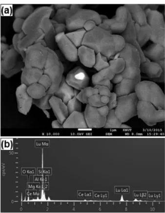
The Ce 3+ doped Lu 3 MgAl 3 SiO 12 (Lu 2.94 Ce 0.06 MgAl 3 SiO 12 )

Corresponding author

Tel: +82-43-841-5384

Fax: +82-43-841-5380

E-mail: [email protected]

(2)

was synthesized by the conventional solid-state reaction method. Lu 2 O 3 (Kanto Chemical, 99.99 %), CeO 2 (Kanto Chemical, 99.99 %), MgO (Kanto Chemical, 99.99 %), and SiO 2 (Kanto Chemical, 99.9 %) were used as start- ing materials. Cerium concentration was fixed at 6.0 mol%. All starting materials were weighted out as the desired stoichiometry and thoroughly mixed using an agate mortar with small amount of ethanol. The mix- ture was first calcined at 800 o C for 2 h using alumina crucible in air then fired at 1450 o C for 5 h in N 2 atmo- sphere. Finally, the phosphor sample was cooled to room temperature in the furnace and ground with an agate mor- tar. The fired phosphor sample (Lu 2.94 Ce 0.06 MgAl 3 SiO 12 ) showed yellow body color.

Powder X-ray diffraction (XRD) data were collected on a Bruker D8 powder X-ray diffractometer with Cu K α radiation ( λ = 1.54178 Å) operating 40 kV and 30 mA.

Step scans were performed from 60 o to 110 o (2 θ) with as step size of 0.01 o . Refined lattice parameters of XRD pat- terns were fit by Rietveld refinements using FULLPROF program [7]. Microstructure and surface morphology of the Lu 2.94 Ce 0.06 MgAl 3 SiO 12 powder were observed by scan- ning electron microscope (Hitachi S-4800, Japan). Photo- luminescence excitation (PLE) and emission (PL) spectra were measured by fluorescence spectrometer (Scinco FS-2) system in a temperature range of 25 and 275 o C using a 450 W xenon lamp as an excitation source.

3. Results and Discussion

Crystallographic information of the Lu 2.94 Ce 0.06 MgAl 3 SiO 12 phosphor is one of the important factors affecting the photoluminescence properties, which closely relates with the crystal field strength surrounding the ions in each crystallographic sites. Crystallographic data of the Lu 2.94 Ce 0.06 MgAl 3 SiO 12 was extracted by the Rietveld refinements of the XRD profile of the Lu 2.94 Ce 0.06 MgAl 3- SiO 12 , as shown in Fig. 1. Approximate structural parameters for Lu 2.94 Ce 0.06 MgAl 3 SiO 12 calculation was obtained from the data, previously reported for Lu 3 Al 5 O 12 (LuAG; JCPDS 73-1368) [8]. The LuAG was decided as the initial structure model for Lu 2.94 Ce 0.06 MgAl 3 SiO 12 calculation due to the similarity of the crystal structure;

both of them belong to the garnet structure family (cubic, space group #230), with the general formula (A 3 )[B 2 ]<C 3 >O 12 , where ( ), [ ] and < > denote dodeca- hedral, octahedral and tetrahedral coordination, respec- tively [9]. In the crystal structure of LuAG, Lu 3+ ions and Al 3+ occupy the dodecahedral and octahedral/tetra-

hedral sites, respectively. On the other hand, in the crys- tal structure of LCMAS, dodecahedral sites are occupied by Lu 3+ and Ce 3+ . Also, Mg 2+ /Al 3+ and Al 3+ /Si 4+ occupy octahedral and tetragonal sites, respectively.

Refined structure parameters of Lu 2.94 Ce 0.06 MgAl 3 SiO 12 are listed in Table 1. The refinement confirmed that Lu 2.94 Ce 0.06 MgAl 3 SiO 12 is cubic with space group Iad, and lattice parameter is refined as 11.942 Å. Wyckoff sites of 24c, 16a, 24d and 96h are occupied by Lu/Ce, Mg/Al1, Si/Al2 and O, respectively. As shown in Table 1, the calculated occupancies of ions from XRD pattern are quite well matched with experimental. The compari- son of lattice parameters and cell volume between LuAG and Lu 2.94 Ce 0.06 MgAl 3 SiO 12 is shown in the Table 2. The deviation of lattice parameter is 0.0359 Å and the deviation of oxygen atomic coordinates (x, y, z) is

−0.0033, −0.0006 and 0.0023, respectively.

Cell volume and lattice parameters of Lu 2.94 Ce 0.06 MgAl 3 SiO 12 are larger than that of LuAG (11.9064 Å) because the smaller Al 3+ is partly substituted by larger Mg 2+ in the octahedral sites. Also, smaller Lu 3+ is partly Fig. 1. Rietveld refinements of XRD patterns of Lu

2.94

Ce

0.06

MgAl

3

SiO

12

phosphor with positions of all the reflection (orange) and a difference profile (blue); Measured (black) and

calculated (red).

Table 1

Refined structural parameters for Lu

2.94

Ce

0.06

MgAl

3

SiO

12

phosphor

Atom Wyckoff x y z Occupancy

Lu 24c 0.125 0 0.25 2.77

Ce 24c 0.125 0 0.25 0.22

Al1 16a 0 0 0 1.01

Mg 16a 0 0 0 1.01

Al2 24d 0.375 0.25 0 1.98

Si 24d 0.375 0.25 0 0.98

O 96h −0.033 0.053 0.153 12.00

(3)

replaced by larger Ce 3+ which results in the increase of the cell parameters and volume of Lu 2.94 Ce 0.06 MgAl 3 SiO 12 . In the same manner, smaller lattice constant of the Lu 2.94 Ce 0.06 MgAl 3 SiO 12 than Y 3 Al 5 O 12 (YAG; 12.066 Å) is likely due to the smaller ionic size of Lu 3+ than Y 3+ in the dodecahedral sites [10]. The lattice parameters of Lu 2.94 Ce 0.06 MgAl 3 SiO 12 between those of LuAG and YAG imply that a solid solution of Lu 2.94 Ce 0.06 MgAl 3 SiO 12 successfully formed. However, small amount of second- ary phases was observed as shown in Fig. 1. The forma- tion of the secondary phases might be possibly due to the reducing conditions during heating process, which leads to the local oxygen deficiency for the formation of the secondary phases.

Fig. 2 displays the crystal structure of Lu 2.94 Ce 0.06 MgAl 3 SiO 12 phosphor based on the results of refine- ments. As explained above, Lu/Ce, Mg/Al1, Si/Al2, and O are randomly distributed on the dodecahedral, octahe- dral, and tetrahedral sites, respectively. Every (Ce/Lu)O 8 dodecahedron is surrounded by four (Ce/Lu)O 8 dodeca- hedra, four (Mg/Al1)O 6 octahedra and six (Si/Al2)O 4 tet- rahedra. The result implies that every coordination O 2−

of Ce 3+ is shared by four cations (two Lu 3+ , one Mg/Al1 and one Si/Al2), which shows that the photoluminescence properties of Lu 2.94 Ce 0.06 MgAl 3 SiO 12 phosphor is affected by (Ce/Lu)O 8 as well as (Mg/Al1)O 6 and (Si/Al2)O 4 [11].

Fig. 2. Simulated crystal structure of Lu

2.94

Ce

0.06

MgAl

3

SiO

12

phosphor.

Fig. 3. (a) SEM photograph and (b) EDX profile for the Lu

2.94

Ce

0.06

MgAl

3

SiO

12

phosphor powder.

Table 2

The lattice parameters and the oxygen atomic coordinates obtained by Rietveld refinement

Sample Lattice Volume

3

)

Oxygen atomic coordinates

R

p

R

wp

x y z

LuAG 11.906 1687.5 −0.0294 0.0538 0.1510 0.046 -

Lu

2.94

Ce

0.06

MgAl

3

SiO

12

11.942 1703.2 −0.0327 0.0532 0.1533 0.104 0.131

For investigation of particle size and morphology, SEM images of the Lu 2.94 Ce 0.06 MgAl 3 SiO 12 phosphor is shown in Fig. 3(a). Fig. 3(a) shows homogeneous and irregular particle shape with diameter of 1~5 μm. The EDX spectra in Fig. 3(b) obtained from Lu 2.94 Ce 0.06 MgAl 3 SiO 12 particles show the presence of Lu, Ce, Mg, Al, Si and O with an approximately correct stoichiome- try and the absence of any impurity. It can be seen the particle surface is not smooth and some pits exist, this is owing to the agglomerate by lots of small particles to form a big particle in the calcination process. In addi- tion, the average particle size is found to be less than 10 µm, which is suitable for the practical application.

The room temperature Photoluminescence excitation

(PLE) and emission (PL) spectra of the Lu 2.94 Ce 0.06 MgAl 3

SiO 12 phosphor powder heat-treated at 1450 o C are

shown in Fig. 4(a). As expected for a garnet host, the

(4)

lowest Ce 3+ 4f 1 ↔ 5d 1 absorption transition is in the blue spectral region, leading to greenish yellow to orange color.

The excitation spectrum of the Lu 2.94 Ce 0.06 MgAl 3 SiO 12 phosphor powder is shown in the left of Fig. 4. The excitation spectrum exhibit two excitation bands of Ce 3+ , one 5d band near at 450 nm, and the other 5d band level near at 330 nm, which correspond to the two low- est 5d levels of Ce 3+ [12].

The electronic configuration of rare-earth element Ce 3+

is [Xe]4f 1 5d 0 . The wide absorption band and the emis- sion peaks of Ce 3+ originate from the 4f ↔ 5d transi- tion. The splitting of the Ce 3+ energy level is induced by the covalency effect (nephelauxetic effect) and the crys- tal field effect, allowing the PL performance of Ce 3+ to be tuned. The energy configuration of Ce 3+ is shown in Fig. 4(b). The PLE spectra show a broad excitation peak at 450 nm, which is attributed to the absorption of the allowed transition from 4f 1 to 5d 1 of Ce 3+ . This makes Lu 2.94 Ce 0.06 MgAl 3 SiO 12 phosphor suitable for fabrication with commercial blue LED chips. The PL spectra show an asymmetric emission peak at around 550 nm under excitation at 450 nm. The broad peak is a combination of two peaks, which derive from the energy transfer of 5d 12 F 5/2 and 2 F 7/2 of Ce 3+ , respectively. The splitting of the 4 f 1 ground state of Ce 3+ into 2 F 5/2 and 2 F 7/2 is due to the spin-orbit coupling effect [13].

Thermal quenching property is one of the most import- ant technological parameters for phosphors applied to white-LEDs. The temperature dependent PL properties of the prepared Lu 2.94 Ce 0.06 MgAl 3 SiO 12 phosphor pow- der was investigated in a temperature range from 300 to

Fig. 4. (a) The room temperature photoluminescence excitation (PLE) and emission (PL) spectra of the Lu

2.94

Ce

0.06

MgAl

3

SiO

12

phos- phor. The inset figure shows the digital photograph for the Lu

2.94

Ce

0.06

MgAl

3

SiO

12

phosphor. (b) Schematic diagram for electronic

energy configuration of Ce

3+

in the Lu

2.94

Ce

0.06

MgAl

3

SiO

12

phosphor.

Fig. 5. (a) The emission spectra measured with increasing tem- perature from 300 to 525 K for the Lu

2.94

Ce

0.06

MgAl

3

SiO

12

phos- phor powder. (b) Temperature dependence of emission intensity in the temperature range. The graph in the inset depicts an Arrhenius fitting of the emission intensity in the measuring temperature range with calculated activation energy for thermal

quenching ( ΔE).

(5)

525 K and shown in Fig. 5(a). At 425 K, the emission intensity was 76 % of those measured at room tempera- ture which was 10 % higher than that of the commer- cial Y 3 Al 5 O 12 : Ce 3+ (YAG : Ce, P46-y3) phosphor. Upon heating the Lu 2.94 Ce 0.06 MgAl 3 SiO 12 phosphor powder, the dwindling of emission intensity and broadening of band- widths (FWHM) are shown clear, which can be explained by the thermal quenching in the configurational coordi- nate model [14]. The low potential curve and the high one represent the total energy of the ground state of 4f and the excited state of 5d 1 , respectively. The equilib- rium positions of the two states are different from each other because of the different spatial distribution of the electron orbitals. The excited luminescent center is ther- mally activated through phonon interaction, and then thermally released through the crossing point between the excited state and the ground state in the configura- tional coordinate diagram. This non-radiative transition induced by thermal activation is strongly dependent on temperature, which results in the thermal quenching of the emission intensity. In addition, the electron-phonon interaction resulted from increased population density of phonon broadens FWHM of emission spectra at high temperature [15].

Fig. 5(b) displays decrease of normalized emission intensity for prepared Lu 2.94 Ce 0.06 MgAl 3 SiO 12 phosphor as a function of measuring temperature. In order to fur- ther understand the temperature dependence of PL inten- sity and to determine the activation energy for thermal quenching, the Arrhenius equation was fitted to the ther- mal quenching data as shown in the inset of Fig. 5(b).

According to the classical theory of thermal quenching, the temperature-dependent emission peak intensity can be described by the expression [16],

(1)

where I 0 is the initial intensity, I(T) is the intensity at a given temperature T, A is a constant, ΔE is the activa- tion energy for thermal quenching, and k B is Boltz- mann’s constant. The graph in the inset of Fig. 6(b) plots ln[(I 0 /I) − 1] versus 1/(k B T) and gives a straight line up to T = 525 K. The best fit following Eq. (1) gives activation energy ( ΔE) of 0.238 eV.

4. Summary

In this work, Ce 3+ doped Lu 3 MgAl 3 SiO 12 (Lu 2.94 Ce 0.06

MgAl 3 SiO 12 ) was successfully synthesized by a conven- tional solid-state reaction at 1450 o C for 5 h. XRD Riet- veld refinement results show that the Lu 2.94 Ce 0.06 MgAl 3 SiO 12 phosphor has a garnet structure, and Ce 3+ ions occupy Lu 3+ sites on the dodecahderal sites, and Si and Al2 are randomly distributed on the octahedral sites, Mg and Al1 are randomly distributed on the tetrahedral sites.

The photoluminescence excitation (PLE) and emission (PL) spectra showed that this novel garnet phosphor can be excited efficiently at 450 nm and it can emit yellow color with high luminescence intensity. Temperature dependence on PL property of the prepared Lu 2.94 Ce 0.06 MgAl 3 SiO 12 phosphor powder was investigated from 300 to 525 K and showed better thermal stability than the commercial YAG : Ce phosphor powder. By fitting the temperature dependent PL data based on the classi- cal thermal quenching theory, the activation energy ( ΔE) for thermal quenching were determined to be 0.238 eV.

Acknowledgement

This research was supported by a grant from the School of Convergence Program of Ministry of Education.

References

[ 1 ] S.M. Hwang, J.B. Lee, S.H. Kim and J.H. Ryu, “A review on inorganic phosphor materials for white LEDs”, J. Kor. Cryt. Growth and Technol. 22 (2012) 233.

[ 2 ] C.C. Lin, Y.S. Zheng, H.Y. Chen, C.H. Ruan, G.W. Xiao and R.S. Liu, “Improving optical properties of white LED fabricated by a blue LED chip with yellow/red phosphors”, J. Electrochem. Soc. 157 (2010) H900.

[ 3 ] A.A. Setlur, W.J. Heward, Y. Gao, A.M. Srivastava, R.G. Chandran and M.V. Shankar, “Crystal chemistry and luminescence of Ce

3+

-doped Lu

2

CaMg

2

(Si,Ge)

3

O

12

and its use in LED based lighting”, Mater. Chem. 18 (2006) 3314.

[ 4 ] J. Zhou, Y. Teng, X. Liu, S. Ye, Z. Ma and J. Qiu,

“Broadband spectral modification from visible light to near-infrared radiation using Ce

3+

-Er

3+

codoped yttrium aluminium garnet”, Phys. Chem. Chem. Phys. 12 (2010) 13759.

[ 5 ] B. Lee, S. Lee, H.G. Jeong and K.-S. Sohn, “Solid-state combinatorial screening of (Sr,Ca,Ba,Mg)

2

Si

5

N

8

: Eu

2+

phosphors”, ACS Comb. Sci. 13 (2011) 154.

[ 6 ] W. Zhou, X. Ma, M. Zhang, Y. Luo and Z. Xia, “Syn- thesis and photoluminescence properties of green-emit- ting Lu

3

(Al,Sc)

5

O

12

: Ce

3+

phosphor”, Ceram. Int. 41 (2015) 7140.

[ 7 ] M.T. Caldes, P. Deniard, X.D. Zou, R. Marchand, N.

Diot and R. Brec, “Solving modulated structures by X- I T ( ) = I 0

1 + A − ΔE k B T ---

⎝ ⎠

⎛ ⎞

exp

---

(6)

ray and electron crystallography”, Micron 32 (2001) 497.

[ 8 ] F. Euler and J.A. Bruce, “Oxygen coordinates of com- pounds with garnet structure”, Acta Cryst. 19 (1965) 971.

[ 9 ] M. Shang, J. Fan, H. Lian, Y. Zhang, D. Geng and J.

Lin, “A double substitution of Mg

2+

-Si

4+

/Ge

4+

for Al

(1)3+

- Al

(2)3+

in Ce

3+

-doped garnet phosphor for white LEDs”, Inorg. Chem. 53 (2014) 7748.

[10] Y. Shi, G. Zhu, M. Mikami, Y. Shimomura and Y.

Wang, “A novel Ce

3+

activated Lu

3

MgAl

3

SiO

12

garnt phosphor for blue chip light-emitting diodes with excel- lent performance”, Dalton Trans. 44 (2015) 1775.

[11] A. Katelnikovas, H. Bettentrup, D. Uhlich, S. Sakirza- novas, T. Jüstel and A. Kareiva, “Synthesis and optical properties of Ce

3+

-doped Y

3

Mg

2

AlSi

2

O

12

phosphors”, J.

Lumin. 129 (2009) 1356.

[12] C.C. Lin and R-S. Liu, “Advances in phosphors for

light-emitting diodes”, J. Phys. Chem. Lett. 2 (2011) 1268.

[13] J.L. Wu, G. Gundiah and A.K. Cheetham, “Structure- property correlations in Ce-doped garnet phosphors for use in solid state lighting”, Chem. Phys. Lett. 441 (2007) 250.

[14] J.H. Ryu, Y.-G. Park, H.S. Won, S.H. Kim, H. Suzuki, J.M. Lee, C. Yoon, M. Nazarov, D.Y. Noh and B.

Tsukerblat, “Luminescent properties of Ca-α-SiAlON : Eu

2+

phosphors synthesized by gas-pressured sintering”, J. Electrochem. Soc. 155 (2008) J99.

[15] Y.-F. Wu, Y.-H. Chan, Y.-T. Nien and I.-G. Chen, “Crys- tal structure and optical performance of Al

3+

and Ce

3+

codoped Ca

3

Sc

2

Si

3

O

12

green phosphors for white LEDs”, J. Am. Ceram. Soc. 96 (2013) 234.

[16] V. Bachmann, C. Ronda and A. Meijerink, “Tempera-

ture quenching of yellow Ce

3+

luminescence in YAG :

Ce”, Chem. Mater. 21 (2009) 2077.

수치

Fig. 2. Simulated crystal structure of Lu 2.94 Ce 0.06 MgAl 3 SiO 12 phosphor.
Fig. 5. (a) The emission spectra measured with increasing tem- tem-perature from 300 to 525 K for the Lu 2.94 Ce 0.06 MgAl 3 SiO 12   phos-phor powder

참조

관련 문서

For job characteristics sub-factors, while the average of functional diversity was high in graduate school graduates, the average of task importance, job

Among the five sub-content categories, only in the communication domain 'listening and speaking', the social relations domain 'living together' and the

This study was conducted to recognize the importance of self - management of dance major students and to understand the relationship between the sub -

Among the sub-factors, confidence, The factors of setting, team harmony, image and will power were higher in the group with national representative

Third, looking at the sub-factors in terms of textbooks and software by priority, they were in the order of development of textbooks and learning tools

Fourth, for the sub-factors of personality traits according to weight lifters’ education, while there was a high difference in neuroticism and agreeableness

For the sub-factors of selection attributes and customer value according to screen golf course handicap, convenience, professionality, facilities, price,

• Inertial sub-layer, viscosity plays a minor role. • Main turbulent