Ⅰ. INTRODUCTION
Leukaemia, which makes up ~30% of all childhood cancers[1], is characterized by malignant transformation of hematopoietic cells followed by proliferation within bone marrow and lymph nodes and subsequent release into peripheral blood and infiltration into various tissues[2]. Chemotherapy and hematopoietic stem cell transplantation (HSCT) are its standard therapies[2],
employed for low- and high-risk patient groups, respectively[3]. In particular, HSCT is preferred in pediatric patients as a curative therapy[4].
Pre-treatment, an integral part of HSCT, fulfils two important functions: removing cancerous cells that may remain in the existing hematopoietic cells, and preparing a favourable condition for the engraftment of the transplanted hematopoietic stem cells by providing sufficient immunosuppression of
<원저>
전신방사선조사 시 선속 스포일러에 따른 선량 분포 및 영향 평가
이동연1)・김정훈2)
1)동남권원자력의학원 방사선종양학과・2)부산가톨릭대학교 방사선학과
Beam Spoiler-dependent Total Body Irradiation Dose Assessment
Dong-Yeon Lee1)・Jung-Hoon Kim2)
1)Department of Radiation Oncology, Dongnam Institute of Radiological & Medical Science
2)Department of Radiology, Catholic University of Pusan
Abstract This study examined the properties of photons and the dose distribution in a human body via a simulation where the total body irradiation(TBI) is performed on a pediatric anthropomorphic phantom and a child size water phantom. Based on this, we tried to find the optimal photon beam energy and material for beam spoiler. In this study, MCNPX (Ver. 2.5.0), a simulation program based on the Monte Carlo method, was used for the photon beam analysis and TBI simulation. Several different beam spoiler materials (plexiglass, copper, lead, aluminium) were used, and three different electron beam energies were used in the simulated accelerator to produce photon beams (6, 10, and 15 MeV).
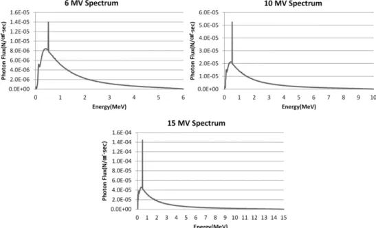
Moreover, both a water phantom for calculating the depth-dependent dosage and a pediatric anthropomorphic phantom for calculating the organ dosage were used. The homogeneity of photon beam was examined in different depths for the water phantom, which shows the 20%–40% difference for each material. Next, the org an doses on pediatric anthro- pomorphic phantom were examined, and the results showed that the average dose for each part of the body was skin 17.7 Gy, sexual gland 15.2 Gy, digestion 13.8 Gy, liver 11.8 Gy, kidney 9.2 Gy, lungs 6.2 Gy, and brain 4.6 Gy.
Moreover, as for the organ doses according to materials, the highest dose was observed in lead while the lowest was ob- served in plexig lass. Plexig lass in current use is considered the most suitable material, and a 6 or 10 MV photon energ y plan tailored to the patient condition is considered more suitable than a higher energy plan.
Key Words : Leukaemia, Total body irradiation, Beam spoiler, MCNPX, Dose assessment 중심 단어 : 백혈병, 전신방사선조사, 빔 스포일러, MCNPX, 선량평가
Corresponding author: Kim Jung Hoon, Department of Radiology, Catholic University of Pusan, 57 Oryundae-ro, Geumjeong-gu, Busan, 46252, Korea / Tel:+82-51-510-0589 / E-mail: [email protected]
Received 27 February 2018; Revised 27 March 2018; Accepted 21 April 2018 Copyright ⓒ2018 by The Korean Journal of Radiological Science and Technology
the existing hematopoietic cells[5,6].
Total body irradiation (TBI) and chemotherapy are used as preparatory regimens either alone or in combination[7]. TBI is easy to administer and has other advantages over chemotherapeutic agents, such as cost-effectiveness and absence of cross-tolerance to other anticancer agents. In addition, it ensures uniform irradiation of the entire body irrespective of the blood flow rate, thus reaching the tissues and organs difficult to penetrate with anticancer agents[7].
However, TBI can have adverse outcomes such as growth retardation, developmental disorders, hormonal imbalance, neurological complications, and secondary malignancies[8]. Pre-intervention dose assessment for each organ is especially important for children because they are considerably more sensitive to radiation than adults are. Currently, the dose-related recommendation for TBI only states that over 90% of prescribed dose should be absorbed into the skin surface without any mention of internal organ doses [4-6,8-9].
In this study, we examined the properties of photons and the dose distribution in a human body via a simulation where the total body irradiation is performed on a pediatric anthropomorphic phantom and a child size water phantom. Based on this, we tried to find the optimal photon beam energy and material for beam spoiler. Therefore, a simulation with water phantom and pediatric anthropomorphic phantom in virtual space is conducted to examine the doses on human organs using the pediatric anthropomorphic phantom and the properties of photons according to water depths. Based on this, we aim to determine whether the existing plexiglass material currently used for the TBI can be replaced and to propose the optimal photon beam energy.
Ⅱ. MATERIALS AND METHODS
MCNPX, as a code using the Monte-Carlo method, was developed by Los Alamos National Laboratory. It can transport a total of 34 particles including electron,
photon, neutron, and quantum while defining various types of desired calculations and source terms for users[10]. Moreover, MCNPX runs under Windows operating systems, which makes the code accessible for users. In particular, the Tally , as a code that represents a way of expressing the resulting values, allows different physical quantity such as fluency, energy distribution, or energy absorption to be printed out. The tallies used for this research are F5 and F6.
F5 is used to represent the number of particles per unit area (㎠) with an imaginary spherical detector installed in a desired space. F6 expresses the energy (MeV) received per unit mass (g) by specifying a region of interest. MCNPX was used for evaluating the dose, and the resulting values were calculated in MeV/g by using Tally 6, which were converted into units of Gy.
The calculation was performed assuming that 16 Gy is prescribed in the AP direction since the dose allowed for total body irradiation is usually 32 Gy.
In this study, MCNPX (Ver. 2.5.0)[10], a simulation program based on the Monte Carlo method, was used for photon beam analysis and TBI simulation. First, photons generated by a simulated linear accelerator head were analy sed, and TBI was performed on a simulated real-size water phantom and pediatric anthropomorphic phantom for dose assessment for different water depths and various organs.
1. Photon beam spectra
The simulation in this study was built on the basis of the shape and material of a linear accelerator borrowed from existing research studies, rather than using a linear accelerator produced by a specific company, in order to obtain standardized data[11-12].
In line with the analysis purpose, i.e. observations of
beam patterns and dose distributions on the water
phantom and the pediatric anthropomorphic phantom
exposed to TBI, respectively, a simplified linear
accelerator structure was used for simulation purposes
focusing on the linear accelerator head where photons
are generated. Fig. 1 shows the geometric structure of
the linear accelerator head. The beam energy for the
dose assessment was varied between 6, 10, and 15
MeV, and a round virtual dosimeter was placed under the 10 ㎝ point immediately after the flattening filter, making calculates at 1 keV intervals. The F5 tally option was used, and the photon flux per electron was expressed as the number of incident photons per unit area (㎠) per second. The reliability of the simulated linear accelerator head was evaluated on the basis of the photon spectra calculated, and the TBI-related dose assessment was performed using the water phantom and the pediatric anthropomorphic phantom.
2. Water phantom
The simulated real-size water phantom model was based on an average 5-year-old child (height: 110 ㎝, weight: 18 ㎏, thickness: 15 ㎝), the age with the highest leukaemia incidence[13-15]. The thickness was partitioned at 1 ㎝ interval to enable thickness- dependent dose assessment (Fig. 2). The F6 tally option was used for calculating the total energy deposition within a 1 cm slice. The deposited energy (MeV/g) calculated was then converted into absorbed dose (Gy).
3. Pediatric anthropomorphic phantom
The pediatric anthropomorphic phantom used in this study is the UF Revised ORNL phantom for pediatric radiology, a medical internal radiation dose
(MIRD)-type phantom. The UF Revised ORNL phantom is a revised version of the whole-body anthropomorphic phantom developed at the Oak Ridge National Laboratory (ORNL) for calculating internal radiation exposure of the organs made of specific materials. The revised ORNL phantom includes the head (containing the brain), kidneys, recto-sigmoid colon, and extrapulmonary airway based on a recently developed anthropomorphic model, in addition to salivary glands, bladder mucosa, a digestive tract, and airway. Fig. 3 schematically illustrates the organs[13] with their respective masses and densities in accordance with the specifications of the International Commission on Radiological Protection (ICRP) 89[14] and International Commission on Radiation Units and Measurements (ICRU) 46[15]. The F6 tally option was used for calculating the deposited energy (MeV/g), which were then converted into the absorbed dose (Gy ). The elemental composition, percentage, density, and volume for materials that make up brain, lung, skin, muscle, liver, kidney, genital gland, and digestion are shown in Tables 1 and 2.
Fig. 1 Schematic of the linear accelerator head in MCNPX
Fig. 2 Water phantom similar to pediatric
Fig. 3 Images of the anthropomorphic phantom in anterior,
diagonal, and lateral position
4. Materials for beam spoiler
Materials used for the beam spoiler were 1.5 ㎝ plexiglass (C
5O
2H
8, density:1.16 g/㎤)[16] widely used in clinical settings as a reference material, 1.5 cm aluminium (
13Al, density: 2.7 g/㎤, Al) with similar atomic number and density, and 0.3 cm copper (
29Cu, density: 8.94 g/㎤, Cu) and lead (
82Pb, density: 11.34 g/㎤, Pb) with higher atomic numbers and densities.
The thicknesses were calculated on the basis of previous studies[17-18], which indicated similar doses were yielded when the thickness of the Cu was about one third the thickness of the Al material. While existing theories recommend the closest possible beam spoiler-to-patient distance (SPD)[4-7], SPD was set at 10 cm, given that gapless application is not implementable. Furthermore, as materials for the head and lungs requiring shielding, we used Lipowitz alloy (
83Bi,
82Pb,
50Sn,
48Cd, density: 9.4 g/㎤), which has a high shielding rate and low melting point due to
the high atomic numbers of the components, and ensured sufficient shielding with a thickness of 7.5 ㎝ corresponding to 5 half value layers that can shield ≥ 95% of the primary beam[6].
5. Simulation methods
Of the two major TBI methods, we used the anterior-posterior method in which radiation is delivered towards the body front with the umbilicus as the midpoint[9]. Photon energy intensity was varied to 6, 10, and 15 MV in the continuous radiation beams analysed in this study, and the source-to-skin distance (SSD) was set at 300 cm to sufficiently cover the height of 110 cm. Simulations were performed as described below and the depth-dependent doses and organ doses were calculated and evaluated in the water phantom and anthropomorphic phantom, respectively.
The simulation values were converted on the basis of the assumption that TBI is administered at 16 Gy.
Element Percent by weight
Brain Lung Liver Digestion Skin Muscle Testis Ovary Kidney
H 10.7 10.3 10.3 10.6 10 10.2 10.6 10.5 10.3
C 14.5 10.5 18.6 11.5 20.4 14.3 9.9 9.3 13.2
N 2.2 3.1 2.8 2.2 4.2 43.4 2.0 2.4 3.0
O 71.2 74.9 67.1 75.1 64.5 71 76.6 76.8 72.4
Na 0.2 0.2 0.2 0.1 0.2 0.1 0.2 0.2 0.2
P 0.4 0.2 0.2 0.1 0.1 0.2 0.1 0.2 0.1
S 0.2 0.3 0.3 0.1 0.2 0.3 0.2 0.2
Cl 0.3 0.3 0.2 0.2 0.3 0.1 0.2 0.2
Ka 0.3 0.2 0.3 0.1 0.1 0.4 0.2 0.2
Mg 0.2
Si 0.2
Table 1 Composition of the tissue for the anthropomorphic phantom.
Organ Density
(g/cm3)
Volume
(cm3) Organ Density
(g/cm3)
Volume (cm3)
Brain 1.04 1194 Lung 0.260 980
Digestion 1.03 439.14 Liver 1.05 562
Skin 1.09 514.74 Muscle 1.03 7547.568
Ovary 1.05 1.66 Testis 1.04 1.57
Kidney 1.05 111.12
Table 2 Volume and density of anthropomorphic Phantom
Ⅲ. RESULTS
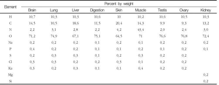
1. Photon spectra
Fig. 4 illustrates the photon beam spectra obtained using electron beams of 6, 10, and 15 MeV in a simulated linear accelerator. The average energies for the calculated photons were 1.44, 2.12, and 2.85 MeV at 6, 10, and 15 MeV incident energies, respectively, and the incident photon energy of 511 keV was calculated for all variants.
2. Thickness-dependent dose calculated in the water phantom
Fig. 5 shows the absorbed dose (Gy) versus the thickness of the water phantom at 1-cm intervals.
Dosimetric comparison between the incident photon beam area (1 ㎝) and the deepest area (15 ㎝) revealed the average dose differences for Al, plexiglass, Cu, and Pb to be 40.2%, 40.9%, 35.5%, and 21%, respectively, showing similar energy-dependent dose differences.
3. Deep organ dose