서 론
아토피 피부염(atopic dermatitis)은 가려움증을 동반한 만성 재발성 습진으로 국내외적으로 증가 추세를 보이고 있으며 우리나라 의 경우 초등학생에서의 아토피 피부염 진단이 1995년 13.7%에서 2005년 29.2%로 증가하였다1). 아토피 피부염은 일반적으로 연력이 증가할수록 유병률이 감소하고 대부분 가벼운 경과를 보이는 것으로 알려져 있으나 2008년 896명에서 2012년 1,367명으로 입원환자는 계속 증가하고 있으며2), 중증도 검사에서 중증 환자 구성비가 지속 적으로 높아지고 있다. 아토피 피부염은 중증화, 난치화 되는 경우가 많아 중증도 이상의 증상을 보이는 경우 30% 정도에서는 성인기까 지 이어져 학업이나 직장 등 사회생활에 지장을 초래한다3). 서양의학에서의 아토피 피부염 치료는 항히스타민제, 스테로이 드 외용제, 국소 면역조절 외용제, 경구 스테로이드제 등을 사용하 고 있으나 원인과 발생기전이 다양하고 호전과 악화를 반복하여 치
료에 어려움을 겪고 있다4). 아토피 피부염 환자는 정상인에 비해 자극에 더 민감하고 다양한 악화요인이 증상의 악화에 영향을 미치 며 질병의 경과는 개인마다 차이가 크므로 이에 적합한 질병관리가 요구되는데 변증(辨證)을 통하여 질병을 인식하는 한의학적 접근이 환자별 차이를 반영하는 중요한 방법론으로 활용될 수 있다5). 아토피 피부염에 대한 변증 분류는 식품의약품안전처의 임상시 험 가이드라인6)을 포함하여 많은 연구7-9)에서 습열(濕熱)과 혈허풍 조(血虛風燥)로 구분하고 있다. 이러한 아토피 피부염의 변증유형 발생에서 비허(脾虛)는 매우 중요한 역할을 하는데 『素問·至眞要大 論』10)에서 “諸濕腫滿 皆屬於脾”라 하여 脾가 정상적 생리기능을 잃으면(脾失健運) 습열이 생겨 피부에 영향을 끼친다고(蘊聚肌膚) 하였고, 『難經·二十二難』11)에서 “血主潤之”라 하여 비허로 생화지 원(生化之源)이 결핍되어 혈허(血虛)가 오래되면 화조생풍(化燥生 風)하여 피부를 자윤(滋潤)하지 못하고 병변을 일으킬 수 있다는 근 거를 제시함으로써 脾와 피부의 밀접한 상호 관계를 설명할 수 있
비허형 아토피 동물모델 개발
양원경1,2·유이란1·김호경3·김승형1,2·박양춘1,2*
1 : 대전대학교 한의과대학 폐계내과학교실, 2 : 대전대학교 동서생명과학연구원, 3 : 한국한의학연구원
Development of Atopic Dermatitis Mouse Model with Spleen Deficiency
Won Kyung Yang
1,2, Yee Ran Lyu
1, Ho Kyoung Kim
3, Seung Hyeong Kim
1,2, Yang Chun Park
1,2*
1 : Division of Respiratory System, Department of Internal Medicine, College of Korean Medicine, Daejeon University, 2 : Institute of Traditional Medicine and Bioscience, Daejeon University,
3 : Korea Institute of Oriental Medicine
Atopic dermatitis (AD) is a common skin disease characterized by chronic and relapsing inflammatory dermatitis with immunological disturbances. Spleen deficiency (脾虛) is one of the major causes of AD, so development of animal model is required for AD research that reflects the pattern identification. The groups that we have used in this study included Senna folium extracts (SFE), 2,4-dinitrochlorobenzene (DNCB), and normal mice. Therefore, the present study was developed to atopic dermatitis mouse model with spleen deficiency in 2,4-dinitrochlorobenzene (DNCB) and senna leaves extracts induced AD in NC/Nga mice. The results demonstrated that senna leaves extract treatment significantly increased the dermatitis clinical score and epidermal thickness in AD-like skin lesions. We also proved beyond doubt that there was occurrence of erythema and skin moisture indices in the senna leaves extract groups. Further, we also found that the level of serum immunoglobulin E (IgE) in the senna leaves extract-treated group was increased. The amount of IL-4, IL-13, TNF-α and TGF-β mRNA determined by real-time PCR was increased remarkably when senna leaves extract groups were treated on dorsal skin. Senna leaves extract groups significantly promoted the number of CD11B+/Gr-1 cell in skin, as well as the number of CD4+/CD8+ cell in dorsal skin compared with control. The review summarizes recent process in our understanding of the immunopathophysiology of spleen deficiency AD and the implications for spleen deficiency mouse models of AD on drug discovery from medical plants.
keywords : Atopic dermatitis, spleen deficiency, leaves of Senna, animal model
* Corresponding author
Yang Chun Park, Korean Medicine Hospital of Daejean Universit,75, Daedeok-daero 176beon-gil, Seo-gu, Daejeon, Republic of Korea
·E-mail : omdpyc@dju.kr ·Tel : +82-42-470-9126
·Received : 2017/07/14 ·Revised : 2016/08/17 ·Accepted : 2017/08/23
ⓒ The Society of Pathology in Korean Medicine, The Physiological Society of Korean Medicine pISSN 1738-7698 eISSN 2288-2529 http://dx.doi.org/10.15188/kjopp.2017.08.31.4.213 Available online at https://kmpath.jams.or.kr
다. 이에 한의학 원리에 더욱 충실한 연구의 기반을 마련하고자 아 토피 피부염의 한의학적 주요 특성인 비허 양상을 반영하는 동물모 델을 개발하고자 하였다.
비허를 유발하기 위하여 NC/Nga 마우스에 곡 등12)의 방법에 따라 센나엽(Senna leaves) 전탕액을 투여하여 사하(瀉下)를 유도 하고 비허점수에 따라 비허 유발을 확인한 다음 제모한 실험동물의 등 피부에 2,4-dinitrochlorobenzene(DNCB) 용액을 도포하여 피 부염을 유발하였다. 피부염 수치, 혈액학적 검사, 혈청생화학검사, 염증세포에 대한 형광유세포분석, 관련 cytokine에 대한 real-time PCR 검사를 시행하여 비허를 유발하지 않은 시험군 및 양성대조군과 비교하여 비허형 아토피 동물모델의 제작 기술을 탐 색함으로써 한의학 특성을 반영하는 아토피 피부염 기초연구에 도 움이 되고자 한다.
재료 및 방법
1. 재료 1) 동물
실험에 사용한 동물은 NC/Nga 7주령 수컷 생쥐(오리엔트바이 오, Korea)로서 물과 고형 사료를 제약 없이 섭취하도록 하였으며, 22-24℃ 의 온도와 50±10%의 습도가 유지되고, 밤낮 주기(12시간 주/야)가 조절되는 조명을 갖춘 실험실 환경에서 사육하였다. 실험 동물의 체중은 24±4 g이었다. 본 실험은 대전대학교 동물실험윤리위 원회가 정한 규정에 따라 시행되었다(승인번호: DJUARB2016-007).
2) 약재
비허유발에 사용된 센나엽은 허브스토리메디신(Korea)에서 구 입하여 사용하였으며, 센나엽 300 g에 70% 에탄올 10배를 가하여 환류 추출기에서 2시간 동안 2회 가열하여 얻은 액을 여과하였다.
이를 감압 증류장치(Buchi B-480, Switzerland)로 농축하고, 다시 동결 건조기(Eyela FDU-540, Japan)를 이용하여 완전 건조한 추 출물을 냉동(-84℃) 보관하면서 사용하였다. 최종적으로 센나엽 초 기 약재로부터 68g의 추출물을 얻어 22.67%의 수율을 나타내었다.
얻어진 분말은 –80℃에 분주하여 보관하였고 사용하기 전 증류수 또는 식염수로 희석하여 필터 후 사용하였다.
3) 시약
본 실험 사용한 IgE ELISA kit는 R&D systems(USA), Real Time PCR 실험시에 사용한 Trizol(Ambion, USA), Chloroform,
isopropanol, ethanol(Sigma-Aldrich), diethylpyrocarbonate-tr eated water(DEPC-water; Invitrogen, USA), PrimeScript™RT reagent kit(TaKaRa, Shiga, Japan), Power SYBR®Green PCR Master Mix와 TaqMan®Gene Expression Master Mix(Applied Biosystems, USA), real-time polymerase chain reaction(real- time PCR) system (Applied Biosystems, USA) 등을 사용하였다.
2. 방법
1) 비허형 동물모델 구축
비허 유발을 위한 적정 농도를 확인하기 위하여 ICR 마우스에 센나엽추출물을 250, 500, 750, 1,000mg/kg의 농도로 1주일간 경 구 투여하였다. 피부염 모델에서 비허를 유발할 농도를 결정하고 치사율 및 간독성 등을 평가하였다.
2) 비허 유발 평가
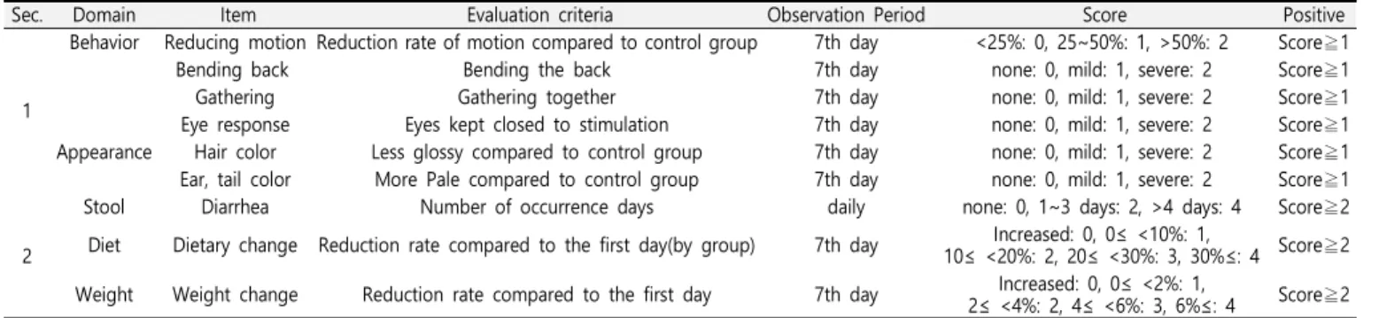
비허 유발여부를 객관적으로 판단하기 위하여 주 등(Zhu HW 등)의 방법을 변형한 평가기준(Table 1)에 따라 비허점수를 부여하 여 대변, 식이, 체중 중 2개 이상 양성이면서 기타 증상이 4개 이 상 양성이면 비허가 유발된 것으로 평가하였다.
3) NC/Nga 아토피 피부염 모델
NC/Nga 마우스의 등을 귀 하단에서부터 꼬리 상단부까지 전 체를 제모하고 24시간 방치 후 1%, 0.2% 2,4-dinitrochlorobenzene (DNCB; Sigma-Aldrich, USA) 용액(아세톤:올리브오일=3:1)을 도 포하여 유발하였다. 실험군은 아무런 처치도 시행하지 않은 정상군 (이하 정상군), 단순 아토피 피부염 대조군(이하 대조군), dexamethasone(Dexa; 3mg/kg) 투여 양성대조군(이하 양성대조 군), 센나엽추출물 1,000mg/kg와 750mg/kg를 1주일간 경구 투여 한 비허 아토피 피부염 고농도군(이하 비허 고농도군), 비허 아토피 피부염 저농도군(이하 비허 저농도군)으로 나누었다(Fig. 1).
Fig. 1. Experimental plan of Atopic Dermatitis Mouse Model with Spleen Deficiency.
The dorsal skin of four of these groups was treated with 1%, 0.2% 2,4-dinitrochlorobenzene (DNCB) diluted in an acetone and olive oil solution (3:1) once each week to shaved skin on the dorsal area to induce dermatitis. SFE: Sennae Folium extract.
Table 1. Evaluation Standard of Spleen Deficiency
Sec. Domain Item Evaluation criteria Observation Period Score Positive
1
Behavior Reducing motion Reduction rate of motion compared to control group 7th day <25%: 0, 25~50%: 1, >50%: 2 Score≧1
Bending back Bending the back 7th day none: 0, mild: 1, severe: 2 Score≧1
Gathering Gathering together 7th day none: 0, mild: 1, severe: 2 Score≧1
Eye response Eyes kept closed to stimulation 7th day none: 0, mild: 1, severe: 2 Score≧1 Appearance Hair color Less glossy compared to control group 7th day none: 0, mild: 1, severe: 2 Score≧1 Ear, tail color More Pale compared to control group 7th day none: 0, mild: 1, severe: 2 Score≧1
2
Stool Diarrhea Number of occurrence days daily none: 0, 1~3 days: 2, >4 days: 4 Score≧2 Diet Dietary change Reduction rate compared to the first day(by group) 7th day Increased: 0, 0≤ <10%: 1,
10≤ <20%: 2, 20≤ <30%: 3, 30%≤: 4 Score≧2 Weight Weight change Reduction rate compared to the first day 7th day Increased: 0, 0≤ <2%: 1,
2≤ <4%: 2, 4≤ <6%: 3, 6%≤: 4 Score≧2
Spleen Deficiency : (the number of positive items in Section 1) ≧ 4 and (the number of positive items in Section 2) ≧ 24) 피부염 임상증상 평가
피부염 수치는 아토피피부염에서 일반적으로 사용되는 임상적 육안 평가방법을 사용하여 아토피피부염의 심각성 정도를 다음 5가 지 항목을 각각 평가한 점수의 총합으로 나타내었다. 평가항목은 홍반(erythema), 가려움과 피부건조(pruritus & dry skin), 부종 과 혈종(edema & excoriation), 짓무름(erosion), 그리고 태선화 (lichenification)이다. 각각의 항목에 대해 중상 없음(0점), 증상 약 함(1점), 보통(2점), 심함(3점)으로 채점한 후 5항목의 점수를 합산 함으로써 최소 0점(아무 증상 없는 상태)에서 최고 15점(모든 항목 의 증상이 심한 상태) 사이의 평가 점수를 부여하였으며, 실험동물 이 긁는 횟수(scratching behavior)는 20분 동안 관찰하였으며, 일주일에 한번, 일정한 시간에 측정하였다.
5) Enzyme-linked immunosorbent assay (ELISA)
NC/Nga 생쥐의 혈장을 분리하여 mouse IgE ELISA kit를 사 용하여 제조사의 지시에 따라 ELISA를 시행하였다. 먼저, coating antibody를 microplate에 100 ㎕ 씩 분주하고 4℃에서 24시간 두 었다. 각 well을 wash buffer로 세 번 세척하고 Assay diluent를 200 ㎕ 씩 넣어서 1시간 동안 실온에서 배양하였다. 배양하는 동 안 표준품과 상등액을 준비하고 완료되면 microplate를 세 번 세 척한 후 각 표준품과 상등액을 100 ㎕ 씩 넣고 2시간동안 실온에 서 배양하였다. Microplate를 세 번 세척하고 detection antibody 를 만들어서 각 well에 100 ㎕ 씩 넣고 1시간 동안 실온에서 배양 하였다. microplate를 다섯 번 세척하고 substrate solution을 만 들어서 각 well에 100 ㎕ 씩 넣고 30분 동안 어두운 곳에서 실온 으로 배양하였다. stop solution을 각 well에 넣고 Microplate spectrophotometer에서 흡광도 450 ㎚로 측정하였다.
6) 형광유세포분석
생쥐로부터 각각 피부를 적출하여 세포를 분리하고, staining buffer에 anti-CD3, CD4, CD8, CD11b, Gr-1를 넣어 각각 4℃에 서 30분 동안 반응시킨 뒤 Cell-Quest software를 사용하여 유세 포 분석기(FACScan analyzer; BD Biosciences, San Jose, CA) 를 통해 분석하였다.
7) Real time PCR
일정량의 NC/Nga 생쥐의 등 피부조직을 분리하여 Trizol을 1
㎖넣고 homogenizer로 잘게 분쇄한 다음 Chloroform을 100 ㎕ 넣었다. 얼음에 10분 동안 두고 13,000 rpm으로 15분 동안 원심 분리 하였다. 층이 분리되면 상등액을 옮겨 담고, 동량으로 isopropanol을 넣어 얼음에 10분 정도 두었다가 13,000 rpm으로 15분동안 원심분리 하였다. 상등액은 버리고 80% ethanol로 세척 하고 7,500 rpm으로 10분 동안 원심분리 하였다. 상등액을 완전히 제거하고 투명해질 때까지 말린 후 diethylpyrocarbonate-treated water (DEPC-water)로 30 ㎕ 씩 넣어 녹였다. 일부는 RNA를 정 량하고 나머지는 PrimeScript™RT reagent kit를 사용하여 제조 사의 지시대로 cDNA를 합성하였다. cDNA와 IL-4, IL-13, TNF- α, TGF-β, glyceraldehyde-3 phosphate dehydrogenase (GAPDH) primer를 적정 농도로 각각 희석하여 넣고 Power SYBR®Green PCR Master Mix와 TaqMan®Gene Expression Master Mix시약을 이용하여 real-time polymerase chain
reaction (real-time PCR) system으로 분석하였다.
3. 통계분석
실험 집단 간 수치 데이터는 각 실험군 결과 값은 mean ± standard deviation (SD)으로 나타내었으며, 동물실험에서는 SPSS 11.0 software (IBM-SPSS Inc., Chicago, IL)를 이용하여 일원배치 분산분석(One-way analysis of variance; ANOVA) 후 에 Duncan’s multiple comparison tests로 유의성을 검증하였 고, 세포실험에서는 독립표본 T-검정(independent samples T-test)을 이용하여 유의성을 검증하였다. p 값이 0.05, 0.01 혹은 0.001 보다 작은 경우를 구분하여 분석하였으며 각 경우에 해당 시 통계적으로 유의적 차이가 있는 것으로 판정하였다.
결 과
1. 농도별 비허평가 결과
비허 판단의 기준으로 정한 평가기준을 토대로 센나엽 추출물 투여에 따른 비허점수를 평가한 결과 정상군의 2.00±0.71에 비하 여 250, 500, 750, 1,000 mg/kg 투여군에서 각각 8.00±2.35, 11.40±1.67, 17.60±0.89, 18.80±1.30으로 나타나 농도의존적으로 증가하였고, 비허 기준에 부합하는 개체수의 비율은 750 및 1,000 mg/kg 투여군에서만 100%로 나타났다(Fig. 2)
A
B
Fig. 2. Score of spleen deficiency(A) and rate of spleen deficiency mouse(B) in SFE induced ICR mice.
A score of spleen deficiency was defined as the sum of the individual scores graded as table 1. ICR mice were divided into five groups and were orally administered PBS, or SFE(250, 500, 750, 1000 mg/kg) for one week. (One-way ANOVA test ***
P <0.001 vs. Normal).
2. 관능평가, 피부염 지수에 미치는 영향
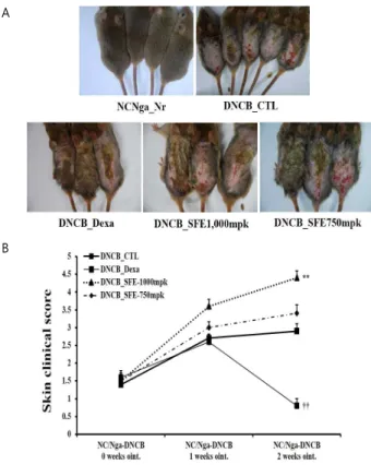
Fig. 3A은 유발 후 피부염유발 정도와 개선 정도를 나타내주 는 사진으로, 대조군, 비허 고농도군 및 비허 저농도군에서 홍반, 피부건조, 부종, 짖무름, 태선화 등의 증상이 확인되었고, 양성대조 군은 대조군에 비해서 그 증상이 현저하게 감소하였음을 확인할 수 있었다. Fig. 3B는 아토피 피부염이 유발된 NC/Nga 생쥐에서 피 부염 지수에 어떠한 영향을 주는지 알아보기 위해 피부염의 중증도 를 점수화하여 측정한 결과로, 유발 후 정상군에 비해 대조군, 비 허 고농도군 및 비허 저농도군의 피부염 지수가 증가하는 것으로 확인되었으며, 비허 고농도군과 비허 저농도군 모두 대조군보다 단 순 아토피 피부염유발군에서 경구투여 농도의존적으로 피부염 지수 가 더 높은 수치를 보였다.
A
B
Fig. 3. Effect on clinical features(A) and Dermatitis index(B) in DNCB, SFE induced NC/Nga mice.
A total clinical index of dermatitis severity was defined as the sum of the individual scores graded as follows:
0(none), 1(mild), 2(moderate),and 3(severe) for each of five signs and symptoms(erythema/hemorrhage, edema/hematoma, excoriation/erosion, itching/dryness, and lichenification(thickness of the skin)). NC/Nga mice were divided into five groups and were orally administered PBS, dexamethasone(3 mg/kg) or SFE(750, 1000 mg/kg) for one week. The dorsal skin of these groups was treated with 1%, 0.2%
2,4-dinitrochlorobenzene(DNCB) diluted in an acetone and olive oil solution(3:1) once each week to shaved skin on the dorsal area to induce dermatitis. The Naïve group was not treated with DNCB and was administered oral doses of PBS. (One-way ANOVA test ** P <0.01 vs.
Control, †† P <0.01 vs. Control).
3. NC/Nga 생쥐의 혈청 내 IgE 생성에 미치는 영향
아토피 피부염이 유발된 NC/Nga 생쥐의 혈청 내 IgE 생성에 영향정도를 알아보기 위해 실험 종료 후 생쥐를 치사하여 혈액을 얻은 뒤 원심 분리하여 혈청에 있는 IgE를 ELISA 방법으로 측정하 였다. 정상군과 비교하여 대조군에서는 IgE의 생산이 5배 가량 증
가하여 생성되었고, 비허 고농도군과 저농도군 모두에서 대조군보 다 유의하게 증가된 양상을 보였다. 또한, dexamethasone을 투여 한 양성대조군에서 IgE의 생성이 유의하게 억제되었음을 알 수 있 었다.
Fig. 4. Effects of DNCB, SFE induced NC/Nga mice on the plasma levels of IgE.
Blood plasmas were isolated from each group of mouse after dermatitis induction. The plasma level of IgE was determined using ELISA. NC/Nga mice were divided into five groups and were orally administered PBS, dexamethasone(3 mg/kg) or SFE(750, 1000 mg/kg) for one week. The dorsal skin of these groups was treated with 1%, 0.2%
2,4-dinitrochlorobenzene (DNCB) diluted in an acetone and olive oil solution(3:1) once each week to shaved skin on the dorsal area to induce dermatitis. The Naïve group was not treated with DNCB and was administered oral doses of PBS. (One-way ANOVA test ** P <0.01 vs.
Control, † P <0.05 vs. Control).
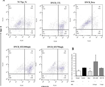
Fig. 5. Effects of DNCB, SFE induced NC/Nga mice on the T cell population of dorsal skins.
FACS analysis of CD4+CD8+ T cells from dorsal skins of DNCB, SFE induced NC/Nga mice. (One-way ANOVA test * P <0.05 vs. Control, †† P <0.01, ††† P <0.001 vs. Control).
4. NC/Nga 생쥐의 dorsal skin 내 면역세포 변화에 미치는 영향 아토피 피부염이 유발된 NC/Nga 생쥐의 dorsal skin 내 면 역세포 변화에 어떠한 영향을 주는지 알아보기 위해 실험 종료 후 생쥐를 치사한 뒤 dorsal skin 내 면역세포를 염색하여 FACS로
분석하였다. 면역기전에 중요한 T세포를 나타내는 CD4+/CD8+세 포와 과립구세포인 CD11b+/Gr-1+ 세포는 정상군에 비해 대조군 에서 증가 하였으며, 양성대조군에서 대조군보다 유의하게 감소를 보였고, 비허 고농도군에서 대조군보다 증가 양상을 보였으나 유의 하지는 않았다(Fig. 5, 6).
Fig. 6. FACS analysis of CD11b+Gr-1+ B cells from dorsal skins of DNCB, SFE induced NC/Nga mice.
(One-way ANOVA test ††† P <0.001 vs. Control).
Fig. 7. Effects of DNCB, SFE induced NC/Nga mice on the mRNA extression in skin.
Total RNAs were isolated and used for synthesis of cDNAs. Levels of IL-4, IL-13, TNF-α, TGF-β mRNA were determined by Quantitative real-time PCR. (One-way ANOVA test *P < 0.05, ** P <0.01,
*** P <0.001 vs. Control. †† P <0.01 vs. Control).
5. NC/Nga 생쥐의 mRNA 발현에 미치는 영향
아토피 피부염이 유발된 NC/Nga 생쥐의 IL-4, IL-13, TNF-
α, TGF-β mRNA 발현에 영향의 정도를 파악하기 위하여 dorsal skin에서 얻은 세포 내 RNA를 분리하고 cDNA를 제조하여 real-time PCR 방법으로 mRNA 발현 정도를 측정하였다. 그 결과 양성대조군에서는 대조군에 비하여 IL-4를 제외한 모든 항목에서 유의하게 발현을 억제하였고, 비허 고농도군과 저농도군에서는 대 조군에 비하여 유의한 발현 증가가 나타났다(Fig. 7).
고 찰
아토피 피부염 변증 분류는 일반적으로 습열과 혈허풍조로 구 분하는데6) 두 기전 모두에서 비허가 기저 병리상태로 작용하고 있
다10,11). 특히 습열형에 손 등13)은 비위기허(脾胃氣虛)로 운화기능
(運化機能)이 실조되어 습열이 발생하는 허증이 포함되어 있다고 하였고, 소화불량, 복부창만의 증상이 있거나 기름진 음식을 좋아 하는 경우, 비만, 음식알레르기 등을 동반하는 아토피 피부염을 비 위기허로 인한 습열로 분류하는 연구들14,15)이 보고되고 있어 아토 피 피부염의 발생에 비위(脾胃)가 중요한 역할을 하고 있음을 알 수 있다. 따라서 비허 양상을 반영하는 아토피 피부염 동물모델을 개발하는 것이 한의학 원리에 기반한 아토피 피부염 연구에 중요한 연구방법을 제공할 것으로 판단되었다.
비허 동물모델의 개발 방법으로는 고한사하법(苦寒瀉下法), 생 화핍원법(生化乏源法), 음식실절법(飮食失節法), 모기파기법(耗氣破 氣法), 편식법(偏食法), 상습법(傷濕法) 등이 제시되고 있으며16) 일 반적으로 고한사하법이 다용되고 특히 센나엽을 이용한 경우가 가 장 많고 임상양상과 가장 근접한 것으로 보고되고 있다17). 본 연구 에서는 센나엽 전탕액을 1주일간 투여한 다음 주 등18)의 방법을 응 용한 비허점수 평가를 통해 비허 유발 여부를 평가하였다. 모든 실 험동물에서 비허기준을 충족시킨 유발 농도로서 저농도 750mg/kg 과 고농도 1,000mg/kg를 도출하여(Fig. 2), 이를 아토피 피부염 모델에 적용한 결과 관능평가에서 대조군, 비허유발군 모두에서 홍 반, 피부건조, 부종, 짖무름, 태선화 증상이 확인되었으며, 피부염 지수는 대조군에 비하여 비허유발군에서 유의하게 증가하였다(Fig.
3). 이러한 결과는 위장관 이상 증상과 아토피 피부염의 중증도와 의 연관성은 밝혀지지 않고 있으나19) 음식 알레르기가 있으면 성인 시기 아토피 피부염 발병의 위험을 높인다고 알려져 있으며20), 아 토피 피부염 환자의 사상체질 분포 중 비위허약(脾胃虛弱)한 체질 인 소음인(少陰人)의 비율이 가장 높이 것으로 나타나21) 아토피 피 부염의 발생에서 비위허약(脾胃虛弱)의 상태가 중요한 조건이 될 수 있음을 알 수 있다. 아토피 피부염은 항원-항체 반응에 의존하 는 전형적인 알레르기 질환으로, IgE 항체와 비만세포와 연관된 면 역 기전이 중요한 역할을 하며 혈청 내 IgE의 함량은 임상적 중증 도에 비례하고 피부장벽의 이상을 초래하는 것으로 알려져 있는데
22), 혈청 내 IgE 생성이 대조군에 비하여 비허유발군에서 유의하게 증가하는 것으로 나타나(Fig. 4) 피부염 지수의 결과와 동일한 양상 을 보였다.
아토피 피부염의 면역 반응에는 수많은 면역세포와 염증세포, 각종 화학매개체들이 관여하며, 이러한 염증 반응에 관여하는 주된 세포로는 T세포, B세포, 비만세포, 호산구, 호중구 등이 있다23). T 세포는 면역반응을 결정하는 중요한 인자로서 B세포에 작용하여
항체 생산을 촉진하고 대식세포를 활성화 시킨다24). T세포의 membrane molecule로 CD(cluster of differentiation) isotype 들은 T세포 수용체에 항원이 결합된 신호를 세포 안으로 전달하는 T세포의 결정표지인자이다25). 면역반응의 조절에 관여하는 Th세포 (T helper cell)는 세포 표면 수용체 외 CD4 분자를 공통적으로 가지고 있으며, 이러한 CD4+ T세포는 Th1과 Th2로 분화 활성화 되고, cytokine을 생산에 관여하여 B세포가 효율적으로 항체를 생 산하도록 도와주고, 면역 반응의 행동세포를 활성화시키는 역할을
한다26,27). 또한 CD8 분자를 가지고 있는 CD8+세포는 세포독성 T
세포(cytotoxic T cell)로서 바이러스 등의 항원에 감염된 세포, 손 상되거나 제 기능을 상실한 세포 등을 제거하는 역할을 한다28). 또 한 아토피 피부염의 알레르기 반응에서 주요한 역할을 하는 호산구 에서 CD11b 분자가 과발현되는 것으로 알려져 있으며29), CD11b+/Gr-1+ 세포로서 호중구는 피부의 기계적 손상에서 leukotrien 증가에 중요한 역할을 한다30). 본 실험의 병변 유발부 위인 dorsal skin에서 T세포의 양상을 보여주는 CD4+/CD8+세포 와 과립구세포인 CD11b+/Gr-1+ 세포는 정상군에 비해 대조군과 비허유발군에서 증가하는 것으로 나타나(Fig. 5, 6) 아토피 피부염 의 이러한 병리기전이 반영됨을 보여주었다.
TNF-α와 Th2 사이토카인은 표피 분화 단백질 및 각질층 지 질에 아토피 피부염과 유사한 특징을 유도하고31), TNF-α는 피부 내피세포에서 유착분자의 발현을 증가시키는 것으로 알려져 있으며
32), TGF-β는 만성적인 아토피 피부염 부위에서 제1형 collagen의 생성을 유발하여 구진과 태선화와 같은 병변을 유발한다33). 본 실 험의 dorsal skin에서 IL-4, IL-13, TNF-α, TGF-β의 mRNA 발 현이 비허유발군에서 대조군에 비하여 유의한 발현 증가가 나타나 (Fig. 7) 비허 조건이 관련 사이토카인의 증가를 통해 피부증상을 더욱 증가시킬 가능성을 시사하였다.
본 연구는 우리나라에서 아토피 피부염을 대상으로 변증유형을 반영하는 동물모델을 개발하고자 하는 첫 시도라는 측면과 일반 아 토피 피부염 동물모델에 비하여 아토피 피부염 양상을 더욱 현저하 게 나타내는 결과를 얻었다는 점에서 의의가 있으나 이러한 결과가 성공적인 비허 모델 제작을 전적으로 입증한다고 보기는 어렵다.
피부염 발생 실험군 중 비허유발군의 저농도군과 고농도군에서 비 허와 관련되는 지표들을 평가하지 못하였다는 한계가 있으며 비허 에 적용할 수 있는 단미 약재 또는 처방을 통하여 추가적인 검증이 필요하다고 판단된다. 향후 이를 보완하여 더욱 확립된 비허형 아 토피 피부염 동물모델이 구축된다면 기존 아토피 치료 한약 효능의 객관적 근거를 더욱 확고히 할 수 있을 뿐만 아니라 한의학 이론에 입각한 새로운 한약치료제의 개발에 유용하게 활용될 것으로 사료 된다.
결 론
비허를 유발하기 위하여 NC/Nga 마우스에 센나엽 전탕액을 투여하고 피부염을 유발하여 다음과 같은 결과를 얻었다.
비허 기준에 부합하는 개체수의 비율은 750 및 1,000 mg/kg 투여군에서 100%로 나타났다.
피부염 점수는 대조군에 비하여 비허유발군에서 유의하게 증가 하였다.
혈청 내 IgE는 대조군에 비하여 비허유발군에서 유의하게 증가 하였다.
등피부조직 내 CD4+/CD8+세포와 CD11b+/Gr-1+세포는 대조 군에 비하여 비허유발군에서 증가하였으나 유의하지 않았다.
등피부조직 세포에서 IL-4, IL-13, TNF-α, TGF-β mRNA 발 현은 대조군에 비하여 비허유발군에서 유의하게 증가하였다.
감사의 글
본 연구는 2015년도 교육과학기술부 재원으로 한국연구재단의 지원(NRF-2015M3A9E3052912)과 보건복지부 한의약선도기술개발 사업의 지원(과제번호: HI15C0006)을 받아 수행된 연구임
References
1. Hong SJ. Korean ISAAC Study Group of Korean Association of Allergy and Respiratory Diseases. Report of Korean ISAAC epidemiologic study for asthma and allergic diseases in children. Pediatr Allergy Respir Dis.
2007;17:Suppl 1:S55-66.
2. National Health Insurance Corporation [Internet]. Seoul:
National Health Insurance Corporation; c2014 [cited 2017 May 31]. Available from:
http://www.nhis.or.kr/menu/retriveMenuSet.xx?menuId=
D4000
3. Oh JW. Recent situation of the management of atopic dermatitis. Korean J Asthma Allergy Clin Immunol.
2012;32:14-5.
4. Park Y. Status of clinical practice on diagnosis and management of atopic dermatitis in Korea: a questionnaire survey of physicians. Allergy Asthma Respir Dis. 2013;1(3):257-65.
5. Han JM, Yang WM. A Review on Korean Medicine and Personalized Medicine: Syndrome-based Personalized Medicine on the Basis of Syndrome Differentiation and Treatment. J Korean Med. 2014;35(3):40-8.
6. Food and Drug Administration. Traditional Korean Medicine Clinical Practice Guideline-Atopic Dermatitis.
Seoul : Food and Drug Administration; 2009. p 13-7.
7. Korean Institute of Oriental Medicine. Korean Medicine Clinical Practice Guideline-Atopic Dermatitis. Seoul. : Elsevier Korea L.L.C.; 2015. p 30-7.
8. Jang JH, Lee JM, Lee SY. A Clinical Study of Atopic Dermatitis for Children. The journal of Korean oriental pediatrics. 2005;19(2):69-84.
9. Yun HJ, Ko WS. Clinical study of atopic dermatitis;
classification of oriental medical type and treatment. J Korean Oriental Med. 2001;22(2):10-21.
10. Institute of Oriental Medical Classics. Lei Bian Huang Di Nei Jing (Class Yellow Emperor’s Inner Canon), Daejeon : Jumin Publishing Company; 2009. p 466-71.
11. Yoon CY, Kim YJ. Study aggregation of "Classic of Difficult Issues(nanjing)". Jumin Publishing Company;
2007. p 472-8.
12. Qu CJ, Liu J, Lin SR, Xia SJ. Comparative Study on Immunological Changes of Spleen Deficiency Mice in Different Modeling Methods. China Journal of Basic Medicine in Traditional Chinese Medicine. 1999;5(4):46-9.
13. Son BK, Choi IH. Research of pattern identification and outcome measurement in atopic dermatitis. J Korean Orient Med Ophthalmol & Otolaryngol & Dermatol.
2008;21(3):150-65.
14. Jung, ARNR. Hong, SU. A Case of Atopic Dermatitis with Nummular Eczema. The journal of Korean Medicine Ophthalmology & Otolaryngology &
Dermatology. 2006;19(2):296-303.
15. Shin SH, Kim JH, Kim M, Yoon HJ, Lyu SA, Lee SY, et al. A Clinical Research about the Effects of Seunggaltang on Patients with Atopic Dermatitis. The journal of Korean Medicine Ophthalmology &
Otolaryngology & Dermatology. 2007;20(2):199-212.
16. Chen MJ. Study on Difference of the Animal's Model about Splenasthenic Syndrome. Heilongjiang Traditional Chinese Medicine University. Master Thesis. 2005.
17. Zong SQ, Chu H. Experimental Study on Rat Model of Spleen Deficiency Syndrome. Journal of China Medical University. 1997;26(2):206-7.
18. Zhu HW, Tian J, Chen JH, Ma XD, Ping CQ, Wang M.
Influence of JianPiYangXueQuFengGang on ICAM-1 and VCAM-1 in Mice with Plenoasthenic Eczema. Chin J Derm Venereol. 2015;29(2):193-6
19. Rokaite R, Labanauskas L. Gastrointestinal disorders in children with atopic dermatitis. Medicina (Kaunas).
2005;41(10):837-45.
20. Yu HS, Tu HP, Hong CH, Lee CH. Lifetime Increased Risk of Adult Onset Atopic Dermatitis in Adolescent and Adult Patients with Food Allergy. Int J Mol Sci.
2016;18(1). pii: E42. doi: 10.3390/ijms18010042.
21. Kim HJ. Analysis of atopic dermatitis patients according to the Sasang constitution. J Oriental Med Ophthalmol &
Otolaryngol & Dermatol. 2003;16(3):200-9.
22. Liu FT, Goodarzi H, Chen HY. IgE, mast cells, and eosinophils in atopic dermatitis. Clin Rev Allergy Immunol. 2011;41(3):298-310.
23. Hardy RR, Hayakawa K. B cell development pathways.
Annu Rev Immunol, 2001;19:595-621.
24. Cooper KD. Atopic dermatitis: recent trends in pathogenesis and therapy. J Invest Dermatol. 1994;102(1):128-37.
25. Robinson DS, Hamid Q, Ying S, Tsicopoulos A, Barkans J, Bentley AM, et al. Predominant TH2-like bronchoalveolar T-lymphocyte population in atopic asthma. N Engl J Med. 1992;326(5):298-304.
26. Hoglund, P. Induced peripheral regulatory T cells : the family grows larger. Eur J Immunol. 2006;36(2):264-266.
27. Laky K, Fleischacker C, Fowlkes BJ. TCR and Notch signaling in CD4 and CD8 T-cell development. Immunol Rev. 2006;209(1):274-283.
28. Bromberg JS. The biology of CD2: adhesion, transmembrane signal, and regulatory receptor of immunity. J Surg Res. 1993;54(3):258-267.
29. Yamada H, Kurashimo S, Chihara J, Matsukura M, Yudate T, Tezuka T. Overexpression of CD11b on eosinophils in atopic dermatitis: downregulation by cyclosporin A and upregulation by interleukin 5. Int Arch Allergy Immunol. 1999;120(Suppl 1):100-3.
30. Oyoshi MK, He R, Li Y, Mondal S, Yoon J, Afshar R, et al. Leukotriene B4-driven neutrophil recruitment to the skin is essential for allergic skin inflammation.
Immunity. 2012;37(4):747-58.
31. Danso MO, van Drongelen V, Mulder A, van Esch J, Scott H, van Smeden J, et al. TNF-α and Th2 cytokines induce atopic dermatitis-like features on epidermal differentiation proteins and stratum corneum lipids in human skin equivalents. J Invest Dermatol.
2014;134(7):1941-50.
32. de Vries IJ, Langeveld-Wildschut EG, van Reijsen FC, Dubois GR, van den Hoek JA, Bihari IC, et al. Adhesion molecule expression on skin endothelia in atopic dermatitis: effects of TNF-alpha and IL-4. J Allergy Clin Immunol. 1998;102(3):461-8.
33. Lan CC, Fang AH, Wu PH, Wu CS. Tacrolimus abrogates TGF-β1-induced type I collagen production in normal human fibroblasts through suppressing p38MAPK signalling pathway: implications on treatment of chronic atopic dermatitis lesions. J Eur Acad Dermatol Venereol. 2014;28(2):204-15.