Dermatofibrosarcoma Protuberans: A Study of Clinical, Pathologic, Genetic, and Therapeutic Features in Korean Patients
Zhenlong Zheng,
1,3,4Junjei Piao,
1,3,4Ji-Hye Lee,
2,3Song-Ee Kim,
2Soo-Chan Kim,
2,3Kee Yang Chung,
1,3and Mi Ryung Roh
2,31Department of Dermatology, Severance Hospital, Seoul, Korea;
2Department of Dermatology, Gangnam Severance Hospital, Seoul, Korea;
3Cutaneous Biology Research Institute, Yonsei University College of Medicine, Seoul, Korea;
4Department of Dermatology, Yanbian University Hospital, Yanbian, China.
Received: January 28, 2014 Revised: May 23, 2014 Accepted: May 28, 2014
Corresponding author: Dr. Mi Ryung Roh, Department of Dermatology,
Yonsei University College of Medicine, Gangnam Severance Hospital, 211 Eonju-ro, Gangnam-gu, Seoul 135-720, Korea.
Tel: 82-2-2019-3363, Fax: 82-2-393-9157 E-mail: [email protected]
∙ The authors have no financial conflicts of interest.
© Copyright:
Yonsei University College of Medicine 2015 This is an Open Access article distributed under the terms of the Creative Commons Attribution Non- Commercial License (http://creativecommons.org/
licenses/by-nc/3.0) which permits unrestricted non- commercial use, distribution, and reproduction in any medium, provided the original work is properly cited.
Purpose: Dermatofibrosarcoma protuberans (DFSP) carries a translocation result- ing in the collagen type I alpha 1 (COL1A1)-platelet-derived growth factor beta (PDGFB) fusion gene, which is responsible for PDGFB activation. The purpose of this study is to evaluate the clinicopathological, genetic, and therapeutic features of DFSP in Korean patients. Materials and Methods: Clinicopathological fea- tures of 37 patients with DFSP were reviewed. Multiplex reverse transcriptase- polymerase chain reaction (PCR) was carried out in 16 patients using formalin- fixed, paraffin-embedded tissues and specific primers for COL1A1 and PDGFB.
Results: The mean age of 37 patients was 37.4 years old. The most common tumor location was the trunk. All patients were treated primarily with surgery: 34 (91.7%) cases with Mohs micrographic surgery (MMS) and 3 (8.3%) cases with wide local excision. The median follow-up time was 33.7 months. Two patients, one in each treatment group, demonstrated local recurrence during the follow-up period. The COL1A1-PDGFB fusion gene was expressed in 14 (87.5%) cases, demonstrated by reverse transcriptase PCR analysis. No association was found among the differ- ent COL1A1-PDGFB fusion transcripts, the various histological subtypes and clinical features. Conclusion: Our results support the effectiveness of MMS in treating DFSP. The COL1A1-PDGFB fusion transcript was observed in 87.5% of patients. Therefore, COL1A1-PDGFB is a useful and accurate tool in diagnosing DFSP in Koreans.
Key Words: Dermatofibrosarcoma protuberans, collagen type I alpha 1-platelet- derived growth factor beta, histological subtype, Mohs micrographic surgery
INTRODUCTION
Dermatofibrosarcoma protuberans (DFSP) is a sarcomatous tumor of the cutane- ous tissue first described by Taylor in 1890.1 It is an uncommon infiltrative dermal and subcutaneous tumor characterized by progressive local growth of CD34+
rence and any subsequent procedures were collected and compiled.
All patients were treated by one of the two surgeons. Pa- tients were treated by WLE or standard MMS using the fresh frozen tissue technique. WLE was performed with a 2-cm margin. For MMS, the visible border of each lesion was demarcated, and a 5-mm margin was drawn around each lesion’s clinical edge. In cases of recurrence, scars were removed en bloc with the recurrent tumor before the MMS layer was excised to identify any residual tumor around the scar tissue. Afterwards, the tissue sections were placed in cassettes and submitted with the MMS map for pathologi- cal examination. If any evidence of tumor was seen within the specimen, further tissue removal was performed. Addi- tional 5-mm-thick layer of tissue was removed after careful mapping of the positive margin, and this layer was patho- logically examined as described above. This process was repeated until all margins were cleared. All sections were embedded in paraffin wax for later confirmation by haema- toxylin and eosin (H&E) staining, and CD34 staining was also used in suspicious cases. For all patients, the central bulk of the tumor was sent to the pathology laboratory to have permanent sections taken to confirm the diagnosis of DFSP. All patients underwent primary reconstruction by the two surgeons.
Immunohistochemistry
Immunohistochemistry was performed on paraffin-embed- ded archival tissue with antibodies against CD34 (1:50;
BioGenex, San Ramon, CA, USA), p53 (1:50; Dako, Car- pinteria, CA, USA), and Ki-67 (1:50; Dako, Carpinteria, CA, USA). Appropriate positive and negative controls were run in parallel. Immunostaining was evaluated separately by two authors. Quantification of the immunohistochemis- try marker CD34 was based on the percentage of immuno- reactive cells as follows: high (≥50%) or low (<50%). The immunohistochemical quantification of nuclear markers was evaluated as follows: positive (>5% immunoreactive nuclei) or negative (<5%).
Identification of COL1A1-PDGFB fusion transcripts Total RNA was extracted from formalin-fixed, paraffin-em- bedded tissue blocks using an RNeasy Mini Kit (QIAGEN, Hilden, Germany) and reverse-transcribed using a Super- script Preamplification System (Gibco-BRL, Gaithersburg, MD, USA). To detect the expression of the PDGFB gene, first-strand cDNA was amplified with the AGexpdgf3 and spindle cells with a highly infiltrative pattern.2 Surgical ex-
cision is the main treatment modality; however, local recur- rence rates after conservative surgical resection have been reported to be 26% to 60%.3,4 Although wide local excision (WLE) has been shown to further reduce the recurrence rate to 0% to 30%,2-7 several reports indicate that Mohs mi- crographic surgery (MMS) results in a lower overall recur- rence rate (0‒8%) compared with WLE.7,8
Cytogenetically, DFSP carries a rearrangement of chro- mosomes 17 and 22, which results in formation of the colla- gen type I alpha 1 (COL1A1)-platelet-derived growth factor beta (PDGFB) fusion gene.9 This gene codes for a fusion protein that overlaps functionally with the mature form of PDGFB.10 It activates PDGFB receptor (PDGFBR), and thus accelerates DFSP growth through an autocrine-para- crine loop. Based on this knowledge, imatinib mesylate has been used to treat patients with inoperable DFSP, and has shown impressive clinical results.9 We previously reported 11 patients with DFSP who were successfully treated by MMS,8 and the purpose of our present study is to evaluate the clinicopathological, genetic (COL1A1-PDGFB), and therapeutic features of DFSP in 37 Korean patients.
MATERIALS AND METHODS
Case selection and clinicopathological data
All cases of DFSP included in the pathological database of Yonsei University Health System in Seoul, Korea from 1997 to 2012 were reviewed. Informed consent was ob- tained in accordance with the ethical committee procedures of the institution (IRB No. 4-2011-0659). The following clinical data were recorded: age, gender, tumor location, tu- mor size (centimeters), tumor type (primary or recurrent), surgical treatment by WLE or MMS, number of micro- graphic stages, type of surgical repair (primary closure, graft, or flap), local recurrence, distant metastasis, and fol- low-up period (months). Patients with both primary and re- current lesions were included. Primary disease was defined as the presence of tumor without previous treatment or re- cent tumor excision with histologically positive margins.
Recurrent disease was defined as that occurring within or juxtaposed to the previous surgical excision site ≥6 months after the initial excision. Histological confirmation of DFSP was done before WLE or MMS in all patients, and all tu- mors were stained for CD34. Patients were contacted to evaluate the surgical sites for recurrence. Data on recur-
AGexpdgf4 primers according to the methods of Greco, et al.10 To detect the presence of COL1A1-PDGFB fusion transcripts, polymerase chain reaction (PCR) was carried out using 17 COL1A1 forward primers and a specific PDGFB reverse primer, as presented in Table 1. Seventeen COL1A1 forward primers were designed for COL1A1 ex- ons 5, 8, 11, 15, 17, 20, 23, 26, 27, 32, 35, 38, 40, 42, 44, 46, and 49, and these primers were considered sufficient to span the various breakpoints within the region encoding the alpha-helical domain of the COL1A1 polypeptide. The PCR products were directly sequenced using an Applied Biosystems 373A automated DNA sequencer to identify the breakpoints.
Statistical analysis
Associations between the clinical, histopathological, and ge- netic aspects were assessed using a χ2 test and Fisher’s ex- act test. The significance level was set at 5%. All tests were performed using a statistical software package (SPSS, Ver- sion 18.0, SPSS Inc., Chicago, IL, USA).
Table 1. Primers for COL1A1 and PDGFB Primer set Exon Sequence 5’ to 3’
COL1A1 (forward)
1 5 GCCGAGATGGCATCCCTGG
2 8 CCCTGGTGAGCCTGGCGA
3 11 TCAGGGTGCTCGAGGATTGC
4 15 GGTGCTCGTGGAAATGATGG
5 17 AAGGTCCCCAGGGTGTGCG
6 20 AACCTGGTGCTCCTGGCAGC
7 23 AAGCTGGTCGTCCCGGTGAAGC
8 26 AAGGCTGGAGAGCGAGGTGTTC
9 27 TGCTGGCAAAGATGGAGAGG
10 32 TAGAACGTGGTGCAGCTGGTCTTC
11 35 TCCCACTGGAGCTCGTGG
12 38 TGCTCCTGGAGCCAAAGGTGC
13 40 TGCTGGCGAGAAAGGATCCCCTG
14 42 AAGGTCCCTCTGGAGCAAGT
15 43 TGGCAAGAGTGGTGATCGTGG
16 46 TGGCTTCTCTGGCCTCCAGGG
17 49 ACCTCAAGAGAAGGCTCACGATGG
PDGFB (reverse)
1 2 ATCAAAGGAGCGGATCGAGTGGTC
COL1A1, collagen type I alpha 1; PDGFB, platelet-derived growth factor beta.
Annealing temperature: 65.7°C.
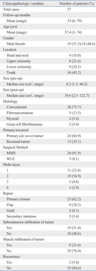
Table 2. Clinicopathologic Characteristics of 37 DFSP Pa- tients
Clinicopathologic variables Number of patients (%)
Total cases 37
Follow-up months
Mean (range) 33 (6‒79)
Age (yrs)
Mean (range) 37.4 (3‒74)
Gender
Male/female 19 (51.3)/18 (48.6) Location
Head and neck 4 (10.8)
Upper extremity 8 (21.6) Lower extremity 9 (24.3)
Trunk 16 (43.2)
Size (pre-op)
Median size (cm2, range) 6.2 (1.2‒44.2) Size (post-op)
Median size (cm2, range) 19.6 (2.5‒122.7) Histology
Conventional 28 (75.7)
Fibrosarcomatous 5 (13.5)
Myxoid 2 (5.4)
Giant cell fibroblastoma 2 (5.4) Primary/recurred
Primary (de novo) tumor 24 (64.9)
Recurred tumor 13 (35.1)
Surgical Method
MMS 34 (91.9)
WLE 3 (8.1)
Mohs layer
1 11 (32.4)
2 19 (54.9)
3 3 (8.8)
4 1 (2.9)
Repair
Primary closure 23 (62.2)
Flap 9 (24.3)
Graft 3 (8.1)
Secondary intention 2 (5.4) Subcutaneous infiltration of tumor
Yes 19 (51.4)
No 18 (48.6)
Muscle infiltration of tumor
Yes 8 (21.6)
No 29 (78.4)
Recurrence
Yes 2 (5.4)
No 35 (94.6)
MMS, Mohs micrographic surgery; WLE, wide local excision; DFSP, der- matofibrosarcoma protuberans.
showed positive margins.
Histopathological findings
H&E-stained slides were reviewed. The tumors were catego- rized histologically as conventional DFSP versus special variants: giant cell fibroblastoma (GCF, combination of spindle cell patterns with myxoid areas, multinucleated giant cells, and distinctive sinusoid-like spaces), myxoid (DFSP with >50% myxoid stromal changes), or the presence of ar- eas with high-grade fibrosarcomatous changes (DFSP-FS) in at least 5% of the lesion. The high-grade fibrosarcoma- tous areas could be recognized by fascicular, herringbone growth patterns at low power and unusually increased cel- lularity and cytologic atypia.12 Conventional DFSP (28 cases, 75.7%) was the most common histologic type in our study.
In 5 cases (13.5%), storiform pattern was admixed with high- grade cellular areas to form a herringbone appearance which is consistent with DFSP-FS. There were 2 cases (5.4%) of myxoid DFSP and 2 cases (5.4%) of GCF. In 19 cases (51.4%), tumors showed tentacle-like projections into the underlying subcutaneous tissue, resulting in a honeycomb appearance. In 8 cases (21.6%), the tumor showed muscle infiltration. Compared with conventional DFSP, DFSP-FS showed significant subcutaneous (p<0.05) and muscle infil- trations (p<0.001). The histologic subtype of the 2 patients who showed recurrence in our study was DFSP-FS. The re- currence rate of DFSP-FS was significantly higher compared to other types of DFSP (p=0.015).
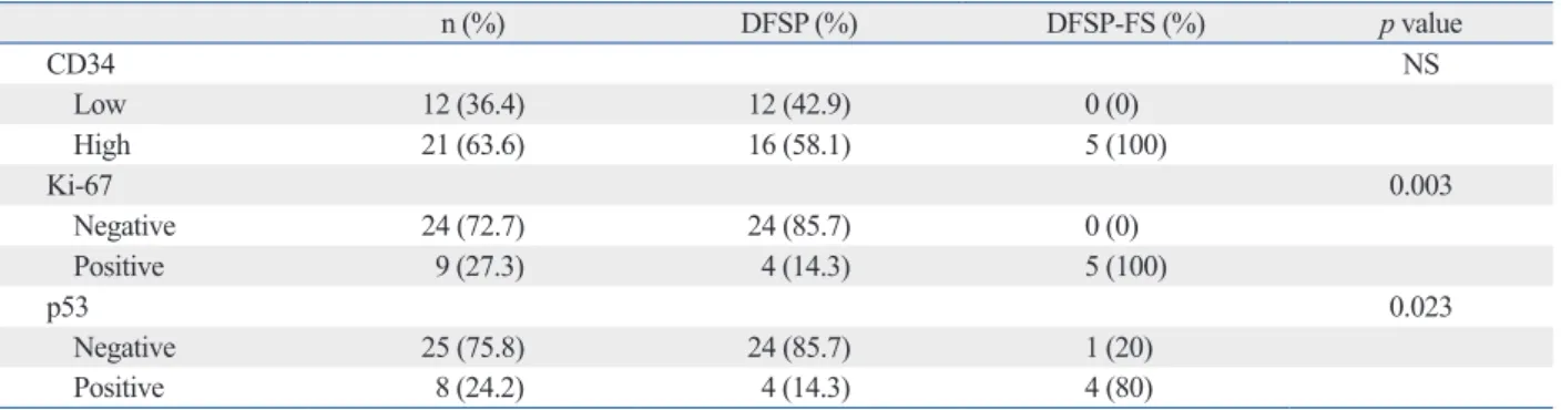
Immunohistochemistry
All of the tumors expressed CD34. In our study, all DFSP- FS demonstrated high CD34 expression. However, there was no significant difference in CD34 expression compared with conventional DFSP. Ki-67 and p53 were expressed in 27.3% and 24.2% of cases, respectively, and were more
RESULTS
Clinicopathological findings
In total, 37 cases of DFSP were reviewed. The clinicopath- ological features are summarized in Table 2. The mean age at diagnosis was 37.4 years, and the ratio of males to fe- males was 1.1:1. The most common tumor location was the trunk (43.2%), followed by the extremities and the head and neck. Of the 37 cases analysed, 24 cases (64.9%) were de novo neoplasms and 13 (35.1%) were recurred cases. All the recurred cases were previously treated by simple exci- sion or WLE. The median preoperative tumor size was 6.2 cm2. All patients were treated primarily by surgery: 34 (91.9%) cases by MMS and 3 (8.1%) by WLE. One patient with recurred DFSP was treated with imatinib mesylate as a neoadjuvant therapy before MMS, because of multiple re- currences and the location of the tumor, which was on the finger web.11 Imatinib 400 mg twice a day for 3 months fol- lowed by reduced dose, 400 mg once a day for 2 months, was prescribed. MMS was performed after reduction of tu- mor size with Imatinib treatment. For those tumors that were treated with MMS, a mean of 1.8 Mohs stages were required. The median postsurgical defect size was 19.6 cm2. Postsurgical defects were reconstructed by primary closure in 23 cases (62.2%), covered by flaps in 9 cases (24.3%), grafts in 3 cases (8.1%), and secondary intention healing in 2 cases (5.4%). None of our patients were treated with ra- diotherapy after surgery. The mean follow-up period was 33 months (range 6–79), and 2 patients (1 MMS and 1 WLE) demonstrated local recurrence during follow-up.
None of the patients showed distant metastasis. Of the 34 cases of DFSP treated by MMS, 1 case recurred. The per- manent pathological slides and CD34 stain of the MMS specimens of this patient were reviewed afterwards, which
Table 3. Immunohistochemical Findings of Conventional DFSP and DFSP-FS
n (%) DFSP (%) DFSP-FS (%) p value
CD34 NS
Low 12 (36.4) 12 (42.9) 0 (0)
High 21 (63.6) 16 (58.1) 5 (100)
Ki-67 0.003
Negative 24 (72.7) 24 (85.7) 0 (0)
Positive 9 (27.3) 4 (14.3) 5 (100)
p53 0.023
Negative 25 (75.8) 24 (85.7) 1 (20)
Positive 8 (24.2) 4 (14.3) 4 (80)
DFSP, dermatofibrosarcoma protuberans; DFSP-FS, dermatofibrosarcoma protuberans-fibrosarcomatous variant.
CD34, high (≥50%) or low (<50%) according to percentage of immunoreactive cells; Ki-67 and p53, positive (>5% immunoreactive nuclei) or negative (<5%).
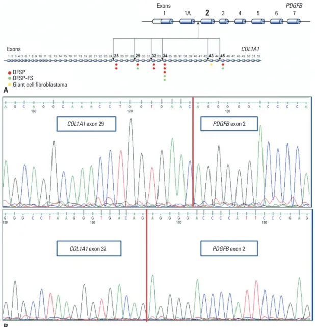
L1A1 (from exon 5 to exon 49) and exon 2 of PDGFB. The COL1A1 breakpoints occurred at the following exons: 25 (two cases), 29 (two cases), 32 (two cases), 34 (five cases), 43 (one case), and 45 (two cases). No association was found among the different COL1A1-PDGFB fusion transcripts, histological subtypes and clinical features. A schematic rep- resentation of our findings is depicted in Fig. 1.
DISCUSSION
Since DFSP is an uncommon tumor, it has not been well- studied in Asia, and there are few reports in the literature.8,13-15 Therefore, we analysed the clinical, pathological, and ge- prevalent in DFSP-FS than in conventional DFSP (Table 3).
All 5 cases of DFSP-FS showed positive immunostaining for Ki-67, compared with 14.3% positivity in conventional DFSP (p=0.003). For p53, 80% of the cases in DFSP-FS stained positive, compared with 14.3% in conventional DFSP (p=0.023).
Analysis of the COL1A1-PDGFB fusion transcripts In 16 of 37 cases, reverse transcriptase-PCR analysis was performed. Only 16 cases out of 37 contained human RNA of sufficient quality/quantity for PCR amplification. The CO- L1A1-PDGFB fusion gene was expressed in 14 (87.5%) cas- es. Sequencing analysis of the different fusion types revealed a high variety of combinations among several exons of CO-
Fig. 1. (A) Schematic presentation of variable positions of collagen type I alpha 1 (COL1A1) breakpoints in the COL1A1-platelet-derived growth factor beta (PDGFB) fusion gene. (B) Some examples of the COL1A1-PDGFB fusion transcript. DFSP, dermatofibrosarcoma protu- berans; DFSP-FS, dermatofibrosarcoma protuberans-fibrosarcomatous variant.
COL1A1 exon 29
COL1A1 exon 32
PDGFB exon 2
PDGFB exon 2
1 1A 2 3 4 5 6 PDGFB7
Exons
COL1A1 Exons
DFSPDFSP-FS
Giant cell fibroblastoma
A
B
87.5% of cases. This percentage is similar to that found by Takahira, et al.21 As reported by other groups,19 we did not find a significant association between the presence of dif- ferent breakpoints and the histologic variants of DFSP or their clinical characteristics.
Advances in the understanding of molecular mechanisms of DFSP have resulted in the implementation of targeted therapy against DFSP based on PDGFR inhibition. Imatinib mesylate is a tyrosine kinase inhibitor rationally developed to target BCR/ABL, KIT, FMS (receptor for colony stimu- lating factor 1), ARG (an ABL-related gene), and PDGFR alpha and beta. It was found to be the first effective system- ic therapy for DFSP. Many studies suggested the usefulness of imatinib in metastatic and locally advanced DFSP. Kérob, et al.22 reported 25 cases of unresectable DFSP treated in a phase II trial with preoperative imatinib at a dose of 600 mg daily for two months, and indicated that some DFSP pa- tients who were initially evaluated as unresectable/meta- static or necessitating mutilating surgery had resectable tu- mors after imatinib therapy. Similarly, we experienced one patient who required neoadjuvant therapy with imatinib to reduce tumor size. The tumor was located at the finger web, where WLE was impossible.
Although WLE has historically been the treatment of choice for DFSP, MMS is another favorable option, as it of- fers the advantage of immediate microscopic examination of the entire surgical margin after excision, which allows for precise tumor mapping. The main purpose of MMS in DFSP is to minimize surgical margins, resulting in less complicated reconstruction with smaller postsurgical defects without jeopardizing the oncologic resection.7,23,24 A meta-analysis by Paradisi, et al.7 of 463 patients with DFSP treated with MMS showed recurrence in 6 cases (1.2%), confirming that the treatment of choice for DFSP is MMS.7,8,25 Our study also supports the use of MMS in DFSP. Thirty-four cases were successfully treated, with only one patient (2.9%) showing recurrence during follow-up. Also, the COL1A1-PDGFB fusion transcript was observed in 87.5% of patients; there- fore, COL1A1-PDGFB may be a useful and accurate tool for diagnosing DFSP in Koreans.
ACKNOWLEDGEMENTS
This study was supported by the Basic Science Research Program through the National Research Foundation of Korea and funded by the Ministry of Education, Science and Tech- netic characteristics, as well as treatment and outcomes in
37 cases of DFSP in Korean patients.
DFSP is an uncommon tumor, with an overall incidence of 0.8 to 5 per million.16 It is most frequently diagnosed in people between 30 and 50 years of age, which is consistent with our results.16 It is found in similar frequencies in men and women, with only a slight male predominance.2 Typi- cally, the tumor initially presents as a slowly growing indu- rated plaque that can eventually transform into a violaceous to red-brown painless nodule, most commonly on the trunk or upper limbs and less often on the head and neck.8 Kim, et al.15 previously reported 65 cases of DFSP in Koreans, which showed similar age range and site distribution.
DFSP has a number of well-described histological vari- ants. Most commonly, conventional DFSP demonstrates pro- liferation of dermal spindle cells that infiltrate into the subcu- taneous fat. These proliferations are made up of monotonous cells with little pleomorphism and a low mitotic index. The spindle cells in the dermis are arranged in storiform pattern, and the infiltrating portion of the tumor is characterized as honeycomb in appearance. DFSP-FS is an another variant which exhibits areas of classic low-grade DFSP with inter- spersed foci of significant cellular atypia and increased mito- ses, and it has been estimated that 7% to 16% of tumors have areas of fibrosarcomatous change.17 Although some authors have reported that these tumors behave more aggressively and are associated with a higher frequency of recurrence and a greater risk of metastasis,12,17 several articles reported that the prognosis of DFSP with fibrosarcomatous transformation has a prognosis similar to that of conventional DFSP.18,19 In our study, 13.5% of cases were DFSP-FS. Compared with other variants, fibrosarcomatous DFSP in our study showed a higher frequency of tumor infiltration into the subcutaneous fat and muscle. Also, the recurrence rate of DFSP-FS was significantly higher compared to other types.
DFSP is characterized by the presence of a distinctive, re- ciprocal rearrangement of chromosomes 17 and 22 in the form of a translocation t(17;22)(q22;q13) or supernumerary ring chromosomes containing material from chromosomal regions of 17q22 and 22q13.20 This rearrangement leads to the fusion of COL1A1, which is localized on 17q22, to the PDGFB, which is localized on 22q13. Confirmation of the presence of the COL1A1-PDGFB fusion gene is a useful tool to identify patients who may be candidates for treatment with imatinib and for the differential diagnosis of DFSP with other CD34+ tumors or CD34-DFSP. In the present study, we indeed identified COL1A1-PDGFB fusion transcripts in
clinicopathologic and immunohistochemical study of a series of 41 cases with emphasis on prognostic significance. Am J Surg Pathol 1998;22:576-87.
13. Tan AW, Tan SH. Dermatofibrosarcoma protuberans: a clinico- pathological analysis of 10 cases in Asians. Australas J Dermatol 2004;45:29-33.
14. Muchemwa FC, Wakasugi S, Honda Y, Ihn H. PDGFB quantifica- tion is a useful tool in the diagnosis of dermatofibrosarcoma protu- berans: a study of 10 cases. Clin Exp Dermatol 2010;35:295-9.
15. Kim M, Huh CH, Cho KH, Cho S. A study on the prognostic val- ue of clinical and surgical features of dermatofibrosarcoma protu- berans in Korean patients. J Eur Acad Dermatol Venereol 2012;
26:964-71.
16. Chuang TY, Su WP, Muller SA. Incidence of cutaneous T cell lymphoma and other rare skin cancers in a defined population. J Am Acad Dermatol 1990;23(2 Pt 1):254-6.
17. Abbott JJ, Oliveira AM, Nascimento AG. The prognostic signifi- cance of fibrosarcomatous transformation in dermatofibrosarcoma protuberans. Am J Surg Pathol 2006;30:436-43.
18. Goldblum JR, Reith JD, Weiss SW. Sarcomas arising in dermato- fibrosarcoma protuberans: a reappraisal of biologic behavior in eighteen cases treated by wide local excision with extended clini- cal follow up. Am J Surg Pathol 2000;24:1125-30.
19. Llombart B, Sanmartín O, López-Guerrero JA, Monteagudo C, Serra C, Requena C, et al. Dermatofibrosarcoma protuberans:
clinical, pathological, and genetic (COL1A1-PDGFB) study with therapeutic implications. Histopathology 2009;54:860-72.
20. Bridge JA, Neff JR, Sandberg AA. Cytogenetic analysis of derma- tofibrosarcoma protuberans. Cancer Genet Cytogenet 1990;49:
199-202.
21. Takahira T, Oda Y, Tamiya S, Higaki K, Yamamoto H, Kobayashi C, et al. Detection of COL1A1-PDGFB fusion transcripts and PDGFB/PDGFRB mRNA expression in dermatofibrosarcoma protuberans. Mod Pathol 2007;20:668-75.
22. Kérob D, Porcher R, Vérola O, Dalle S, Maubec E, Aubin F, et al.
Imatinib mesylate as a preoperative therapy in dermatofibrosarco- ma: results of a multicenter phase II study on 25 patients. Clin Cancer Res 2010;16:3288-95.
23. Ratner D, Thomas CO, Johnson TM, Sondak VK, Hamilton TA, Nelson BR, et al. Mohs micrographic surgery for the treatment of dermatofibrosarcoma protuberans. Results of a multiinstitutional series with an analysis of the extent of microscopic spread. J Am Acad Dermatol 1997;37:600-13.
24. Gloster HM Jr. Dermatofibrosarcoma protuberans. J Am Acad Dermatol 1996;35(3 Pt 1):355-74.
25. Snow SN, Gordon EM, Larson PO, Bagheri MM, Bentz ML, Sa- ble DB. Dermatofibrosarcoma protuberans: a report on 29 patients treated by Mohs micrographic surgery with long-term follow-up and review of the literature. Cancer 2004;101:28-38.
nology (2011-0022376) and by 2012 14th GSK (Stiefel)/
KDA Research Award.
REFERENCES
1. Schiff BL, Tye MJ, Kern AB, Moretti G, Ronchese F. Dermatofi- brosarcoma protuberans. Review of the literature and report of four cases. Am J Surg 1960;99:301-6.
2. Bowne WB, Antonescu CR, Leung DH, Katz SC, Hawkins WG, Woodruff JM, et al. Dermatofibrosarcoma protuberans: a clinico- pathologic analysis of patients treated and followed at a single in- stitution. Cancer 2000;88:2711-20.
3. Rutgers EJ, Kroon BB, Albus-Lutter CE, Gortzak E. Dermatofi- brosarcoma protuberans: treatment and prognosis. Eur J Surg On- col 1992;18:241-8.
4. Lemm D, Mügge LO, Mentzel T, Höffken K. Current treatment options in dermatofibrosarcoma protuberans. J Cancer Res Clin Oncol 2009;135:653-65.
5. DuBay D, Cimmino V, Lowe L, Johnson TM, Sondak VK. Low recurrence rate after surgery for dermatofibrosarcoma protuberans:
a multidisciplinary approach from a single institution. Cancer 2004;100:1008-16.
6. Fiore M, Miceli R, Mussi C, Lo Vullo S, Mariani L, Lozza L, et al. Dermatofibrosarcoma protuberans treated at a single institu- tion: a surgical disease with a high cure rate. J Clin Oncol 2005;
23:7669-75.
7. Paradisi A, Abeni D, Rusciani A, Cigna E, Wolter M, Scuderi N, et al. Dermatofibrosarcoma protuberans: wide local excision vs.
Mohs micrographic surgery. Cancer Treat Rev 2008;34:728-36.
8. Roh MR, Bae B, Chung KY. Mohs’ micrographic surgery for der- matofibrosarcoma protuberans. Clin Exp Dermatol 2010;35:849- 9. Rubin BP, Schuetze SM, Eary JF, Norwood TH, Mirza S, Conrad 52.
EU, et al. Molecular targeting of platelet-derived growth factor B by imatinib mesylate in a patient with metastatic dermatofibrosar- coma protuberans. J Clin Oncol 2002;20:3586-91.
10. Greco A, Fusetti L, Villa R, Sozzi G, Minoletti F, Mauri P, et al.
Transforming activity of the chimeric sequence formed by the fu- sion of collagen gene COL1A1 and the platelet derived growth factor b-chain gene in dermatofibrosarcoma protuberans. Onco- gene 1998;17:1313-9.
11. Jeon IK, Kim JH, Kim SE, Kim SC, Roh MR. Successful treat- ment of unresectable dermatofibrosarcoma protuberans on finger with imatinib mesylate and Mohs microsurgery. J Dermatol 2013;
40:288-9.
12. Mentzel T, Beham A, Katenkamp D, Dei Tos AP, Fletcher CD. Fi- brosarcomatous (“high-grade”) dermatofibrosarcoma protuberans: