211
Uterine Adenomyosis in Beagle Dogs
Hak Soo Kim
1,2, Sang-Chul Kang
2,3, Hu-Song Zhang
2, Jin Seok Kang
4,5, Jae-Hoon Kim
3, Kap-Ho Kim
2, Boo-Hyon Kang
2and Byung-IL Yoon
1,5,*
1
College of Veterinary Medicine & Institute of Veterinary Science, Kangwon National University, Chuncheon, Korea
2Preclinical Research Center, Chemon Inc., Yongin, Korea
3
College of Veterinary Medicine, Jeju National University, Jeju, Korea
4
Department of Biomedical Laboratory Science, Namseoul University, Chonan, Korea
5
Peer Review Working Group of Preclinical Research Center, Chemon Inc., Yongin, Korea
Adenomyosis is a nonneoplastic hyperplastic lesion, characterized by invagination of proliferating endometrial glands into myometrium. In dogs, uterine adenomyosis is relatively rare and it is important in Toxicologic Pathology to differentiate other non-neoplastic and neoplastic lesions in uterus. In the present study, we report two cases of adenomyosis in the female beagle dogs used for a chemical toxicity test. Clinically, one out of the two female beagle dogs, 15 months of age, had vaginal bleeding for 2 weeks and the other one, 11 months of age, showed swelling of vulva for a week. At necropsy, the weight of uterus was markedly increased to 27.9 g and 15.8 g, compared with the mean value (4.01
±2.37, n=6) of that of other normal dogs, respectively. The parameters of hematology and serum chemistry were ranged normal in both of the dogs with enlarged uterus. For differentiation of connective tissue with muscle fibers, Van Gieson stain was also performed in the serial tissue sections. Histopathologically, the lesions of the enlarged uteruses were characterized by proliferating endometrial glands into myometrium, surrounded by connective tissue. The endometrial glands were proliferating downward to myometrium or embedded in multiple clustered glands in deeper myometrium without compressing the adjacent muscle fibers. The gland epithelial cells are uniformly cuboidal shape with a dense and bottom-located nucleus.
These gross and histological findings were consistent with adenomyosis.
Key words: Adenomyosis, endometrial hyperplasia, beagle, uterus.
Received 6 May 2010; Revised version received 14 June 2010; Accepted 15 June 2010
Adenomyosis is a nonneoplastic hyperplastic lesion, characterized by invagination of proliferating endometrial glands into myometrium. Adenomyosis can occur in many species including dogs, cats, rodents and non-human primates (Tamada et al., 2005; Bulman-Fleming, 2008; Hur et al., 2008; Schlafer and Gifford, 2008; Graham et al., 2009). This altered change is relatively frequently found in the mouse and less frequently in the rat (Greaves, 1990). In dogs, uterine adenomyosis is relatively rare and usually found as an incidental lesion and it is important in Toxicologic Pathology to differentiate other non-neoplastic and neoplastic lesions in uterus (Nielsen and Kennedy, 1990; MacLachlan and Kennedy,
2002). The invasive glands consist of uniform, normal appearing epithelial cells, surrounded by endometrial stroma, within the myometrium (Nielsen and Kennedy, 1990;
MacLachlan and Kennedy, 2002; Schlafer and Gifford, 2008).
The glands may be small and have the appearance of an endometrial gland, or they may be multiple, or larger with more proliferative endometrial epithelial cell lining (Schlafer and Gifford, 2008). In dogs, an extreme case of uterine adenomyosis may produce
“Swiss cheese
”pattern in the uterine wall (Schlafer and Gifford, 2008).
The importance of uterine adenomyosis has not yet been defined in terms of the safety assessment. Adenomyosis is classified as Type IIE response with cystic endometrial hyperplasia, squamous metaplasia, endometrial polyp and endometriosis, all associated with the estrogenic effects (Haschek and Rousseaux, 1998). However, its underlying mechanism is still unclear.
In the presents study, we report two cases of adenomyosis
*Corresponding author: Byung-IL Yoon, College of Veterinary Medicine & Institute of Veterinary Science, Kangwon National University, 192-1 Hyoja-dong, Chuncheon, Gangwon 200-701, Republic of Korea
Tel: +82-33-250-8679 Fax: +82-33-244-2367 E-mail: byoon@kangwon.ac.kr
Lab. Anim. Res. 2010: 26(2), 211-213
Case Report
212 Hak Soo Kim
et al.Lab. Anim. Res. | June, 2010 | Vol. 26, No. 2
spontaneously found in the beagle dog which has been commonly used for preclinical safety assessment and discuss its clinical and histopathological characteristics, compared to other non-neoplastic and neoplastic lesions in uterus.
Clinically, one out of the two female beagle dogs, 15 months of age, had vaginal bleeding for 2 weeks and the other one, 11 months of age, showed swelling of vulva for a week.
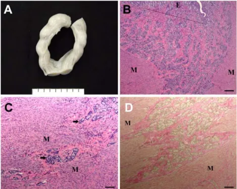
At necropsy, the uteri of the dogs were markedly enlarged (Figure 1A); the weight of uterus was increased to 27.9 g and 15.8 g, compared with the mean value (4.01
±2.37, n=6) of that of other normal dogs, respectively. The parameters of hematology and serum chemistry were ranged normal in both of the dogs with enlarged uterus (data not shown). The enlarged uterus of each dog was fixed in 10% neutral buffered formalin and, after routine tissue processing, tissues sections were prepared for hematoxylin and eosin stain and examined under light microscope. For differentiation of connective tissue with muscle fibers, Van Gieson stain was also performed in the serial tissue sections. Briefly, after deparaffinization and hydration, they were stained by Weigert’s hematoxylin and Van Gieson’s solution for 10 min and 15 min, respectively.
Histopathologically, the lesions of the enlarged uteruses were characterized by proliferating endometrial glands into myometrium, surrounded by connective tissue (Figure 1B- 1D). The endometrial glands were proliferating downward
to myometrium or embedded in the multiple clustered glands in deeper myometrium without compressing the adjacent muscle fibers (Figure 1C). The gland epithelial cells are uniformly cuboidal shape with a dense and bottom-located nucleus (Figure 1C). These gross and histological findings were consistent with adenomyosis.
Despite of some confusion in cases, adenomyosis may usually be differentiated from other non-neoplastic lesions such as endometriosis, cystic endometrial hyperplasia and pseudo-plancentational endometrial hyperplasia and benign uterine tumors such as leiomyoma, in particular adenomyoma.
Endometriosis is defined as the presence of endometrial tissue in locations distant to the uterus, usually in the peritoneal cavity (Leininger and Jokinen, 1990). In general, endometriosis has the tissue resembling endometrium lying outside the endometrial cavity. Cystic endometrial hyperplasia is limited in endometrium and it can be induced by estrogen (Type IIE response) or more frequently progesterone in the dog (Type IIP response) (Nielsen and Kennedy, 1990). Cystic endometrial hyperplasia generally arises from endometrial glandular epithelium, but it also develops from villous folds covered by luminal epithelium. Since this condition with exudates accumulation is frequently associated with inflammation and bacterial infection, it may progress to development of pyometra, namely cystic hyperplasia/
Figure 1.
Gross and microscopic findings of adenomyosis. The uteri of the dogs were markedly enlarged (A). Note the proliferating
endometrial glands into myometrium (M) from endometrium (E, upper of line) (B). Some multiple clustered endometrial glands were
also found with being embedded in deeper myometrium without compressing the adjacent muscle fibers (C). The glandular
epithelial cells are uniformly cuboidal shape without cellular atypia. The glands were surrounded by connective tissue (deep red in
D). H&E stain for B and C and Van Gieson stain for D. Bars=100
µm for C and D and 200
µm for B.
Uterine adenomyosis in Beagle dogs
213Lab. Anim. Res. | June, 2010 | Vol. 26, No. 2
pyometra complex (Schlafer and Gifford, 2008). Pseudo- plancentational endometrial hyperplasia is also a kind of endometrial hyperplasia, resembling maternal placental tissues. It has been known to occur primarily during the luteal phase of the cycle when the endometrium is highly sensitive (Schlafer and Gifford, 2008). However, the histological features, as they have the normal histology of the endometrium at placentation sites in normal pregnancy, are very much different from adenomyosis (Schlafer and Gifford, 2008). Another confusing lesion in uterus with adenomyosis is leiomyoma.
But unlike leiomyoma, adenomyosis has indistinct, poorly delimited margins from the adjacent myometrium (Bergeron
et al