1)
본 연구는2008년도 대한소아알레르기호흡기학회Outstanding Fellow Award를 위한 건일제약 연구지원금에 의해 일부 이루어진 연구임. 접수: 2011년 월5 17 ,일 수정:2011년 월 일 승인6 2 , :2011년 월 일6 6
책임저자 : 김진택 경기도 의정부시 금오동, 65- 가톨릭대학교 의정부성모병원 소아청소년과1 Tel : 031)820-3107 Fax : 031)821-3108 E-mail : jintackk@catholic.ac.kr
활성화에 의한 호산구 자멸사에서 CD30
caspase- 의 매개작용9
가톨릭대학교 의과대학 소아과학교실1, 서울대학교 의과대학 소아과학교실2
이혜진1ㆍ이근영1ㆍ김유진1ㆍ장필상1ㆍ윤종서1ㆍ김현희1ㆍ고영률2ㆍ김진택1ㆍ이준성1
CD30 Activation Induced Eosinophil Apoptosis is Mediated by Caspase-9
Hye Jin Lee, MD1, Keun Young Lee, MD1, Yoo Jin Kim, MD1, Pil Sang Jang, MD1, Jong Seo Yoon, MD1, Hyun Hee Kim, MD1, Young Yull Koh, MD2,
Jin Tack Kim, MD1, Joon Sung Lee, MD1
1Department of Pediatrics, The Catholic University of Korea School of Medicine, Seoul,
2Department of Pediatrics, Seoul National University College of Medicine, Seoul, Korea
Purpose : Although CD30 is known to be expressed more on eosinophils undergoing apoptosis, it is still not known how CD30 activation leads to eosinophil apoptosis. In this study, we evaluated whether ligation of CD30 incites apoptosis and investigated whether the mechanisms of CD30 indu- ced eosinophil apoptosis are dependent on caspase activation.
Methods : We drew 90 mL of peripheral blood from healthy donors and then purified eosinophils using a MACS system. Expression of CD30 on eosinophils was measured, and eosinophils were cultured in wells pretreated with anti-CD30 mAb, isotype control immunoglobulin G1, interleukin (IL)-5, and dexamethasone in Roswell Park Memorial Institute 1640 media supplemented with 10%
fetal bovine serum. Their rates of apoptosis were then compared using flow cytometry. To evaluate whether caspase-9 is involved in CD30-induced eosinophil apoptosis, the apoptotic rate was evaluated after the addition of caspase-9 inhibitor. The expression of procaspase-9 was also measured using Western blot.
Results : Expression of CD30 molecules on eosinophils increased steadily as the culture time lapse. The apoptotic rates of eosinophils cultured in the presence of anti-CD30 mAb were signifi- cantly increased to 29.1 6.1% and 47.3 4.7% compared to 17.1 6.7% and 29.4 9.2% of the ± ± ± ± control at 4 and 24 hours, respectively (both P <0.05). The apoptotic rates of eosinophils treated with anti-CD30 mAb were even faster than those of eosinophils treated with dexamethasone, and the mAb also suppressed the IL-5-induced enhancing effect of eosinophil survival. Caspase-9 inhibitor suppressed mAb induced eosinophil apoptosis from 54.8 6.9% and 71.5 11.6% to 24.5 ± ± ± 6.0% and 47.8 11.4% at 18 and 36 hours, respectively (both ± P <0.001). We also demonstrated that the expression of procaspase-9 with mAb was diminished compared to that of the control and of IL-5.
Conclusion : This study showed CD30 activation enhances eosinophil apoptosis, and the effect
is mediated by caspase-9 activation. [Pediatr Allergy Respir Dis(Korea) 2011;21:115-122]
서 론
호산구는 알레르기염증을 일으키는 중요한 작동세포
이며 과립단백 사이토
(effector cell) (granular proteins), 카인(cytokine),지질매개체(lipid mediators)등 알레르기 염증반응의 시작과 유지에 관여하는 다양한 세포독성물질 들(cytotoxic substances)을 가지고 있다.1)호산구의 수 명 연장과 사멸 감소는 알레르기 염증반응을 지속시키는 것 으로 알려져 있으며2)호산구 생존 및 제거와 관련된 요소들 에 많은 관심이 집중되고 있다 호산구 생존은. interleukin (IL)-3, IL-5, granulocyte-macrophage colony-stimula- ting factor (GM-CSF)에 의해 주로 야기되며 특히IL- 는5 호산구 세포 내 생존친화성 신호전달체계를 활성시킨다.3) 세포사멸은 괴사와 자멸사(apoptosis)가 있으며 전자는 급성 세포손상으로 인해 세포사멸이 일어나므로 염증반응 을 일으키지만 자멸사는 염증매개물질을 분비하지 않고 세 포가 수축하여 대식세포에 의해 염증의 동반 없이 제거되며
4,5)caspase가 인체세포의 자멸사에 중요한 역할을 하는 것 으로 알려졌다6).호산구 자멸사는 조직손상 없이 알레르기 염증반응을 종결시키는 중요한 과정이며 CD95,7)CD69,8) CD459)등의 호산구의 세포막 표면 수용체에 의해 자멸사 가 유도되는 것으로 알려져 있다. CD30은tumor necro- 에 속하며 호지킨림프 sis factor receptor (TNFR) family
종의Hodgkin and Reed Sternberg세포에서 처음 관찰되 었다.10)
은 주로 자연세포독성세포
CD30 activated T cells, (NK 인체면역결핍바이러스
cells), (human immunodeficiency 나 엡스타인
virus) -바 바이러스(Epstein-Barr virus)에 감 염된 림프구 표면에서 발현되며11)CD30가 활성화 될 때 세 포 종류에 따라 세포가 증식되거나 한편으로 자멸사 하기도 하는 다양한 생물학적 효과를 나타낸다.12-16)과거에 CD30 은 호산구에 존재하지 않는 것으로 알려졌으나17,18)자멸사 가 진행중인 호산구에서는 발현이 증가된다는 보고들도 있
다.19,20)그러나 실제CD30활성화가 어떻게 호산구 자멸사
를 일으키는지는 아직 명확하지 않다 본 연구에서는 호산구. 의CD30발현여부를 확인하고CD30활성화에 의해 호산 구 자멸사가 유도되는지 여부와 그 자멸사 기전에 caspase 가 관여하는지를 밝히고자 하였다.
대상 및 방법
대 상 1.
가톨릭대학교 의정부성모병원 임상시험심사위원회의 공 인 동의서 작성에 동의하고 알레르기질환의 기왕력이 없는 건강한24명 성인들의 말초혈액90 mL를 채취하였다.
방 법 2.
호산구 분리 1)
혈액을 Percoll (GE health care, Uppsala, Sweden) 용액을 이용하여 밀도차에 의해 원심분리한 후 과립구들을 anti-CD16 Ab coated immunomagnetic beads (Milten- 와 시간 배양 yi Biotec, Bergisch Gladbach, Germany) 1
하여MACS magnetic system을 통해 호산구를 선택적으 로 분리하였다21).분리된 호산구 수와 생존능(viability)의 측정은 Randolph 염색하여 hemocytometer와 trypan 로 각각 평가하였으며 순도 blue dye exclusion method
와 생존능은 모두 이상이었다 호산구는
(purity) 98% . 10%
이 첨가된
fetal bovine serum (FBS) Roswell Park Me- morial Institute (RPMI) 1640, streptomycin (100 g/μ
에
mL) 1 10× 6cells/mL의 농도로 재현탁하고 모든 실험은 각각 회 시행하였다4 .
호산구의 발현
2) CD30
호산구 표면에 발현되는CD30표현정도는flow cyto- 를 이용하여 측정하였다
metry .22) 분리한 호산구를 0.1%
bovine serum albumin (BSA; Sigma-Aldrich Co., St.
와
Louis, MO, USA) 4 mg/mL human immunoglobulin G (IgG; Sigma-Aldrich Co.)가 함유된Dulbecco s phos’ - phate-buffered saline (PBS)에서 포화농도의 항CD30항 체 또는 동형의 대조항체와 각각4℃에서30분간 표지하였 다. 세포를 세척한 후 적정농도의 FITC-conjugated F(ab )’2 goat anti-mouse IgG (BD Biosciences, San
와 함께
Jose, CA, USA) PBS-BSA에서4 , 30℃ 분간 다시 배양하였다. 1% paraformaldehyde로 고정 후FACScan 유세포분석기(flow cytometry, BD, Franklin Lakes, NJ,
을 이용하여 형광분석하였다 형광강도는
USA) . logarith--
을 이용하여 각 검체의 개 세포를 mic amplification 20,000 대상으로 결정하였고 Becton Dickenson lysis II soft-
Key Words : CD30, Eosinophil apoptosis, Caspase-9
를 통하여 로 전환시켰다 ware linear equivalent .
호산구 배양 3)
활성화가 호산구 자멸사에 미치는 영향은
CD30 CD30
단클론항체인 Ber-H8 (BD Biosciences)처리군과 대조군 인 동형의 IgG1 (Santa Cruz Biotechnology Inc., Santa 및 순수배양액의 자멸사율을 각각 비교 Cruz, CA, USA)
평가하였다.
각 항체들을PBS용액에20 g/mLµ 로 부유시킨 후flat bottom 24 well plates (Costar group Inc., Boston, MA,
에 각 당 씩 분주하였다 각
USA) well 500 Lµ . plate well 들을4℃에서24시간 단클론항체로 미리 코팅하여 고정시 킨 후well들을1% BSA함유PBS로 회 세척하고 이어서3
로 에서 시간 하였다 이어서
1% BSA 1 mL 4℃ 24 block . 각well에 같은 수의 호산구를 분주하고10% heat inacti-
이 함유된 에
vated fetal bovine serum RPMI 1640 20 g/µ 의
mL Ber-H8,대조항체인 동형의20 g/mLµ 의IgG1, 10 의
ng/mL IL-5, 10 M/mLµ 의dexamethasone (Sigma- 을 각각 투여한 후
Aldrich Co.) 37 , 5% CO℃ 2배양기에서 시간 또는 시간 배양하였다 또한 호산구를
4 24 . IL- 또는5
caspase- 억제제9 (BD)와 함께18시간 및36시간 동안 별 도 배양한 후 유세포분석기로 자멸사 여부를 측정하였다.
호산구 자멸사 4)
호산구 생존능은 FITC-conjugated annexin V와pro- 을 이용한
pidium iodide (PI) apoptosis detection kit 으로 검사하였다
(BD) .
배양된 호산구를 부드럽게 피펫팅한 후 수거하여PBS로 회 세척하여
1 Ca2+함유 완충액에 현탁하고 실온에서 15 분간 FITC-conjugated annexin V와 반응시키고10 g/μ
로 염색하여 로 최소 개 이상
mL PI flow cytometry 5,000 의 세포들을 측정하였다.
에 의한 호산구 발현
5) Western blot caspase
호산구를 66시간 배양한 후 수거하여 radioimmuno-
를 이용하여 단백을
precipitation assay buffer caspase 추출하였다. 4 , 13,000 rpm℃ 으로20분간 원심분리 후 단 백 농도를 Brad-ford 방법으로 측정하고 5배 sodium 로 희석 후 희석된 단백을 dodecyl sulfate sample buffer
에서 분간 비등하였다 농도의 단백을
100℃ 5 . 1 g/ Lμ μ 12.5
에 투여 후
% polyacrylamide gel polyvinylidine difluo-
에 옮겨 에서 시간 동안 분리하
ride membrane 100 mV 1
였다 위의. Membrane을Tween 20 Tris완충액과 무지방 분유가 섞인 용액에서 실온에 1시간 고정하였다. Mem-
을
brane caspase- 항체9 (BD)와4℃에서24시간 반응시
켜 결합시킨 후 다시membrane을 실온에서tris buffered 으로 회 세척하고 이차항체인
saline/Tween 4 Goat anti-
rabbit IgG-HRP conjugate (Bio-Rad Laboratories Inc, 와 실온에서 시간 반응시켰다 이후
Hercules, CA, USA) 2 .
을 이용하여 Western blotting luminal reagent kit proca- spase- 단백을 검출하고9 Kodak BioMax Light film에 노 출시켜 현상하였다.
통계 분석 6)
결과는 mean SEM± 로 표시하였으며 각 군의 차이는 one-way ANOVA로 분석하였으며
P
값이0.05미만인 경 우를 통계적으로 유의한 것으로 하였다.결 과
호산구의 발현
1. CD30
호산구의CD30발현은 배양시간이 경과함에 따라 지속 적으로 증가하였다. 10% FBS RPMI 배양액 배지에서0, 시간 배양 후 양성을 보인 호산구는 4, 24, 66, 90 CD30
에서 로
flow cytometry 1.2%, 3.1%, 5.5%, 9.4%, 17.6%
측정되었다 각각.(
P
<0.05,P
<0.05,P
<0.01,P
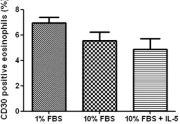
<0.001, 에Fig. 1) 10% FBS 10 ng/mL IL- 첨가군과5 1% FBS군 의24시간 배양 후 발현율은 각각4.8%, 6.9%로10% FBS
Fig. 1. Expression of CD30 on human blood eosi- nophils. Isolated eosinophils were incubated in Roswell Park Memorial Institute 1640 media supplemented with 10% fetal bovine serum and then analyzed CD30 expression in a time depen- dent manner by flow cytometry using anti-CD30 mAb and fluorescein isothiocyanate-conjugated F(ab )’ 2 goat anti-mouse immunoglobulin G (*
P
<0.05,†
P
<0.01,‡P
<0.001 compared with the 0 hour group).군과 유의한 차이가 없었다.(
P
=0.49, Fig. 2)항 단클론항체에 의한 호산구 자멸사 2. CD30
항CD30단클론항체인Ber-H8투여군의 호산구 자멸사 율은 시간4 , 24시간 배양 시29.1 6.1%, 47.3 4.7%± ± 를 보여 동형의 대조항체 투여군의 17.1 6.7%, 29.4 9.2%± ± 에 비해 각각 유의한 증가를 보였으며 모두(
P
<0.05, Fig.대조항체군과 군은 서로 유사한 자멸사
3A, B), 10% FBS
율을 보였다. Ber-H8군의 자멸사율은 4시간 배양 시
투여군 에 비해서 높았고
dexamathasone (14.2 5.0%)±
시간 배양 에서는 유사하였다 또한
24 (48.3 4.3%)± . IL-5
존재하의 4시간, 24시간 배양 자멸사율이 12.5 4.5%,± 이었으나
16.1 11.0%± Ber-H8을 첨가하면 29.4 6.4%,± 로 각각 유의하게 증가하여 모두
47.6 2.7%± (
P
<0.05, Fig.3A, B) IL- 가5 CD30활성화로 인한 자멸사 증가를 억제하 지 못하였다.
활성화의 호산구 자멸사 유도에 의 관련여
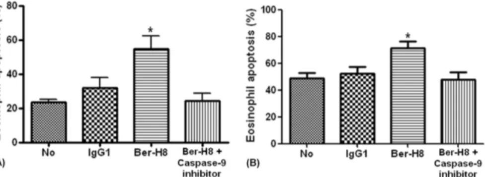
CD30 caspase
부를 알기 위해 caspase- 억제제를 투여한 후 자멸사 증가9 가 억제되는지를 측정하였다. Caspase- 억제제 첨가 후의9 자멸사율은18시간 배양은54.8 6.9%± 에서24.5 6.0% ,± 로
시간 배양은 에서 로 각각 유
36 71.5 11.6%± 47.8 11.4%±
의하게 감소하였으며 모두(
P
<0.05, Fig. 4A, B)대조군 의 자멸사율과 유사하였다 (23.6 3.5%, 49.1 9.0%)± ± .항 단클론항체에 의한 호산구의 3. CD30
caspase- 9 발현
Ber-H8배양군의 호산구를 수거 후Western blotting 을 이용하여 procaspase- 의 발현을 관찰하였다9 . Ber- H8 군의36시간 배양 후procaspase- 의 발현율이9 IL- 군에5 비해 현저히 감소하고(
P
<0.05)대조군에 비해서도 통계적 으로 유의하지는 않으나 낮은 소견을 보였다.(Fig. 5)고 찰
본 연구의 목적은CD30활성화에 따른 호산구 자멸사 여부와 자멸사 발생 기전을 알아보고자 하였다. CD30발현 율은 시간경과에 따라 지속적으로 증가하였고 IL- 첨가에5 의해서도 영향을 받지 않았으며 호산구 자멸사율은 CD30 Fig. 2. Expression of CD30 on human blood eosi-
nophils. Isolated eosinophils were incubated for 24 hours in Roswell Park Memorial Institute 1640 media supplemented with either of 1% fetal bovine serum (FBS), 10% FBS and 10% FBS with 10 ng/
mL of interleukin (IL)-5. The rate was not signi- ficantly different each other (
P
=0.49).Fig. 3. Effect of immobilized anti-CD30 mAb on eosinophil apoptosis. Eosinophils were cultured in Roswell Park Memorial Institute 1640 media supple mented with 10% fetal bovine serum of 24 well culture plates previously coated with 20 g/mL of antiµ -CD30 mAb or control immunoglobulin G (IgG)1 mAb for 4 hours (A) and 24 hours (B). 10 ng/mL of interleukin (IL)-5, and 10 µM/mL of dexamethasone were added in other plates. Values represent the mean SEM of 4 separate experi± ments (*
P
<0.05 compared with isotype matched control or media alone).발현 정도에 따라 증가하였다 또한 본 연구에서 처음으로. caspase- 가9 CD30 활성화에 의한 호산구 자멸사기전에 연관되어 있음을 규명하였다.
지속적인 호산구 증가증은 만성 알레르기질환 염증조직 에서 관찰되는 특징적 소견으로 호산구 생성이 증가하고 생 존기간도 연장되어 나타난다 호산구 성장인자인. IL-5, IL-3, GM-CSF는 골수의 호산구 생산증가와 말초혈액의 호
산구 증식을 통해 조직 내 호산구 축적을 일으킨다 이전에. 호산구수와 천식의 연관성에 대해 의문을 가진 보고가 있었 으나23),항IL- 제 투여 후 기관지세척액 내 호산구 수가 감5 소한 천식환자들의 기관지생검 소견에서 major basic pro- 양성 호산구가 다량 관찰되어 조직 내 호산구 활성화에 tein
의해 기도과민성이 지속됨이 알려졌다.24)
이처럼 기도염증의 지속은 호산구 활성화가 중요한 역할 Fig. 4. Effect of caspase-9 inhibitor on immobilized anti-CD30 mAb induced eosino-
phil apoptosis in Roswell Park Memorial Institute 1640 media supplemented with 10
% fetal bovine serum. Eosinophils were cultured in 24 well culture plates previously coated with 20 g/mL of antiµ -CD30 mAb and 20 g/mL of control immunoglobulin Gµ (IgG)1 mAb for 18 hours (A) and 36 hours (B). Caspase-9 inhibitor was added in other plate. Values represent the mean SEM of 4 separate experiments (± *
P
<0.05 compared with isotype matched control, media alone or with inhibitor).Fig. 5. Expression of procaspase-9 protein in eosinophils. Eosinophils were cultured in Roswell Park Memorial Institute 1640 media supplemented with 10 fetal bovine serum of 24 well culture plates previously coated with 20 g/mL ofµ anti-CD30 mAb, 20 µg/mL of control immunoglobulin G (IgG)1 mAb and with addition of 10 ng/mL of interleukin (IL)-5 for 36 hours. The expression was determined by Western blot (*
P
<0.05 compared with eosinophils cultured in the presence of IL-5).을 하기 때문에 호산구 자멸사에 의해서 효과적으로 억제 될 수 있다 그러나 호산구 자멸사가 순조롭게 이루어지지. 않으면 기도 내 호산구 증가증은 지속되기 때문에 호산구 자멸사를 유도하는 것이 알레르기 질환의 새로운 치료 목표 가 되고 있다 본 연구에서 호산구 배양기간을. 96시간까지 지속한 결과CD30발현이 계속 증가하는 추세를 보였으며
배양조건을 달리한 시간 배양에서는
(Fig. 1), 24 1% FBS
에서의CD30발현이10% FBS또는10% FBS에IL- 를5 첨가한 경우보다 통계적으로 유의하지는 않으나 높게 나타 났다 이는 호산구. CD30발현이 세포 사멸 시까지 계속 증 가하고 호산구 생존이 열악한 조건에서 증가함을 보여준다.
또한 IL- 를 첨가한 전후의5 CD30발현율에 차이가 없는 것 으로 보아 IL- 가5 CD30발현에 영향을 미치지 않음을 알 수 있었다.
본 연구에서 호산구 자멸사율은 모든 군에서 시간에서4 시간으로 배양시간이 경과함에 따라 증가하였는데 특히
24 ,
항CD30단클론항체 투여군에서 가장 높은 자멸사율을 보 이고 스테로이드 투여군보다 높거나 유사하였다 이는 항. 단클론항체의 배양농도에 따라 호산구 생존율이 감 CD30
소하였다는 최근의 보고25)와 유사한 소견이다 또한 항. 항체 투여군에
CD30 IL- 를 첨가한 후에도 자멸사율에 변5 화를 보이지 않는 흥미로운 소견을 보였다 즉. , CD30활성 화는 스테로이드에 비해 더 빠르게 호산구 자멸사를 유도하 며 IL- 에 의해서도 자멸사 진행이 억제되지 않는 것으로5 보아 혈청결핍 등의 조건에서 보이는 자멸사와는 다른 경로 에 의해 호산구 자멸사를 유도하는 것으로 보인다 이와 같. 은CD30의 활성화에 의해 유도되는 빠르고 강력한 호산구 자멸사는 호산구 생존이 지속적으로 유지되는 상태를 호전 시킬 수 있는 방안으로 생각되나 실제 임상에서 알레르기염 증 질환의 치료수단으로 이용되기 위해서는 앞으로 많은 연 구를 통한 검증이 필요하다.
은 계의 시토카인 수용체로서 세포질 내에 CD30 TNFR
사멸영역(death domain, DD)을 갖는 일반적인TNFR계와 달리 세포질에 DD는 없지만 TNFR-associated factor 영역을 가지고 있다 그러므로 수용체는 배
(TRAF) . CD30
위자와 결합하면 삼합체(trimerization)를 형성하고 여기 에TRAF1, TRAF2/5, TRAF3등이 결집하여 상호작용 함으로서caspase를 활성화 시킨다.26,27)
본 연구에서 나타난CD30단클론항체 투여 시의 호산구 자멸사 증가도 CD30와 결합한 후 TRAF들과 연계하여 caspase- 이 활성화되기 때문에 유발되는 것으로 생각된9 다 세포 자멸사는 사멸수용체. (death receptor)를 통해 이
루어지는지 여부에 따라 자멸사에 이르는 신호전달경로가 다르다 즉. , caspase- 과 같이 사멸수용체에 의존하여 활성8 화되는 경로와28,29)사멸수용체에 의존하지 않고 미토콘드 리아에 의해 조절되는caspase활성 경로가 있는데 후자는 procaspase- 이 자멸사 초기에 미토콘드리아에서 유리된9
에 의해 활성화된 와 결합하면
cytochrome c Apaf1 caspa-
se- 으로 활성화된다9 .30)
본 연구에서 caspase- 억제제가 항9 CD30단클론항체 에 의한 호산구 자멸사를 현저히 감소시키고(Fig. 4A, B) procaspase- 의 발현은 항9 CD30단클론항체와 배양한 호 산구에서 낮았으나 IL-5존재 하에서는 증가하였다 이는. 앞의 결과에서 보인 바와 같이 IL- 가 호산구5 CD30발현에 영향을 미치지 않아 procaspase- 의 발현율이 높게 나타난9 것으로 생각되며 한편 항CD30 단클론항체 투여 후에는
의 교차결합에 의해
CD30 procaspase- 이 활성상태의9 caspase- 으로 전환되어 호산구 자멸사를 항진시킨 것으9 로 보인다 더구나 최근의 연구에서. caspase-3, - 억제제8 투여 후에도 항CD30단클론항체에 의한 호산구 자멸사에 영향을 미치지 않았다고31) 하였는데 본 연구에서도 항
단클론항체와 배양한 호산구에서
CD30 caspase- 이8
에서 발현되지 않아 서로 유사한 소견을 보였 Western blot
다 즉. , CD30에 의한 호산구 자멸사는 사멸수용체에 의존 하지 않고 미토콘드리아에 의해 직접 조절되는 별도의 자멸 사 신호전달경로에 따라 caspase- 이 활성화되어 일어나9 는 것으로 추정된다 그러나 호산구 자멸사 기전을 명확히. 밝히기 위해서는 향후TRAF단백질과 같은 광범위한 신호 전달분자에 대한 연구가 필요하다.
본 연구는 알레르기질환이 없는 건강한 공여자의 호산구 만을 이용하였기 때문에 제한적 결과가 나올 수 있는 제한 점이 있으며 정상인과 알레르기 질환자의 호산구 자멸사에 차이가 있는지 아직 명확히 알려져 있지 않으나 알레르기 환자의 호산구를 이용한 자멸사 연구도 추가적으로 필요하 다고 생각된다.
이상의 연구에서 결론적으로 CD30활성화로 유발되는 호산구 자멸사에 caspase- 가 매개작용을 할 것으로 추정9 되었다.
요 약
목 적 :CD30은 자멸사가 진행중인 호산구에서 발현이 더 증가되는 것으로 알려져 있으나 실제CD30활성화가 어 떻게 호산구 자멸사를 일으키는지는 명확하지 않다 본 연구.
에서는 인체세포 자멸사의 중요한 요소인caspase가CD30 활성화에 의한 호산구 자멸사에 미치는 역할을 규명하고자 한다.
방 법 : 건강한 성인의 말초혈액90 mL를 채취하여 혈액 을Percoll밀도차와MACS magnetic system으로 호산구 를 분리한 후 호산구 표면CD30발현율과 자멸사율을flow
로 측정하였다 호산구 배양은 단클론항
cytometry . CD30
체와 동형의 immunoglobulin G1을 대조항체로 하여24 에 미리 코팅하고 고정한 후 호산구를 분주하고 well plates
interleukin (IL)-5, Dexamethasone을 첨가하여 시간4 , 시간 배양한 후 자멸사 정도를 각각 비교하였다 또한
24 .
에 의한 호산구 자멸사에서 의 역할을 규명하
CD30 caspase
기 위해 caspase- 억제제를 첨가한 후 배양하여 자멸사율9 변화를 측정하고 Western blotting을 이용하여 proca- spase- 단백을 검출하였다9 .
결 과 : 호산구 표면CD30발현은 배양시간이 연장됨에 따라 점점 증가하였다 호산구 자멸사율은 항. CD30단클론 항체에서 시간4 , 24시간 배양 시29.1 6.1%± 와47.3 4.7±
으로 대조항체의 와 에 비해 유의
% 17.1 6.7%± 29.4 9.2%±
하게 증가하였다(
P
<0.05).항CD30항체투여군의 자멸사 율은 스테로이드 투여군에 비해 빠르게 중가하였으며 IL-5 투여군의 호산구 생존능을 현저히 감소시켰다 또한 항.항체투여군에
CD30 caspase- 억제제를 첨가하면9 18시간 과36시간 배양 후 자멸사율이 첨가전의54.8 16.9%± 와
에서 와 로 각각 유
71.5 11.6%± 24.5 6.0%± 47.8 11.4%±
의하게 감소하였으며(
P
<0.05) 항CD30 항체투여군의 procaspase- 단백의 발현이9 IL- 투여군과 대조항체군에5 비해 저하됨을Western blot을 통해 관찰하였다.결 론 : 본 연구에서CD30활성화는 호산구 자멸사를 촉 진하며 caspase- 활성화가 자멸사 과정에서 중요한 역할9 을 할 것으로 보인다.
참 고 문 헌
1. Gleich GJ. The eosinophil and bronchial asthma:
current understanding. J Allergy Clin Immunol 1990;85:422-36.
2. Gleich GJ. Mechanisms of eosinophil-associa- ted inflammation. J Allergy Clin Immunol 2000;
105:651-63.
3. Sanderson CJ. Interleukin-5: an eosinophil growth and activation factor. Dev Biol Stand 1988;69:23-9.
4. Kerr JF, Wyllie AH, Currie AR. Apoptosis: a basic biological phenomenon with wide-ranging implications in tissue kinetics. Br J Cancer 1972;26:239-57.
5. Allen RT, Hunter WJ 3rd, Agrawal DK. Mor- phological and biochemical characterization and analysis of apoptosis. J Pharmacol Toxicol Me- thods 1997;37:215-28.
6. Alnemri ES, Livingston DJ, Nicholson DW, Sal- vesen G, Thornberry NA, Wong WW, et al.
Human ICE/CED-3 protease nomenclature. Cell 1996;87:171.
7. Matsumoto K, Schleimer RP, Saito H, Iikura Y, Bochner BS. Induction of apoptosis in human eosinophils by anti-Fas antibody treatment in vitro. Blood 1995;86:1437-43.
8. Walsh GM, Williamson ML, Symon FA, Willars GB, Wardlaw AJ. Ligation of CD69 induces apop- tosis and cell death in human eosinophils cultured with granulocyte-macrophage colony-stimulating factor. Blood 1996;87:2815-21.
9. Blaylock MG, Sexton DW, Walsh GM. Ligation of CD45 and the isoforms CD45RA and CD45RB accelerates the rate of constitutive apoptosis in human eosinophils. J Allergy Clin Immunol 1999;104:1244-50.
10. Dürkop H, Latza U, Hummel M, Eitelbach F, Seed B, Stein H. Molecular cloning and ex- pression of a new member of the nerve growth factor receptor family that is characteristic for Hodgkin's disease. Cell 1992;68:421-7.
11. Stein H, Mason DY, Gerdes J, O'Connor N, Wainscoat J, Pallesen G, et al. The expression of the Hodgkin's disease associated antigen Ki- 1 in reactive and neoplastic lymphoid tissue:
evidence that Reed-Sternberg cells and histio- cytic malignancies are derived from activated lymphoid cells. Blood 1985;66:848-58.
12. Jung W, Krueger S, Renner C, Gause A, Sahin U, Trümper L, et al. Opposite effects of the CD30 ligand are not due to CD30 mutations:
results from cDNA cloning and sequence com- parison of the CD30 antigen from different sources. Mol Immunol 1994;31:1329-34.
13. Masuda M, Ishida C, Arai Y, Okamura T, Ohsa- wa M, Shimakage M, et al. Dual action of CD30 antigen: anti-CD30 antibody induced apoptosis and interleukin-8 secretion in Ki-1 lymphoma cells. Int J Hematol 1998;67:257-65.
14. Mir SS, Richter BW, Duckett CS. Differential effects of CD30 activation in anaplastic large cell lymphoma and Hodgkin disease cells.
Blood 2000;96:4307-12.
15. Gruss HJ, Boiani N, Williams DE, Armitage RJ, Smith CA, Goodwin RG. Pleiotropic effects of the CD30 ligand on CD30-expressing cells and lymphoma cell lines. Blood 1994;83:2045-56.
16. Muta H, Boise LH, Fang L, Podack ER. CD30 signals integrate expression of cytotoxic effec- tor molecules, lymphocyte trafficking signals, and signals for proliferation and apoptosis. J Immunol 2000;165:5105-11.
17. Pinto A, Aldinucci D, Gloghini A, Zagonel V, Degan M, Improta S, et al. Human eosinophils express functional CD30 ligand and stimulate proliferation of a Hodgkin's disease cell line.
Blood 1996;88:3299-305.
18. Chiarle R, Podda A, Prolla G, Gong J, Thor- becke GJ, Inghirami G. CD30 in normal and neoplastic cells. Clin Immunol 1999;90:157- 64.
19. Berro AI, Perry GA, Agrawal DK. Increased expression and activation of CD30 induce apoptosis in human blood eosinophils. J Immu- nol 2004;173:2174-83.
20. Matsumoto K, Terakawa M, Miura K, Fukuda S, Nakajima T, Saito H. Extremely rapid and in- tense induction of apoptosis in human eosi- nophils by anti-CD30 antibody treatment in vitro. J Immunol 2004;172:2186-93.
21. Hansel TT, De Vries IJ, Iff T, Rihs S, Wand- zilak M, Betz S, et al. An improved immuno- magnetic procedure for the isolation of highly purified human blood eosinophils. J Immunol Methods 1991;145:105-10.
22. Simon HU, Yousefi S, Schranz C, Schapowal A, Bachert C, Blaser K. Direct demonstration of delayed eosinophil apoptosis as a mechanism causing tissue eosinophilia. J Immunol 1997;
158:3902-8.
23. Leckie MJ, ten Brinke A, Khan J, Diamant Z, O'Connor BJ, Walls CM, et al. Effects of an interleukin-5 blocking monoclonal antibody on
eosinophils, airway hyper-responsiveness, and the late asthmatic response. Lancet 2000; 356:
2144-8.
24. Flood-Page PT, Menzies-Gow AN, Kay AB, Robinson DS. Eosinophil's role remains uncer- tain as anti-interleukin-5 only partially depletes numbers in asthmatic airway. Am J Respir Crit Care Med 2003;167:199-204.
25. Matsumoto K, Terakawa M, Fukuda S, Saito H.
Rapid and strong induction of apoptosis in human eosinophils by anti-CD30 mAb-coated microspheres and phagocytosis by macropha- ges. Int Arch Allergy Immunol 2007;143 Suppl 1:60-7.
26. Gedrich RW, Gilfillan MC, Duckett CS, Van Dongen JL, Thompson CB. CD30 contains two binding sites with different specificities for members of the tumor necrosis factor recep- tor-associated factor family of signal trans- ducing proteins. J Biol Chem 1996;271:
12852-8.
27. Horie R, Aizawa S, Nagai M, Ito K, Higashihara M, Ishida T, et al. A novel domain in the CD30 cytoplasmic tail mediates NFkappaB activation.
Int Immunol 1998;10:203-10.
28. Dempsey PW, Doyle SE, He JQ, Cheng G. The signaling adaptors and pathways activated by TNF superfamily. Cytokine Growth Factor Rev 2003;14:193-209.
29. Muzio M, Stockwell BR, Stennicke HR, Salve- sen GS, Dixit VM. An induced proximity model for caspase-8 activation. J Biol Chem 1998;
273:2926-30.
30. Zou H, Henzel WJ, Liu X, Lutschg A, Wang X.
Apaf-1, a human protein homologous to C. ele- gans CED-4, participates in cytochrome c- dependent activation of caspase-3. Cell 1997;
90:405-13.
31. Matsumoto K, Terakawa M, Fukuda S, Saito H.
Analysis of signal transduction pathways invol- ved in anti-CD30 mAb-induced human eosino- phil apoptosis. Int Arch Allergy Immunol 2010;152 Suppl 1:2-8.