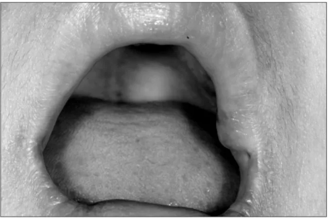
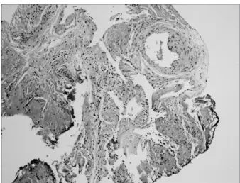
Treatment of oral hemangioma with decompression using CO2 laser: Case report
3
0
0
전체 글
(2)
(3)
수치
관련 문서