Basiliximab-Induced Non-Cardiogenic Pulmonary Edema in a Kidney Transplant Patient
Yoo Jin Lee, M.D.
1, Bong Soo Park, M.D.
1, Sihyung Park, M.D.
1, Kang Min Park, M.D.
2, Jin Han Park, M.D.
1, Il Hwan Park, M.D.
1and Yang Wook Kim, M.D.
1Departments of Internal Medicine
1, Neurology
2, Inje University Haeundae Paik Hospital, Inje University College of Medicine, Busan, Korea
Induction therapy with basiliximab is widely administered after kidney transplantation to prevent acute rejection. Herein, we report a case of non-cardiogenic pulmonary edema induced by basiliximab. To the best of our knowledge, such case has not been reported to date in Korea. A 54-year-old man with polycystic kidney disease received kidney transplantation. As induction therapy, he was prescribed basiliximab. On day 4, the second dose of basiliximab was administered. The patient complained of acute hypo- xia 23 hours later, which led to circulatory collapse. He was discharged 3 weeks later with stable renal function. Pulmonary edema was presumed to have been caused by increased pulmonary capillary permeability. A possible hypothesis for this event occurring after the second basiliximab injection is steroid-related effects. Non-cardiogenic pulmonary edema is a complication that might occur after basiliximab induction therapy. Physicians should be aware of this potentially life-threatening complication.
Key Words: Basiliximab, Pulmonary edema, Kidney transplantation
중심 단어: 바실릭시맙, 폐부종, 신장이식Received April 11, 2018 Revised July 3, 2018 Accepted July 3, 2018
Corresponding author: Yang Wook Kim
Department of Internal Medicine, Inje University Haeundae Paik Hospital, Inje University College of Medicine, 875 Haeun-daero, Haeundae-gu, Busan 48108, Korea
Tel: 82-51-797-3324, Fax: 82-51-797-3282
E-mail: [email protected] Fig. 1. There are numerous cysts in both kidneys.
INTRODUCTION
Acute rejection is one of the most important predictors of long-term prognosis after renal transplantation(1).
Induction therapy, defined simply as the short-term use of an immunosuppressive agent, most commonly a T-lympho- cyte‐depleting rabbit‐derived anti-thymocyte globulin or an interleukin‐2 receptor antagonist (IL2RA), is administered to reduce acute rejection and improve long-term graft survival. The use of selective induction agents, such as IL2RA, basiliximab, and daclizumab, has reduced the fre-
quency of acute rejection(2). Because of the relatively few- er adverse effects and higher convenience than those of oth- er induction therapy agents, basiliximab is widely used in
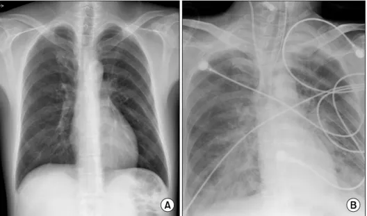
Fig. 2. The chest radiograph showed no active lung lesion (A), and acute pulmonary infiltration in both lung fields (B).
immunologically low-risk recipients(3). Pulmonary edema as an adverse effect of basiliximab has not been reported in Korea. Herein, we report a case of non-cardiogenic pul- monary edema induced by basiliximab during the induction therapy.
CASE REPORT
A 54-year-old male patient with autosomal dominant pol- ycystic kidney disease and hypertension was advised a pre- emptive kidney transplantation. The donor was his wife, a 54-year-old woman, with human leukocyte antigen mis- match 4/6, ABO compatibility, complement-dependent cyto- toxicity cross-match negativity, and 0% panel reactive antibodies. Pre-operative evaluation of the patient was un- remarkable, except for his kidney enlargement (Fig. 1).
Pre-operative laboratory findings were as follows; hemoglo- bin 8.6 g/dL, platelets 269×109/L, protein 6.9 g/dL, white blood cells 6.30×10⁹/L, albumin 3.7 g/dL, aspartate trans- aminase 15 U/L, alanine transferase 15 U/L, blood urea ni- trogen 61.5 mg/dL, creatinine 5.02 mg/dL, sodium 143 mmol/L, potassium 4.9 mmol/L, and chloride 111 mmol/L.
There was no active lung lesion on pre-operative plain chest radiograph (Fig. 2). Preoperative echocardiography results were as follows: ejection fraction 66%, no regional wall mo- tion abnormality with normal global left ventricular (LV) systolic function. He received 20 mg of basiliximab as in-
duction therapy and prednisone, tacrolimus, and mycophe- nolate mofetil as maintenance therapy. A 1,000 mg of methylprednisolone was to be administered on the day of surgery, and the dose was then to be reduced by half each day thereafter, until a maintenance dose of 1 mg/kg was reached. Due to limited abdominal space, his enlarged kid- neys were removed by laparoscopic simple nephrectomy on the left side and hand-assisted laparoscopic nephrectomy on the right side before the kidney transplantation. The surgery lasted 7 hours and 30 minutes. The transplanted kidney had 104 minutes of cold ischemic time and 33 minutes of warm ischemic time. On the first post-operative day, his vital signs were stable and the urine output was preserved at >10.1 mL/kg/hour. Post-operative 24-hour creatinine was 2.43 mg/dL. On the 4th post-operative day, the second dose of basiliximab was administered as per the treatment schedule.
The patient was administered 1 g of methylprednisolone during the first dose of basiliximab and 60 mg at the second administration. Twenty-three hours later, he suddenly com- plained of shortness of breath and required more than 15 L/min of oxygen via a reservoir mask. Additionally, circu- latory collapse was noticed with a systolic pressure of 50 mmHg. Arterial blood analysis results were as follows: pH 7.08, PaCO2 45 mmHg, PaO2 58 mmHg, O2 saturation 76%, and bicarbonate 13.3 mmol/L; and his laboratory findings were as follows: white blood cells 8.90×10⁹/L, eosinophil 1.7%, and lactate dehydrogenase 155 IU/L. The chest radio-
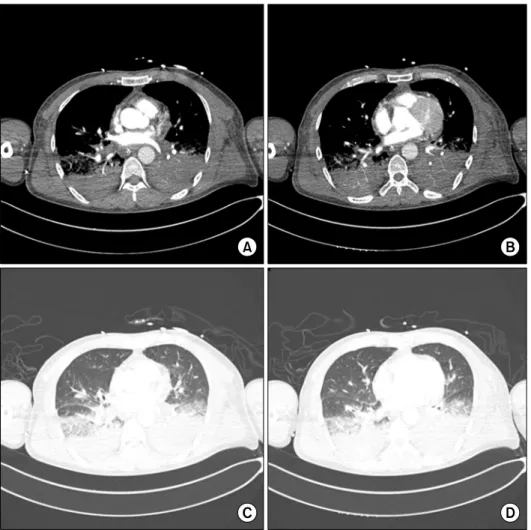
Fig. 4. (A-D) Chest computed to- mography showed bronchovascular bundle thickening and increased interlobular septal thickening in both lung and there was no evi- dence of acute pulmonary throm- boembolism.
Fig. 3. Results from electrocardiography were within normal limits.
graph showed acute pulmonary infiltration in both lung fields (Fig. 2). The central venous pressure was 8 cmH2O.
Results from electrocardiography were within normal limits (Fig. 3). Chest computed tomography showed increased in- terstitial infiltrates in the dependent portion of both lower
lung fields (Fig. 4). As he failed to respond to usual thera- pies, including high doses of inotropics and vasopressors with mechanical ventilation, he was put on extracorporeal membrane oxygenation (ECMO, V-A mode) to maintain his oxygen saturation and blood pressure. There was no evi- dence of infection (C-reactive protein 0.98 mg/dL, procalci- tonin 0.5 ng/mL, blood and urine culture were negative).
However, except for methylprednisolone, tacrolimus and mycophenolate mofetil were temporarily stopped and em- pirical antibiotics were administrated as septic shock. With an ejection fraction of 50%, global hypokinesia on echo- cardiography, no definite findings on electrocardiography, and normal cardiac enzyme levels (creatine kinase-MB [CKMB] 2.8 ng/mL, troponin I <0.0041 ng/mL), cardio- genic shock was not suspected. Two days later, his blood pressure returned to normal and he was weaned off the ECMO. Tacrolimus and mycophenolate mofetil were ad-
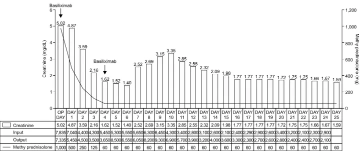
Fig. 5. This is a graph showing the clinical course of the patient. Abbreviation: OP, operative.
ministered again. After 4 days of supportive care, he recov- ered from the acute pulmonary event and was weaned off the ventilator. The follow-up echocardiography showed an ejection fraction of 60% and no regional wall motion abnor- mality with normal LV systolic function and the follow-up cardiac enzyme levels (CKMB 4.1 ng/mL, troponin I
<0.0039 ng/mL) were normal. Three weeks later, he re- turned home safely. His last serum creatinine was 1.59 mg/dL and spot urine protein/creatinine ratio was 114.9 mg/gCr. Two months later, his serum creatinine 1.76 and spot urine protein/creatinine ratio was 68.4 mg/gCr (Fig. 5).
DISCUSSION
Acute rejection plays an important role in determining the outcome of kidney transplants(1). Induction therapy is administered to prevent acute rejection and improve long-term graft survival. The choice of induction therapy depends on the immunological risk of the recipient, the un- derlying disease, or the choice of maintenance im- munosuppression therapy. Basiliximab and chimeric mono- clonal antibodies are used in combination with other im- munosuppressants to prevent acute rejection reactions in adult and pediatric kidney transplant patients. It blocks the binding of serum interleukin‐2 (IL-2) to CD25, inhibiting the proliferation of activated T-cells and subsequently re- leasing cytokines. Basiliximab (20 mL) is administered
twice, on day 1 after transplantation and on day 4 after transplantation(4). It reduces the frequency of acute re- jection and has been proven effective in reducing the se- verity of acute rejections in kidney transplant patients(2).
Because of the relatively fewer side effects and its ease of use compared with other induction therapy agents, basi- liximab is widely used in immunologically low-risk recipi- ents(3). Unlike OKT-3, basiliximab was initially thought to be free from immune-mediated side effects(4-6). However, basiliximab-related hypersensitivity was recently reported.
Type 1 anaphylactic reactions to basiliximab have been de- scribed in young women who successfully received human- ized monoclonal antibodies as induction agents for renal transplantation(7). In a similar report by the Mayo Clinic, infliximab, a chimeric human/murine monoclonal antibody (mAb) against tumor necrosis factor (TNF-), was found to be involved in acute respiratory distress syndrome trig- gered by type IV anaphylaxis(8). Patients suffering an ana- phylactic reaction usually develop hypotension and allergic signs such as pruritic rash, swelling of the tongue, and flush- ing of the skin within minutes or hours following basilix- imab injection(9,10). However, our patient had no allergic signs and symptoms. Moreover, there was a delay of several hours between basiliximab injection and the onset of symptoms. The mechanism of pulmonary edema in our pa- tient is presumed to be an increase in pulmonary capillary permeability resulting from cytokine release. Unlike that of
type 1 hypersensitivity reactions, the expression of respira- tory symptoms is rather insidious, developing within 48 hours. The pathogenesis of acute lung injury in transplant recipients administered monoclonal antibodies might include cytokine release from the activated T-cells and sequestration of neutrophils within the pulmonary capillary beds. The smaller diameter of the pulmonary capillaries might increase the possibility of neutrophil deformation and increase the regulation of adherent capillaries to neutrophils and vascular endothelium, leading to leakage of neutrophils into the al- veolar tissue(10,11). The activation of neutrophils by an in- flammatory mediator results in the release of L-selectin from their surface and in the upward regulation of B2 in- tegrins, which interact with endothelial intercellular adhe- sion molecule-1 to cause translocation of neutrophils into the alveoli(12).
A possible hypothesis for the occurrence of this event af- ter the second basiliximab injection is the use of steroids.
Glucocorticoids inhibit the expression and action of most cytokines(13). Mitsuta et al.(14) have demonstrated that pre-operative corticosteroid therapy significantly suppressed both IL-5 and TNF-α production. The first basiliximab in- jection would have been accompanied by high concen- trations of methylprednisolone, which would have led to suppression of cytokine production. With the relatively small doses of methylprednisolone being administered dur- ing the second basiliximab injection, cytokine production would have not been adequately suppressed, which could have led to pulmonary edema.
We reported a life-threatening episode of non-cardio- genic pulmonary edema and shock in a 54-year-old patient following administration of the second dose of basiliximab.
To our knowledge, non-cardiogenic pulmonary edema as an adverse effect of basiliximab has not been reported in Korea. This report does not show a clear causal relationship between basiliximab and pulmonary edema. However, we were able to exclude other causes of pulmonary edema or infiltration, such as cardiogenic and infectious causes.
Non-cardiogenic pulmonary edema, although rare, might occur as a complication after basiliximab induction therapy.
Physicians need to be aware of this potentially life-threat- ening complication. This might improve graft survival in kidney transplantation.