197
Immune Network
MHC-restricted Exogenous Antigen Presentation
Hyunjin Kim1, Kyung-Mi Cho1, Turmunkh Gerelchuluun1, Ji-Seon Lee2, Kyeong-Soo Chung2 and Chong-Kil Lee1
1College of Pharmacy, Chungbuk National University, Cheongju, 2College of Pharmacy, Chungnam National University, Daejeon, Korea
ABSTRACT
Background: Immunomodulators enhancing MHC-restricted antigen presentation would affect many cellular immune reactions mediated by T cells or T cell products. However, modulation of MHC-restricted antigen presentation has received little attention as a tar- get for therapeutic immunoregulation. Here, we report that lectins isolated from mush- room Fomitella fraxinea enhance MHC-restricted exogenous antigen presentation in pro- fessional antigen presenting cells (APCs). Methods: Lectins, termed FFrL, were isolated from the carpophores of Fomitella fraxinea, and its effects on the class I and class II MHC-restricted presentation of exogenous ovalbumin (OVA) were examined in mouse dendritic cells (DCs) and mouse peritoneal macrophages. The effects of FFrL on the ex- pression of total MHC molecules and the phagocytic activity were also examined in mouse DCs. Results: DCs cultured in the presence of FFrL overnight exhibited enhanced capacity in presenting exogenous OVA in association with class I and class II MHC molecules. FFrL increased slightly the total expression levels of both class I (H-2Kb) and class II (I-Ab) MHC molecules and the phagocytic activity of DCs. Antigen pre- sentation-enhancing activity of FFrL was also observed in macrophages isolated from mouse peritoneum. Conclusion: Lectins isolated from the carpophores of Fomitella frax- inea increase MHC-restricted exogenous antigen presentation by enhancing intracellular processing events of phagocytosed antigens. (Immune Network 2007;7(4):197-202) Key Words: Fomitella fraxinea, lectin, MHC-restricted antigen presentation, dendritic
cell, macrophage
Correspondence to: Chong-Kil Lee, College of Pharmacy, Chungbuk National University, 12, Gaesin-dong, Heungdeok-gu, Cheongju 361- 763, Korea (Tel) 82-43-261-2826, (Fax) 82-43-268-2732, (E-mail) cklee@chungbuk.ac.kr
This work was supported by the research grant of the Chungbuk National University in 2006.
Introduction
Two distinct pathways have been known for the MHC-restricted presentation of exogenous antigens (1).
In the classical paradigm of exogenous antigen pre- sentation by professional APCs, antigens internalized by phagocytosis or endocytosis are processed and loaded on class II MHC molecules in a post-Golgi compart- ment. Professional APCs, however, have been shown to process exogenous antigens for the presentation on class I MHC molecules. This process, termed cross-pre-
sentation, was first demonstrated in the generation of CTL responses to minor histocompatibility antigens (2).
Cross-presentation is now recognized as a more general mechanism for the generation of CTL responses against tumor cells, transplanted cells, bacteria, and even vi- ruses (3-7). In the absence of such a mechanism, viral or tumor antigens expressed in nonprofessional APCs could escape immunosurveilance because cytotoxic T cell (CTL) responses can only be induced efficiently for the antigens presented via class I MHC molecules on professional APCs (8-10).
Since T cells can only recognize antigens presented on MHC molecules, modulators of MHC-restricted an- tigen presentation would affect many cellular immune reactions mediated by T cells or T cell products. How- ever, modulation of MHC-restricted antigen presenta- tion has received little attention as a target for thera-
Figure 1. Effects of FFrL on the cross-presentation of exogenous OVA in DC2.4 cells. DC2.4 cells were incubated with the indica- ted amounts of FFrL for 18 h, and then added with OVA-micro- spheres. After 2 h incubation, the cells were fixed, and the amo- unts of OVA peptides presented on class I MHC molecules were assessed using B3Z cells, which expresses β-galactosidase upon recognition of the OVA peptide, SIINFEKL, complexed with H- 2Kb molecule. The amount of β-galactosidase expressed in B3Z cells was determined by an enzymatic assay using chlorophenolred β-D-galactopyranoside as a substrate.
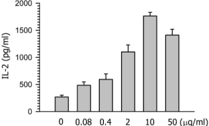
Figure 2. Effects of FFrL on the class II MHC-restricted presen- tation of exogenous OVA in DC2.4 cells. Indicated amounts of FFrL were added to cultures of DCs generated from mouse BM cells with GM-CSF for 18 h, and then added with OVA-micro- spheres. After 2 h incubation, the cells were fixed, and the amo- unts of OVA peptides presented on class II MHC molecules were assessed using OVA-specific CD4 T cell hybridoma, DOBW. The amounts of IL-2 produced from OVA-specific CD4 T cells were assayed by a commercial IL-2 ELISA kit.
peutic immunoregulation. Recently, we showed that cyclosporin A and tacrolimus, but not rapamycin, inhi- bit MHC-restricted exogenous antigen presentation in professional APCs in vivo (11) as well as in vitro (12).
These results demonstrate that modulation of MHC- restricted antigen processing may provide novel phar- macological targets for the regulation of T cell responses.
In an effort to search for and characterize activators of MHC-restricted antigen presentation, we examined the effects of lectins and polysaccharides isolated from several species of Basidiomycetes, and found that the lec- tins isolated from mushroom Fomitella fraxinea enhan- ces efficiently MHC-restricted exogenous antigen pre- sentation in a dendritic cell line, DC2.4 cells, and also in normal macrophages. Our study demonstrates that modulation of MHC-restricted antigen presentation may provide novel pharmacological targets for the reg- ulation of T cell responses.
Materials and Methods
Cells and cell lines. T cell hybridomas, B3Z86/90.14 (B3Z) and DOBW, were kindly provided by Dr. Nilabh Shastri (University of California, Berkeley, CA) and by Dr. Clifford V. Harding (Case Western Reserve Univer- sity, Cleveland, OH), respectively (13,14). The DC cell line (DC2.4) was obtained from the Dana-Farber Can- cer Institute, Boston, MA, USA (15). Thioglycollate-eli-
cited mouse peritoneal macrophages were obtained from C57BL/6 mice.
Isolation of lectins. The lectins were isolated from the carpophores of Fomitella fraxinea as described previously (16). Briefly, two kilograms of the frozen fruiting bod- ies were homogenized in 20 liters of 50 mM Tris-HCl buffer (pH 8.0) with a blender and then extracted for 18 hours at 4oC with frequent gentle swirling. The resulting suspension was filtered and ammonium sulfate was added to the filtrate to 95% saturation. After stan- ding overnight at 4oC, the resulting precipitate was separated by centrifugation at 8,000 ×g at 4oC for 20 min. This precipitate was dissolved in 50 mM NaCl, 50 mM Tris-HCl buffer (pH 8.0) and then dialyzed against distilled water for 5 days at 4oC with frequent changing of distilled water. After dialysis, the dialysate was freeze-dried to yield crude FFrL.
Preparation of microencapsulated OVA. Microspheres con- taining OVA were prepared using a solvent-evaporation method, as described previously (12), using OVA dis- solved in 3% polyvinyl alcohol (4 mg/ml) and poly (DL-lactide-co-glycolide) (PLGA; lactide:glycolide=
50:50; Sigma-Aldrich, St. Louis, MO, USA) dissolved in a mixture of acetone and ethanol (9:1) (5%). The concentration of OVA was determined by micro-bicin- choninic acid assay kit (Pierce, Rockford, IL) according to the manufacturer’s instructions after lysing the mi-
Figure 3. Effects of FFrL on the expression of MHC molecules. DC2.4 cells were cultured with 10 μg/ml of FFrL for 18 h, and then the cells were harvested by gentle pipetting after cooling on ice. The expression levels of class I and class II MHC molecules were assessed using anti-H-2Kb and anti-I-Ab monoclonal antibodies. Shaded histogram represents the expression levels of H-2Kb and I-Ab molecules in DC2.4 cells cultured in the presence of FFrL, and thin line histogram represents the expression levels of H-2Kb and I-Ab molecules in DC2.4 cells cultured in the absence of FFrL. Isotype control was shown as thin histograms (left).
crospheres in a lysis buffer containing 0.1 % SDS and 0.1 N NaOH. For phagocytosis assays, micropheres containing both OVA and fluorescein isothiocyanate (FITC) were prepared by adding FITC (final, 5 mg/ml) to a mixture of acetone and ethanol (9:1) together with PLGA (final, 5%).
Cross-presentation assay. LacZ T cell activation assays were used to assess the amounts of cross-presented OVA peptides, as previously described (12). Briefly, APCs were cultured in the presence of different con- centrations of FFrL for 18 h in 96-well plates (1×10⁵/ well), and then added with OVA-microspheres (50 μg /ml as OVA). After 2 h incubation at 37oC, the plate was washed twice with 300 μl/well of pre-warmed PBS, and then fixed with 100 μl/well of ice-cold 1.0%
paraformaldehyde for 5 min at room temperature. The plate was washed 3 times with 300 μl/well of PBS, and B3Z cells were added (2×10⁵/well). After incu- bating for 4 h at 37oC, lacZ activity was measured ei- ther by colorimetric analysis after incubating freeze-thaw lysed cells with β-galactosidase substrate, chlorophe- nol red β-D-galactopyranoside (Calbiochem, Darms- tadt, Germany), as described previously (12).
Generation of bone marrow-derived DCs (BM-DCs). DCs were generated from total BM cells as described pre- viously (17). Briefly, BM cells obtained from femurs of BALB/c mouse were cultured in a 6-well plate (5×
106/well) in a culture medium supplemented with 200 U/ml rmGM-CSF. At days 3 and 4 from the initiation of the culture, nonadherent cells were discarded by re- placing the culture medium with fresh medium con- taining the cytokines after gentle shaking. DCs were harvested by gentle pipetting at day 6.
MHC class II-restricted presentation assay. BM-DCs were cultured in the presence of different concentrations of FFrL for 18 h in 96-well plates (1×10⁵/well), and then added with OVA-microspheres (50 μg/ml as OVA).
After 2 h incubation at 37oC, unphagocytized OVA- microspheres were removed by suction, and then fixed with ice-cold 1.0% paraformaldehyde for 5 min at room temperature. The plate was then washed twice with 300 μl/well of pre-warmed media, and added with DOBW cells (1×10⁵/well). After 24 h incubation at 37oC, the plate was centrifuged at 1,800 rpm, and the culture supernatant was collected and assayed for IL-2 content using an IL-2 ELISA kit (BD Biosciences).
Phagocytosis assay. DCs were cultured in the presence of different concentrations of FFrL for 18 h in 6-well plates (2×10⁶ cells/well), and then added with micro- spheres (average diameter, 300 nm) containing both ovalbumin (OVA) and fluorescein isothiocyanate (FITC).
After 2 h, unphagocytozed microspheres were removed by washing with pre-warmed PBS. The plate was chil- led on ice for 20 min, then the cells were harvested
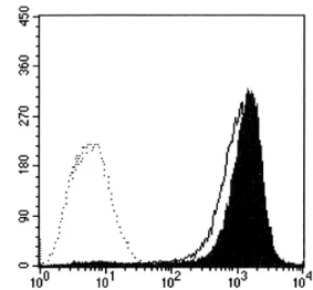
Figure 4. Effects of FFrL on the phagocytic activity. DC2.4 cells were cultured with 10 μg/ml of FFrL for 18 h, and then added with microspheres containing both OVA and FITC. After 2 h incubation, unphagocytized microspheres were washed, and the cells were harvested by gentle pipetting after cooling on ice, and then analyzed by flow cytometry. Shaded histogram represents the phagocytic activity of DCs cultured in the presence of FFrL, and thin line histogram represents the phagocytic activity of DCs cultured in the absence of FFrL. DCs that were not incubated with FITC-labeled microspheres were shown as dotted histo- grams (left).
by treating with Cell stripper solution (Cellgro Media- tech, Herndon, VA) as suggested in the manufacturer’s instruction, fixed in 1% paraformaldehyde in PBS, and flow cytometric analysis was performed on a FACS Cali- ver flow cytometer (Becton Dickinson).
Phenotypic analysis. DCs were cultured in the presence of different concentrations of FFrL for 18 h in 6-well plates (2×10⁶ cells/well). The plate was then chilled on ice for 20 min, and the cells were harvested by treating with Cell stripper solution (Cellgro Mediatech).
The cells were stained with monoclonal antibodies rec- ognizing murine MHC molecules after blocking of FcR-binding anti-CD16/CD32 monoclonal antibody (clone 2.4G2) as described previously (17), and flow cytometric analysis was performed on a FACS Caliver (Becton-Dickinson). The monoclonal antibodies, H-2Kb (clone AF6-88.5) and I-Ab (clone AF6-120.1, and an isotype-matched control antibody were purchased from BD Biosciences.
Results
FFrL enhances cross-presentation of exogenous antigen in DCs. To examine the effects of the lectins, FFrL, on the presentation of exogenous OVA in association with class I MHC molecules, DC2.4 cells were cultured in the presence of FFrL for 18 h, and then allowed to phagocytose OVA-microspheres for 2 h. The DC2.4 cells were then washed, fixed with paraformaldehyde, and the amount of OVA peptide-class I MHC com- plexes was measured using a T cell hybridoma, B3Z, which recognizes OVA peptide (SIINFEKL)-H-2Kb com- plex and expresses β-galactosidase [14]. As shown in Fig. 1, DC2.4 cells stimulated with FFrL exhibited en- hanced capability in cross-presenting exogenous OVA.
The optimal dose of FFrL for maximum augmentation of cross-presentation capability appeared to be 10 μg /ml in DC2.4 cells.
FFrL enhances class II MHC-restricted presentation of exoge- nous antigen in DCs. To examine the effects of FFrL on class II MHC-restricted presentation of exogenous antigen, DCs generated from bone marrow cells of Balb /c mice (H-2d) were cultured with different concentra- tions of FFrL for 18 h, and then allowed to phagocytize OVA-microspheres for 2 h. The DCs were fixed with paraformaldehyde, and then the amount of OVA pepti- de-class II MHC complexes was measured using OVA- specific CD4 T cell hybridoma, DOBW cells. As shown
in Fig. 2, FFrL also enhanced class II MHC restricted presentation of exogenous OVA significantly and dose dependently in BM-DCs.
FFrL enhances slightly the total expression levels of both class I and class II MHC molecules. To examine whether the enhanced capacity of FFrL-stimulated DCs to present OVA peptides in association with MHC molecules was due to increased expression of MHC molecules on the cell surface, DC2.4 cells were cultured with 10 μg/ml FFrL for 18 h, harvested, and then the expression levels of class I and class II MHC molecules were determined by anti-H-2Kb and anti-I-Ab monoclonal antibodies.
FFrL increased slightly the total expression levels of both class I and class II MHC molecules (Fig. 3).
FFrL slightly enhances the phagocytic activity. To examine whether the enhanced capacity of FFrL-stimulated DCs to present OVA peptides in association with MHC molecules was due to increased phagocytic activity, DC2.4 cells were cultured with 10 μg/ml FFrL for 18 h, and then added with microspheres containing both OVA and FITC. After 2 h incubation, unphago- cytized microspheres were washed, and the cells were harvested by gentle pipetting after cooling on ice. Flow cytometric analysis of the harvested cells showed that
Figure 5. Effects of FFrL on the cross-presentation of exogenous OVA in macrophages. Thioglycollate-elicited macrophages were incubated with the indicated amounts of FFrL for 18 h, and then added with OVA-microspheres. After 2 h incubation, the cells were fixed, and the amounts of OVA peptides presented on class I MHC molecules were assessed using B3Z cells, as described in Fig. 1.
FFrL slightly increased the phagocytic activity of DC2.4 cells (Fig. 4).
FFrL also enhances cross-presentation of exogenous antigen in peritoneal macrophages. Cross presentation-enhancing activity of FFrL was also examined in macrophages.
In this experiment, peritoneal macrophages were ob- tained from thioglycollate-injected mouse peritoneum, and were cultured in the presence of FFrL for 18 h, fixed with paraformaldehyde, and the amount of OVA peptide-class I MHC complexes was measured using a T cell hybridoma, B3Z. As shown in Fig. 5, macro- phages stimulated with FFrL also exhibited enhanced capability in cross-presenting exogenous OVA. The optimal dose of FFrL for maximum augmentation of cross-presentation capability appeared again to be 10 μg/ml.
Discussion
The present study demonstrates that the lectins iso- lated from the carpophores of Fomitella fraxinea increase MHC-restricted exogenous antigen presentation. FFrL enhanced both class I MHC and class II MHC-restric- ted presentation of exogenous antigen in professional APCs. FFrL appeared to increase MHC-restricted exo- genous antigen presentation by activating intracellular processing events of the phagocytosed antigens, because treatment of DCs with FFrL increased only slightly the total level of expression of H-2Kb and I-Ab molecules and the phagocytic activity.
Lectins are glycoproteins which have been known to induce cell agglutination (18). Lectins have been found from diverse living organisms including animals, plants and microorganisms (19,20). Some of the lectins isolated from mushrooms have been shown to possess immunomodulatory and antitumor activity (21-25), al- though the mechanism of action is not clear. Our results suggest that immunomodulatory and antitumor activity activities of mushroom lectins may at least in part due to activation of antigen presenting capability of profe- ssional APCs. Since T cells can only recognize antigens presented on MHC molecules, the impact of the aug- mentation of MHC-restricted antigen presentation must be far-reaching.
In all of the experiments described in the present study, APCs were exposed with FFrL for 18 h, and then allowed to phagocytose OVA-microspheres for 2 h. The APCs were washed to remove unphagocytosed OVA-microspheres, fixed with paraformaldehyde, and then washed thoroughly again to remove paraformal- dehyde, before functional assays with OVA-specific CD4 or CD8 T cells. Thus, the activation of OVA- specific T cells must be due to enhanced expression of OVA peptides on APCs, and not due to the carry- over of the lectins to T cell cultures.
The precise mechanisms by which FFrL enhances MHC-restricted antigen processing pathways remain to be determined. Because FFrL was shown to exert mito- genic activity when added to splenic lymphocytes, we examined the effects of FFrL on the proliferation of DCs and peritoneal macrophages. We found that FFrL did not exert mitogenic activity on these cells (data not shown). Recently, we showed that DCs efficiently phagocytose OVA-microspheres encapsulated with PLGA, and then process the phagocytosed OVA in a vacuolar alternate mechanism to present OVA peptides in asso- ciation with class I MHC molecules to T lymphocytes (26). The vacuolar alternate pathway involves partial digestion of phagocytosed antigens within the acidic phagolysomes and binding of the peptides to class I MHC molecules in the same compartment, much like the classical class II MHC pathway of exogenous anti- gen (27). Because FFrL increased both class I and class II MHC-restricted exogenous antigen presentation pa- thways, we are tempted to speculate that FFrL activates molecular machinery in the vacuolar alternate pathway of antigen processing.
Although the precise mechanism of action in not elucidated, the present study demonstrates that the lectins isolated from mushroom Fomitella fraxinea en- hances MHC-restricted exogenous antigen presentation in professional APCs. Based on the present study as well as our recent studies which demonstrated that MHC-restricted antigen presentation can be blocked by immunosuppressive calcineurin inhibitors (11,12), we would like to propose that modulation of MHC-re- stricted antigen presentation could be a novel means for the regulation of T cell responses.
References
1. Harding CV: Phagocytic processing of antigens for presenta- tion by MHC molecules. Trend Cell Biol 5;105-108, 1995 2. Bevan MJ: Cross-priming for a secondary cytotoxic response
to minor H antigens with H-2 congenic cells which do not cross-react in the cytotoxic assay. J Exp Med 143;1283-1288, 1976
3. Huang AY, Golumbek P, Ahmadzadeh M, Jaffee E, Pardoll D, Levitsky H: Role of bone marrow-derived cells in presenting MHC class I-restricted tumor antigens. Science 264;961-965, 1994
4. Heath WR, Carbone FR: Cross-presentation, dendritic cells, tolerance and immunity. Annu Rev Immunol 19;47-64, 2001 5. Sigal LJ, Crotty S, Andino R, Rock KL: Cytotoxic T-cell im- munity to virus-infected non-haematopoietic cells requires presentation of exogenous antigen. Nature 398;77-80, 1999 6. Heath WR, Carbone FR: Cross-presentation, dendritic cells,
tolerance and immunity. Annu Rev Immunol 19;47-64, 2001 7. Yewdell JW, Haeryfar SM: Understanding presentation of vi- ral antigens to CD8+ T cells in vivo: the key to rational vaccine design. Annu Rev Immunol 23;651-682, 2005 8. Sigal L, Rock KL: Bone marrow-derived antigen-presenting
cells are required for the generation of cytotoxic T lympho- cyte responses to virus and use transporter associated with antigen presentation (TAP)-dependent and –independent pa- thways of antigen presentation. J Exp Med 192;1143-1150, 2000
9. Carbone FR, Kurts C, Bennett SR, Miller JF, Heath W: Cross- presentation: a general mechanism for CTL immunity and to- lerance. Immunol Today 19;368-373, 1998
10. Guermonprez P, Valladeau J, Zitvogel L, Thery C, Amigorena S: Antigen presentation and T cell stimulation by dendritic cells. Annu Rev Immunol 20;621-667, 2002
11. Lee YR, Yang IH, Lee YH, Im SA, Song S, Li H, Han K, Kim K, Eo SK, Lee CK: Cyclosporin A and tacrolimus, but not rapamycin, inhibit MHC-restricted antigen presentation pathways in dendritic cells. Blood 105;3951-3955, 2005 12. Lee YH, Lee YR, Im SA, Park SI, Kim KH, Gerelchuluum
T, Song S, Kim K, Lee CK: Calcineurin inhibitors block MHC-restricted antigen presentation in vivo. J Immunol 179;
5711-5716, 2007
13. Karttunen J, Sanderson S, Shastri N: Detection of rare anti- gen-presenting cells by the lacZ T-cell activation assay sug- gests an expression cloning strategy for T-cell antigens. Proc Natl Acad Sci 89;6020-6024, 1992
14. Harding CV, Collins DS, Kanagawa O, Unanue ER: Liposome- encapsulated antigens engender lysosomal processing for class II MHC presentation and cytosolic processing for class I pre- sentation. J Immunol 147;2860-2863, 1991
15. Shen Z, Reznikoff G, Dranoff G, Rock KL: Cloned dendritic cells can present exogenous antigens on both MHC class I and class II molecules. J Immunol 158;2732-2730, 1997 16. Lee JS: Purification and characterization of a lectin with potent
antitumor and immunomodulatory activity from a Korean wild mushroom Formitella fraxinea. A PhD thesis. Graduate School of Pharmacy, Chungnam National University, 2005.
17. Lee JK, Lee MK, Yun YP, Kim Y, Kim JS, Kim YS, Kim K, Han SS, Lee CK: Acemannan purified from Aloe vera in- duces phenotypic and functional maturation of immature den- dritic cells. Int Immunopharmacol 1;1275-1284, 2001 18. Goldstein IJ, Heys CE: The lectins: Carbohydrate-binding pro-
teins of plants and animals. Adv Carbohydr Chem Biochem 35;127-340, 1978
19. Cammue B, Stinissen HM, Peumans WJ: A new type of ce- real lectin from leaves of couch grass (Agropyrum repens).
Eur J Biochem 148;315-322, 1985
20. Avichezer D, Gilboa-Garber N: Antitumoral effects of Pseudo- monas aeruginosa lectins on Lewis lung carcinoma cells cul- tured in vitro without and with murine splenocytes. Toxicon 29;1305-1313, 1991
21. Licastro F, Morini MC, Kretz O, Dirheimer G, Creppy EE, Stirpe F: Mitogenic activity and immunological properties of bolesatine, a lectin isolated from the mushroom Boletus sata- nas Lenz. Int J Biochem 25;789-792, 1993
22. Wang HX, Liu WK, Ng TB, Ooi VE, Chang ST: The im- munomodulatory and antitumor activities of lectins from the mushroom Tricholoma mongolicum. Immunopharmacol 31;
205-211, 1996
23. She QB, Ng TB, Liu WK: A novel lectin with potent immu- nomodulatory activity isolated from both fruiting bodies and cultured mycelia of the edible mushroom Vovariella volvacea.
Biochem Biophys Res Commun 247;106-111, 1998 24. Wang H, Gao J, Ng TB: A new lectin with highly potent
antihepatoma and antisarcoma activities from the oyster mush- room Pleurotus ostreatus. Biochem Biophys Res Commun 275;810-816, 2000
25. Wang H, Ng TB, Liu Q: Isolation of a new heterodimeric lectin with mitogenic activity from fruiting bodies of the mu- shroom Agrocybe cylindracea. Life Sci 70;877-885, 2002 26. Gerelchuluum T, Lee YH, Lee YR, Im SA, Park JS, Song S,
Kim K, Lee CK: Dendritic cells process antigens encapsulated in a biodegradable polymer, poly (D,L-lactide-co-glycolide), via an alternate class I MHC processing pathway. Arch Pharm Res 30;1440-1446, 2007
27. Harding CV, Song R: Phagocytic processing of exogenous par- ticulate antigens by macrophages for presentation by class I MHC molecules. J Immunol 153;4925-4933, 1994