Chemotherapy
Received: April 21, 2015 Revised: May 29, 2015 Accepted: June 5, 2015 Corresponding Author : Hyun Hee Kwon, MD
Department of Internal Medicine, Daegu Catholic University Medical Center, 33 Duryugongwon-ro 17-gil Nam-gu, Daegu 705-718, Korea
Tel: +82-53-650-4708, Fax: +82-53-621-4106 E-mail: heeya0035@naver.com
This is an Open Access article distributed under the terms of the Creative Commons Attribution Non-Commercial License (http://creativecommons.org/licenses/by-nc/3.0) which permits unrestricted non-commercial use, distribution, and repro- duction in any medium, provided the original work is properly cited.
Copyrights © 2015 by The Korean Society of Infectious Diseases | Korean Society for Chemotherapy
www.icjournal.org
Toxocariasis: A Rare Cause of Multiple Cerebral Infarction
Hyun Hee Kwon
Department of Internal Medicine, Daegu Catholic University Medical Center, Daegu, Korea
Toxocariasis is a parasitic infection caused by the roundworms Toxocara canis or Toxocara cati, mostly due to accidental in- gestion of embryonated eggs. Clinical manifestations vary and are classified as visceral larva migrans or ocular larva migrans according to the organs affected. Central nervous system involvement is an unusual complication. Here, we report a case of multiple cerebral infarction and concurrent multi-organ involvement due to T. canis infestation of a previous healthy 39-year- old male who was admitted for right leg weakness. After treatment with albendazole, the patient’s clinical and laboratory results improved markedly.
Key Words: Toxocara canis; Cerebral infarction; Larva migrans, visceral
Introduction
Toxocariasis is a parasitic infection caused by infection with the roundworm species Toxocara canis or less frequently Toxocara cati whose hosts are dogs and cats, respectively [1].
Humans become infected accidentally by ingestion of embry- onated eggs from contaminated soil or dirty hands, or by in- gestion of raw organs containing encapsulated larvae [2].
Keeping dogs and ingestion of raw cow liver are associated with increased risk of toxoriasis. A cross-sectional study of Ko- rean patients reported that a recent history of eating raw cow liver was associated with an increased risk of toxocariasis [3].
Clinical manifestation range from asymptomatic infection to fulminant disease, and the lungs, livers, and eyes are the most
commonly involved organs [4]. Central nervous system (CNS) involvement is relatively rare in toxocariasis, especially CNS presenting as multiple cerebral infarction. We report a case of multiple cerebral infarction with lung and liver involvement due to T. canis infection in a previously healthy patient who was admitted for right leg weakness.
Case Report
A 39-year-old right-handed man with no significant past medical history was admitted to the hospital with a 3-day his- tory of right leg weakness. He had undergone discectomy for herniated nucleus pulposus at the L5-S1 level 8 years prior Infect Chemother 2015;47(2):137-141
ISSN 2093-2340 (Print) · ISSN 2092-6448 (Online)
and had not experienced any complications. He smoked a pack of cigarettes per day for 20 years. He habitually ate un- dercooked meat, including 2 weeks prior to admission. He had no recent history of contact with pet animals and no past medical history of allergy.
On admission, his blood pressure was 120/80 mmHg, his pulse was regular at 88 beats per minute, and his temperature was 36.8℃. There were neither audible carotid bruits nor car- diac murmurs. Neurological examination revealed alert men- tal status and grade 3 weakness of the right leg. Deep tendon reflexes were increased in his right leg. Fundoscopy results were normal.
His white blood cell count was 11,900/mm3 with 26.7% eo- sinophils (2,600/mm3). Laboratory data were as follows: he- moglobin, 14.5 g/dL; platelets, 160,000/mm3; aspartate ami- notransferase, 37 IU/L; alanine aminotransferase, 20 IU/L;
alkaline phosphatase, 61 IU/L; creatinine 0.7 mg/dL; C-reac- tive protein, 32.8 mg/L; and erythrocyte sedimentation rate, 38 mm/h. Renal function and electrolytes were normal. The Venereal Disease Research Laboratory (VDRL) test and hu- man immunodeficiency virus (HIV) antibody test in serum were negative. Electrocardiogram showed normal sinus rhythm and transthoracic echocardiogram showed no evi- dence of mural thrombi or vegetations. Diffusion-weighted brain MRI revealed multifocal small embolic acute infarctions in the internal border zone of both cerebral hemispheres,
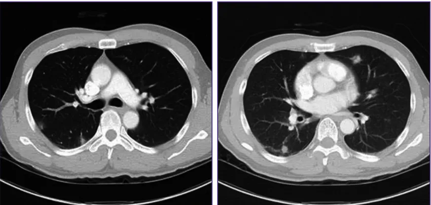
temporooccipital lobes of both PCA territory, and left cerebel- lar hemisphere (Fig. 1). Chest CT showed multiple peripheral ground glass opacities in both lungs that were considered eo- sinophilic pneumonia (Fig. 2). Abdominal CT on portal phase showed small ill-defined hypodense lesions in both lobes of the liver that were considered eosinophilic infiltrations (Fig. 3). His CSF contained 0/mm3 WBC, 63 mg/dL protein, and 49 mg/dL sugar; no bacteria or viruses were detected.
Because he had marked eosinophilia and multiple eosino- philic infiltrations in both lungs and liver, serologic testing for parasitic infections was performed. Serologic test result (per- formed at Seoul Clnical Laboratory (SCL), Seoul, Korea) for T.
canis IgG antibody via enzyme-linked immunosorbent assay (ELISA, Bordier Affinity Products SA, Crissier, Switzerland) was positive with an antibody titer of 3.12 (reference range
<1.00). This method has 91% sensitivity and 86% specificity [5].
Results of serologic tests for other helminthes (cysticercus, Paragonimus westermani, Clonorchis sinensis sparaganum, Entamoeba histolytica) were all negative. Any larva was not found in the CSF and CSF ELISA assay for T. canis IgG anti- body was negative.
While biopsy could be performed to find Toxocara larva in the affected tissue and confirm the diagnosis, toxocariasis is usually diagnosed based on a combination of clinical symp- toms, exposure history, marked eosinophilia, characteristic imaging findings and positive toxocariasis ELISA results. Thus, Figure 1. Diffusion-weighted brain magnetic resonance imaging showing multifocal small acute infarctions in the internal border zone of both the cerebral hemisphere (A) and left cerebellar hemisphere (B).
A B
the patient was diagnosed with multiple cerebral infarction and concurrent multi-organ involvement due to T. canis in- fection without tissue biopsy. He was treated with albendazole (400 mg twice daily for 2 weeks) and steroid (prednisolone 30mg twice daily for 7 days). Prednisolone was gradually ta- pered for one month. On day 14 of albendazole administra- tion, his leukocytosis was 14,300 /mm3 with 2.7% eosinophils.
In subsequent weeks, the patient’s leg weakness improved to grade 4. Two months after treatment, the follow-up tests in-
cluding CT scan and brain MRI were planned but the patient didn’t visit our hospital.
Discussion
Toxocariasis is a zoonotic parasitic infection contracted from dogs and cats. This disease occurs in developed coun- tries as well as in the tropics and sub-tropics where dog treat- Figure 2. Chest computed tomography shows multiple peripheral ground opacities in both lungs.
Figure 3. Abdomen computed tomography on portal phase shows small ill-defined hypodense lesions (arrows) in both lobes of the liver.
ment and population control are limited [3]. Human infection occurs by ingestion of embryonated eggs in contaminated soil or on unwashed hands, therefore high exposure to dogs and cats and contaminated soil are associated with toxocariasis [2]. Occasionally, toxocariasis is caused by consumption of raw liver and meat contaminated with larvae of T. canis. In South Korea, chops of raw cow liver and meat are popular dishes and are thought to have health benefits; toxocariasis, including subclinical toxocariasis, is therefore prevalent in South Korea [3, 6]. We report here a case of toxocariasis pre- senting as multiple embolic infarction with concurrent viscer- al migrans involving the lungs and liver. The patient frequently consumed raw cow liver and meat, and this was likely the source of infection.
Toxocara can infect any organ, but is commonly recognized in the lungs, liver, and eyes [4]. Although T. canis can cross the blood-brain barrier, CNS infection is rarely reported, but epi- lepsy, eosinophilic meningitis, meningo-encephalitis, enceph- alitis, arachnoiditis, vasculitis, meningo-myelitis, meningo-ra- diculitis, and optic neuritis due to Toxocara infection have been reported [7-10]. Until recently, however, there has only been a few report of cerebral infarction due to toxocariasis [11, 12]. Toxocara larvae are metabolically active and produce an array of enzymes and waste products that cause tissue dam- age, necrosis, and a marked inflammatory reaction, with eo- sinophils as the major component [13]. The mechanism of ce- rebral infarction is not well known in toxocariasis, but could be derived from direct invasion of larva particles and second- ary hypereosinophilia. Larva particles can accidentally invade the brain parenchyme or block cerebral vessels, similar to mi- croemboli [14]. In hypereosinophilia, eosinophils could induce mural thrombus in the left ventricle or endomyocardial fibrosis by infiltrating the endocardium [15]. Direct eosinophilic toxici- ty to the vascular wall has also been hypothesized [16].
A definitive diagnosis of toxocariasis is based on finding lar- vae in the affected tissue by histologic examination. Clinically, the diagnosis is based on medical history, clinical presentation, eosinophilia in the serum, and/or high serum titer of T. canis antibodies as assessed by ELISA or western blotting [1]. Our patient had multiple cerebral infarctions concurrent with vis- ceral migrans on CT, hypereosinophilia, positive serology for T. canis IgG, and a history of frequently eating raw cow liver and meat. He had no risk factors for multiple cerebral infarc- tion and no cerebral vessel stenosis on cerebral CT angiogra- phy. We therefore diagnosed cerebral infarction caused by toxocariasis.
There is no proven effective therapy for patients with neuro-
toxocariasis due to the rarity of the disease. Albendazole is the most commonly used drug as it can penetrate the cerebrospi- nal fluid and has acceptable toxicity. Other antihelmintics in- clude thiabendazole, mebendazole, oxibendazole, and flubendazole. Concurrent administration of corticosteroids has been used to suppress intense allergic responses [1, 9]. In some studies, corticosteroids were effective at relieving seri- ous neurologic symptoms brought on by an intensive inflam- matory response [17] and increased plasma levels of albenda- zole by approximately 50% [18].
Because ingestion of uncooked cow liver and meat in South Korea is common, toxocariasis is prevalent. This infection has various clinical manifestations including asymptomatic eosin- ophilia and eosinophilic infiltration of the lungs, liver, and eyes. Central nervous system (CNS) involvement is relatively rare in toxocariasis, especially CNS presenting as multiple ce- rebral infarction. However, physicians should consider that cerebral infartion could occur simultaneously to the patient with toxocariasis.
Conflicts of Interest
No conflicts of interest.
ORCID
Hyun Hee Kwon http://orcid.org/0000-0002-8509-3968
References
1. Despommier D. Toxocariasis: clinical aspects, epidemiol- ogy, medical ecology, and molecular aspects. Clin Micro- biol Rev 2003;16:265-72.
2. Chang S, Lim JH, Choi D, Park CK, Kwon NH, Cho SY, Choi DC. Hepatic visceral larva migrans of Toxocara canis: CT and sonographic findings. AJR Am J Roentgenol 2006;187:
W622-9.
3. Choi D, Lim JH, Choi DC, Lee KS, Paik SW, Kim SH, Choi YH, Huh S. Transmission of Toxocara canis via ingestion of raw cow liver: a cross-sectional study in healthy adults.
Korean J Parasitol 2012;50:23-7.
4. Abdel Razek AA, Watcharakorn A, Castillo M. Parasitic diseases of the central nervous system. Neuroimaging Clin N Am 2011;21:815-41.
5. Jacquier P, Gottstein B, Stingelin Y, Eckert J. Immunodiag- nosis of toxocarosis in humans: evaluation of a new en- zyme-linked immunosorbent assay kit. J Clin Microbiol
1991;29:1831-5.
6. Kwon NH, Oh MJ, Lee SP, Lee BJ, Choi DC. The prevalence and diagnostic value of toxocariasis in unknown eosino- philia. Ann Hematol 2006;85:233-8.
7. Dauriac-Le Masson V, Chochon F, Demeret S, Pierrot-De- seilligny C. Toxocara canis meningomyelitis. J Neurol 2005;252:1267-8.
8. Duprez TP, Bigaignon G, Delgrange E, Desfontaines P, Hermans M, Vervoort T, Sindic CJ, Buysschaert M. MRI of cervical cord lesions and their resolution in Toxocara ca- nis myelopathy. Neuroradiology 1996;38:792-5.
9. Finsterer J, Auer H. Neurotoxocarosis. Rev Inst Med Trop Sao Paulo 2007;49:279-87.
10. Liao CW, Cho WL, Kao TC, Su KE, Lin YH, Fan CK. Blood- brain barrier impairment with enhanced SP, NK-1R, GFAP and claudin-5 expressions in experimental cerebral toxo- cariasis. Parasite Immunol 2008;30:525-34.
11. Kim YB, Ko YC, Jeon SH, Park HM, Shin WC, Lee YB, Ha KS, Shin DJ, Lim YH, Ryu JS, Chung MS. Toxocariasis: an unusual cause of cerebral infarction. J Korean Neurol As- soc 2003;21:651-4.
12. Han WH, Kim JE, Do JK, Jung BW, Kwon HH. Cerebral in-
farctions associated with toxocariasis-induced secondary hypereosinophilia. Korean J Stroke 2010;12:109-11.
13. Xinou E, Lefkopoulos A, Gelagoti M, Drevelegas A, Diakou A, Milonas I, Dimitriadis AS. CT and MR imaging findings in cerebral toxocaral disease. AJNR Am J Neuroradiol 2003;24:714-8.
14. Torvik A. The pathogenesis of watershed infarcts in the brain. Stroke 1984;15:221-3.
15. Grigoryan M, Geisler SD, St Louis EK, Baumbach GL, Da- vis PH. Cerebral arteriolar thromboembolism in idiopath- ic hypereosinophilic syndrome. Arch Neurol 2009;66:528- 31.
16. Prick JJ, Gabreëls-Festen AA, Korten JJ, van der Wiel TW.
Neurological manifestations of the hypereosinophilic syn- drome. Clin Neurol Neurosurg 1988;90:269-73.
17. Vidal JE, Sztajnbok J, Seguro AC. Eosinophilic meningoen- cephalitis due to Toxocara canis: case report and review of the literature. Am J Trop Med Hyg 2003;69:341-3.
18. Jung H, Hurtado M, Medina MT, Sanchez M, Sotelo J.
Dexamethasone increases plasma levels of albendazole. J Neurol 1990;237:279-80.