대한치주과학회지 : Vol. 35, No. 4, 2005
탈단백 우골의 골이식 대체재로서의 특성에 대한 평가 : 세 종류의 골 대체재의
기본 특성에 대한 비교분석
박진우
경북대학교 치과대학 치주과학교실
Ⅰ. 서론
질환이나 외상 등에 의한 골결손을 회복하기 위해 골이식을 이용한 재건수술이 시행되어진다. 결손된 골조직을 회복하기 위해 사용되는 여러 종류의 이식 재중 자가골이 골형성, 골유도, 골전도의 성질을 모 두 가진 이상적인 이식재로 평가되지만, 채득을 위 한 부가적인 수술부위가 필요하고, 이로 인한 환자 불편감과 합병증의 가능성이 증가하는 등의 단점을 가진다.1-3) 이러한 문제점으로 인해 자가골을 대체 하기 위한 다른 종류의 골이식 재료의 개발과 사용 이 광범위하게 이루어져 왔으며, 이 중 부분적으로 골유도능을 가지는 것으로 알려진 탈회냉동건조동종 골 이식이 많이 시행되어져 왔다.4-8) 하지만 골유도 능을 가지는 것으로 알려진 골형성 단백질의 존재와 양에 대해 의문이 제기되고 있으며, 질병전염의 가 능성을 전혀 배제할 수 없다는 문제점이 있고, 특히 임상사용시 부적절한 흡수율 또한 단점으로 작용한
다.9-11)또한 이와 더불어 감마선조사를 통한 멸균과
정이 이식재의 기계적 성질을 저하시키는 것으로 알
려졌다.12,13) 동종골 이식재와 더불어 최근에는 우골
에서 단백질을 제거한 이종골 이식재,14)생체유리와
15) 여러 조성의 인산칼슘염과16,17) 탄산칼슘염18) 등 으로 이루어진 합성골 이식재에 대한 연구가 광범위 하게 이루어져 왔다.1)
이 중 우골에서 단백질 성분을 제거한 이종골 이 식재가 현재 임프란트 치료와 연관된 골재건 수술에 서 광범위하게 사용되고 있다. 우골을 이용한 이종 골 이식재는 1960년대에 대중적으로 사용되었으나, 시술 후 자가면역 질환의 발생이 보고된 이래로 그 사용이 감소하였다.19) 그 이후로 단백질을 분리해 내는 기술적인 사항이 개선됨에 따라 1990년대 들 어 그 사용이 점차 증가하고 있다. 이러한 탈단백 우 골의 사용과 관련하여 많은 성공적인 결과가 보고되 고 있다.20-24) 상악동 골이식술과 치조제 증강술을 포함한 골재생 수술에 있어서 자가골과 혼합하거나, 단독 사용시의 우골 유래의 이종골 이식재의 비교적 장기간의 높은 성공률이 보고되었다.25-28)유기질 성 분이 제거된 후의 탈단백 우골은 인간의 골조직에서 의 망상골 구조와 다공성, 높은 표면적의 구조적 특
*교신저자 : 박진우, 대구광역시 중구 삼덕동 경북대학교 치과대학 치주과학교실, 우편번호: 700-412 E-mail : [email protected]
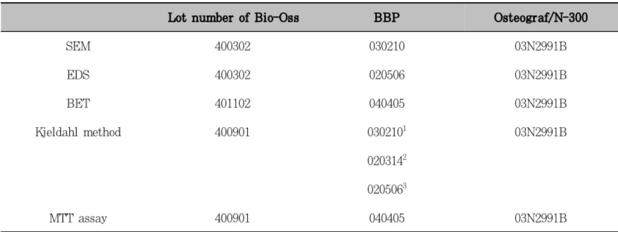
Table I. Characteristics of the investigated deproteinized bovine bone grafts
Lot number of Bio-Oss BBP Osteograf/N-300
SEM 400302 030210 03N2991B
EDS 400302 020506 03N2991B
BET 401102 040405 03N2991B
Kjeldahl method 400901 0302101 03N2991B
0203142 0205063
MTT assay 400901 040405 03N2991B
1,2,3)Repeated measurements were performed for BBP due to unexpected higher % of crude protein.
성과, 인간의 골조직과 유사한 화학적 조성과 미소 결정(crystallite)의 미세구조로 인해 넓은 표면적을 제공하고, 혈관과 골형성 세포의 내부성장을 촉진하 여 결과적으로 골형성을 증가시키는 것으로 알려졌
다.29,30) 가공하지 않은 원재료로부터 면역학적 문제
를 유발하는 단백질을 포함한 유기질 성분을 제거하 기 위한 처치방법과 연관하여, 탈단백 우골은 Bovi- ne spongiform encephalopathy(BSE)의 전염과 연관하여 안전한 것으로 알려져 있으나,31) 현재까지 잔존 단백질의 완전한 제거에 관해서는 명확히 밝혀 진 것이 없으며 이와 관련된 상반된 견해가 보고되 고 있다.31-33) 우골 유래 이종골 이식재내의 잔존 단 백질의 존재여부에 대해서는 차후 더 연구가 필요한 실정이다.
현재까지의 많은 임상보고를 살펴볼 때, 탈단백 우골은 골재생을 위한 재건수술에 사용시 비교적 성 공적인 임상결과를 나타내는 골대체재이나, 이러한 많은 수의 임상적 연구보고에 비해 현재 시판되는 제품의 기본적 특성에 대해서는 대부분 제조사의 부 족한 광고에 의존하는 실정이다. 이에 본 연구는 골 이식 대체재로 현재 시판되는 세 종류의 탈단백 우 골의 기본적인 특성을 조사하고 이를 상호 비교하기 위한 목적으로 시행하였다.
Ⅱ. 재료 및 방법
1. 탈단백 우골
아래와 같이 시판되는 세 종류의 탈단백 우골의 기본적 특성을 조사하였으며, 각기 분석에 사용된 제품의 일괄 번호는 표 1에 나타내었다. 각기 분석 에 사용된 골이식재는 세 가지 이상의 다른 일괄 번 호의 제품을 이용하였으나, Osteograf/N-300의 경 우 동일한 일괄번호의 제품을 분석에 사용하였다.
a) Bio-Oss(Geistlich-Pharma, 스위스); 250- 1000 µm입자크기, 망상골 이식재
b) BBP(오스코텍, 한국) ; 400-600 µm, 600- 1000 µm입자크기, 망상골 이식재
c) Osteograf/N-300(Dentsply Friadent Ce- ramed, 미국); 250-420 µm입자크기, 피질골 이식재
2. 평가 항목
1) 주사전자현미경 관찰
주사전자현미경(Scanning electron microscope (SEM); S-4200, Hitachi, 일본)을 이용하여 미세한 형 태학적 특성을 관찰하였다.
2) 시료의 성분 분석
SEM에 부착된 Energy dispersive X-ray spectrometer(EDS)를 이용하여 칼슘과 인을 포함 한 시료 구성 성분의 정성, 정량 분석을 시행하였다.
각기 3회씩 시행하였다.
3) 표면적 측정
시료의 표면적을 측정하기 위해 비표면적/기공도 분석기(BET)(High Speed Gas Sorption Analyzer;
Nova 2000-version 7.02; Quantachrome, 미국)를 이 용하여 고온, 진공하에서 표면에 부착된 불순물을 제거한 시료에, 질소가스를 흡착시킨 후 흡착등온선 으로부터 단위 질량당 표면적을 측정하였다.
4) 잔존 단백질량 측정
질소 분석의 국제적인 표준분석법으로 질소화합물 인 단백질 분석에 사용되는 Kjeldahl법을 이용하여 잔존 단백질량을 측정하였다. 1회 분석을 위해 0.5g 씩의 탈단백 우골을 이용하였으며, 필요한 경우 재 측정 시행하였다. 시료를 진한 황산과 분해촉매제를 이용하여 고온에서 가열하여 완전히 분해한 후 시료 내 질소를 황산 암모니아로 전환하여 이를 수산화나 트륨을 이용하여 증류하여 암모니아로 분리하여 산 과 발색시약을 이용하여 총질소를 정량하여 질소계 수 6.25를 사용하여 계산된 조 단백질(crude pro- tein)량을 측정하였다. 측정은 Kjeldahl 단백질/질 소 분석기(FOSS 1035 analyzer; Tecator, 스웨덴)을 이용하여 시행하였다.
5) 세포 활성도 평가
Rat osteosarcoma에서 유래한 ROS 17/2.8 cell line을 이용하여 각기 이식재에 대한 세포 활성 도를 평가하기 위해 MTT(3-(4,5-dimethylthia- zol-2yl)-2,5 diphenyltetrazolium bromide;
thiazol blue) assay를 시행하였다. 배양된 세포를 well당 30 mg의 이식재가 함유된 24 well plate에 well당 2×10⁴개로 접종하여 CO2 배양기에서 37℃
에서 1, 4 일간 배양하였다. 각기 해당일에 배양액
을 제거하고 인산완충생리식염수로 2회 세척하였다.
각기 well당 37℃의 MTT용액 250 µl를 첨가하고 같은 조건에서 3시간동안 배양하였으며, 750 µl의 dimethyl sulfoxide와 250 µl의 glycin buffer를 첨가하였다. 용액을 이식재를 포함하지 않은 well로 옮기고 ELISA reader(Precision Microplate Rea- der, Molecular Devices, USA)를 이용하여 570 nm 에서 흡광도를 측정하였다. 각기 이식재에 대한 세 포 활성도의 유의성을 analysis of variance(ANO- VA)를 이용하여 평가하였다.
Ⅲ. 결과
1. 이식재의 주사전자현미경 관찰소견
세 종류의 이식재의 미세 형태학적 관찰에서 이식 재 표면은 서로 다른 형태학적 특성을 나타내었다 (Figure 1)
.
다른 두 종류의 이식재와 비교시 고온에 서 처리된 Osteograft/N-300의 경우 sintering에 의해 형성된 특징적인 표면 형태를 관찰할 수 있었 다(Figure 1g-i). 동일한 방법으로 제조된 것으로 알 려진 Bio-Oss와 BBP의 표면은 형태학적 관찰에서 서로 다른 양상을 보였다. Bio-Oss의 경우 유기성 분 제거후의 마이크론 단위의 미세한 표면구조가 특 징적으로 고배율에서 관찰되는 반면(Figure 1a ,b), BBP의 표면은 동일한 배율에서 이러한 미세구조의 특징이 관찰되지 않았고, 비교적 평활한 양상을 보 였다(Figure 1d, e).2. 시료의 성분분석 결과
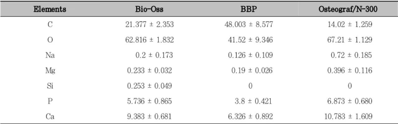
EDS분석을 통한 이식재 표면 구성성분의 정성, 정량분석을 위하여 시료당 3회 분석후의 평균값을 측정한 결과, 칼슘과 인의 원소비율(Ca/P)은 Bio- Oss는 1.636, BBP는 1.665, Osteograf/N-300 은 1.569로 나타났으며, 탄소, 산소, 칼슘, 인이 많 은 비율을 차지하였고, 그 외 나트륨, 마그네슘, 실 리콘이 미량 존재하였다. 이 중 탄소의 비율은 다른
Figure 1. SEM pictures of Bio-Oss(a,b,c), BBP(d,e,f) and Osteograf/N-300(g,h,i) at magnifications of ×10,000(a,d,g), ×5,000(b,e,h) and ×1,000(c,f,i).
Table 2. EDS results of the bone substitutes (Atomic %; Mean ± SD)
Elements Bio-Oss BBP Osteograf/N-300
C 21.377 ± 2.353 48.003 ± 8.577 14.02 ± 1.259
O 62.816 ± 1.832 41.52 ± 9.346 67.21 ± 1.129
Na 0.2 ± 0.173 0.126 ± 0.109 0.72 ± 0.185
Mg 0.233 ± 0.032 0.19 ± 0.026 0.396 ± 0.116
Si 0.253 ± 0.049 0 0
P 5.736 ± 0.865 3.8 ± 0.421 6.873 ± 0.680
Ca 9.383 ± 0.681 6.326 ± 0.892 10.783 ± 1.609
이식재에 비해 BBP에서 두 배 이상 높게 나타났다 (Table 2, Figure. 2).
3. 표면적 측정
BET분석결과 이식재의 표면적은 Bio-Oss는
55.3726m2/g, Osteograf/N-300은 40.9978 m2/g 으로 나타났으며, BBP의 경우 측정을 위한 온도 상 승시 시료의 변성이 발생하여 정확한 측정을 시행할 수 없었다.
Figure 2. EDS spectra of Bio-Oss (a), BBP (b), Osteograf/N-300 (c).
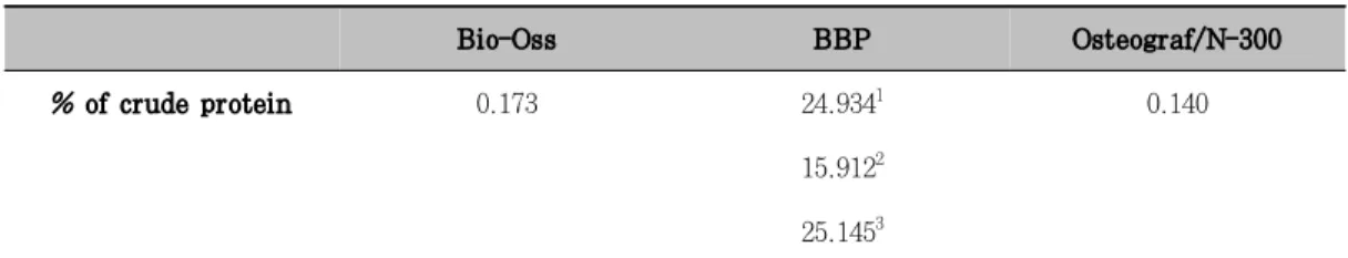
Table 3. Results of Kjeldahl protein analysis of the bone substitutes (% of crude protein)
Bio-Oss BBP Osteograf/N-300
% of crude protein 0.173 24.9341 0.140
15.9122 25.1453
1,2,3)Repeated measurements were performed due to high % of crude protein, samples of different lot
number were used
4. 잔존 단백질량 측정
Kjeldahl법을 이용한 잔존 단백질량의 측정에서 Bio-Oss의 조 단백질(crude protein)량은 0.173%, OsteoGraf/N-300은 0.140%로 나타났으며, BBP 는 3회의 측정에서 모두 15% 이상의 높은 수치를 나타내었다(Table 3).
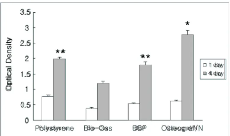
5. 세포 활성도 평가
MTT assay를 이용한 세포 활성도 평가에서 모 든 군에서 1일에 비해 4일에 세포수가 시간이 지남 에 따라 증가하는 양상을 관찰할 수 있었다(Figure 3). 4일째 세포수는 Osteograf/N-300이 다른 모든
군에 비해 현저히 높았고(p < 0.01), polystyrene dish와 BBP에서의 세포수는 Bio-Oss에 비해 더 높은 양상을 관찰할 수 있었다(p < 0.01).
Ⅳ. 고찰
상실된 치아를 회복하기 위한 임프란트 시술의 증 가와 더불어 손상되거나 위축된 치조제에서의 임프 란트 식립을 위해 골재건수술의 필요성 또한 증가된 다. 자가골 이식이 가장 이상적인 이식재이지만, 골 채취를 위한 부가적인 수술부위가 필요하며 이로 인 한 합병증의 가능성도 배제할 수 없다. 자가골 이식 을 대체하기 위한 여러 종류의 골 대체재에 대한 연 구와 성공적인 임상 결과에 대해 보고되어져 왔다.
Figure 3. The optical density measured after culture for 1, 4 days at a wavelength of 570nm by ELISA reader(n=4 per each group).
* statistically significant difference compared to polystyrene, Bio- Oss and BBP at 4 days(p < 0.01)
** statistically significant difference compared to Bio-Oss at 4 days(p < 0.01)
이중 우골에서 단백질 성분을 제거한 이종골 이식재 를 이용한 골재건 수술에서 비교적 성공적인 결과가 많은 연구를 통해 보고되어지고 있다.20-28) 망상골 구조와 넓은 표면적은 생체 내에서 사용 시 혈관과 골형성 세포의 이식재로의 성장을 촉진함으로써 결 과적으로 골결손부의 성공적인 골화를 이루는 것으 로 알려졌다. 골재건수술에 사용된 이식재의 흡수에 대한 보고에서 자가골 이식재를 이용 시 상당한 양 의 이식재 흡수에 대해서는 잘 알려진 사실이며,
34-36)4개월에 최대 56%의 cortical bone graft의
흡수가 일어난 경우도 보고되었다. 이에 비해 이종 골 이식재인 Bio-Oss의 생체 내 흡수는 매우 더딘 것으로 알려져 있고, 6년까지도 거의 흡수가 되지 않는다는 보고도 있다.37,38) 이식후 3-7개월동안 점 진적으로 흡수가 일어난다는 동물실험에서의 결과와
39,40) 달리 인간에서의 결과는 이식후 6개월에서는
osteoclastic activity를 관찰할 수 없었고,41) 장기 간의 관찰에서 Bio-Oss입자의 흡수와 연관된 osteoclast의 출현이 보고되었다.37) 이러한 특성은 골재건수술 시행후 remodeling의 결과로 발생하는 자가골의 흡수를 보상하는 지지체로서 임상에서 사
용 시 장점을 가지는 것으로 보고되었다.42,43) 이종 골 이식재로 사용되는 탈단백 우골은 약 10nm의 결 정크기를 가지는 calcium-deficient carbonate apatite로 인간의 골과 유사한 화학적, 물리적 성질 을 보인다.44) 골내 이식되는 생체재료의 미세한 구 조적 표면 특성은 골조직 반응에 결정적인 영향을 미치는 것으로 알려져 있으며, 특히 망상골을 원재 료로 이용하여 가공된 이종골 이식재는 골의 이식재 내부로의 성장에 적합한 것으로 알려진 pore 크기를 가지는 것으로 알려졌으며 큰 내부면적을 가지는 것 으로 알려졌다.29)
본 연구에서 측정된 이식재의 표면적은 Bio-Oss 에 있어서 다른 문헌에서 보고된 수치인 79.7m2/g과 비교시에는45낮은 수치인 55.3726 m2/g를 나타내었 으며 Osteograf/N-300은 40.9978 m2/g의 표면적 을 나타내었다. 문헌을 통해 제공되는 생체재료의 특 성에 관한 자료는 사용된 측정법과 또는 저자에 따라 간혹 편향된 결과를 제시할 수도 있으므로, 이에 관해 서는 보다 많은 연구자의 평가가 필요할 것으로 사료 된다. 예를 들어 골이식재의 표면적에 대한 Algipore (Friadent, Manheim, 독일)의 수치는 두 가지 문헌을
45,46) 통해 각기 4.9-14.6m2/g과 50m2/g으로 현격 한 차이가 있는 것으로 보고되었다.
본 연구에서 MTT assay를 이용한 세포 생활력 평가에서 세 종류의 이식재 모두 시간이 지남에 따라 세포수가 증가하였고, 이는 생체적합성에는 별 문제 가 없는 것으로 나타났다. 현재 탈단백 우골을 골대 체로 사용함에 있어서, 문제가 될 수 있는 부분은 합 성골과 달리 질병전염의 가능성이 있는지의 여부이 다. 현재 우골을 골이식 대체재로 사용하는데 있어서 잔존 단백질의 존재여부에 대해서는 확실히 밝혀지지 는 않았으며, 상반된 보고가 존재한다. Schwartz 등은33) 이종골 이식재인 Bio-Oss의 osteopromo- tive ability가 이식재 내에 존재하는 변형성장인자 베타(TGF-β)와 골형성단백질(BMP-2)의 역할일 수도 있다고 제시하여, 이식재 내의 단백질의 존재에 대해 서 보고하였다. 이에 반해 다른 연구에서는 Bio-Oss 내 측정가능한 단백질은 포함되지 않는 것으로 보고
되었다.31,47)우골을 이용한 이종골 이식재는 화학적
인 처리방법과 열처리과정을 통해 원재료 내 존재하 는 단백질을 변성, 제거하고 다른 유기성분을 제거하 는 단계를 통해 제조된다. Bio-Oss의 경우 강알칼리 처리후, 300℃에서 최소 15시간 동안 열처리 과정과 이후 유기용매를 이용한 부가적인 처리 과정 후 감마 선 멸균을 시행하여 제조되는 것으로 알려진다. 이식 재를 제조하기 위해 사용되는 원재료는 BSE의 가능 성이 없는 국가에서 기원한 우골을 사용하며, BSE 의 감염가능성에 관한 조직분류에 관한 WHO recommendation에 따르면 골은 감염 잠재력이 낮 은 것으로 분류된 골수조직과 달리 감염 가능성이 감 지되지 않는 조직인 ‘범주 4’에 포함된다.48)
이종골 이식재의 제조과정에서 사용되는 여러 단 계중 알칼리 처리는 1-2 M농도의 NaOH를 2시간 동안 처치시 프리온의 감염력이 최소 10만배 감소하 는 것으로 알려졌으며, BSE 프리온은 거의 불활성 화되는 것으로 보고되었다.49) Bio-Oss나 BBP와는 달리 Osteograf/N은 600℃를 초과한 온도에서 6 시간 이상, 최고 온도 1000℃를 초과하는 열처리 과 정을 통해 제작되는 것으로 알려져 있으며, 이러한
열처리 과정이 화학용매의 사용과 관계없이 BSE 병 원체를 불활성화하는데 가장 효과적인 것으로 보고 되었다.50) 고온의 열처리 과정은 표면을 소결하여 pore를 포함한 미세한 표면구조를 변경할 수도 있는 것으로 알려졌으며, 실제 SEM 관찰에서 이러한 소 견이 관찰되며(Figure 1g-i), Bio-Oss에 비해 비교 적 낮은 표면적의 결과로 본 실험에서 나타났다.
조직내 존재하는 잔존 단백질량을 측정하는데 있 어서 여러 종류의 방법이 사용될 수 있으며,32,33,47,50)
본 실험에 사용된 Kjeldahl법은 현재 질소 함량을 이용한 단백질 분석법으로 널리 이용되는 방법으로 다른 보고에서 사용된 Lowry법 등에 비해 시료내 잔존 지질성분을 포함한 구성성분에 의한 간섭을 받 지 않고, 분석에 사용되는 모든 단백질의 질소를 암 모늄으로 분해할 수 있는 장점을 가지는 것으로 알려 졌다.51~53)
인간의 골조직은 유기기질과 광물질로 구성되는 데, 건조된 비탈회상태의 전체성분의 약 30%를 type I collagen이 차지하며 약 5%를 차지하는 비 교원성 단백질에는 bone sialoprotein, osteopon- tin, osteonectin 등이 여기에 포함되며, 전체 성분 의 2-4%이하를 차지하는 지질 등이 주요구성 성분 이며 나머지는 광물질이 차지한다.54-56)
Kjeldahl 법을 이용한 조 단백질량의 측정은 유 기질소를 중화 적정법에 의하여 총질소를 정량하여 질소계수(nitrogen coefficient)를 이용하여 산출하는 데 아미노산, purine염기, amide 화합물 등도 질소 화합물을 함유하므로, 총질소량에 질소계수를 이용 하여 산출한 값이 순수한 단백질 양을 나타내지는 않는다. 하지만 골조직의 구성 성분의 종류와 비율 을 고려하면, 본 실험에서 측정된 조 단백질의 수치 는 Bio-Oss와 Osteograf/N-300의 0.1%대의 수 치에 포함되는 기타 구성 성분에 대해서는 향후 추 가적인 분석을 통한 고려와 더불어, 이보다 상당히 높은 15%이상의 수치를 보인 BBP의 경우 이를 대 부분 잔존 조 단백질량으로 보아야 할 것이다. 본 실 험의 결과에서 나타난 예상치 못한 높은 잔존 조 단 백질량에 대해서는 향후 다른 종류의 분석방법을 통
해서도 확인이 필요한 것으로 사료된다. 하지만 이 러한 잔존 단백질량과 본 실험에 사용된 다른 분석 법의 결과에 대한 상호연관성에 대해서도 고려해야 될 필요가 있다. 유사한 제조과정을 거치는 것으로 알려진 Bio-Oss와 BBP의 SEM소견의 비교에서 Bio-Oss에서 관찰되는 수 µm폭의 mineral fibril 의 network의 특징적인 구조30)를 BBP에서는 관찰 할 수 없었다(Figure 1a, d). 이는 잔존 유기성분의 영향인 것으로 보이며, 또한 BET를 이용한 표면적 분석에서의 온도상승시의 시료의 변성과도 연관이 있는 것으로 추측된다.
현재 우골을 이용하여 제작된 이종골 이식재는 한 국을 포함하여 BSE에 비교적 안전한 것으로 알려진 미국을 기원국으로 하는 원재료를 사용하고 있고, 골수 부위를 제외한 골조직은 감염가능성이 감지되 지 않는 ‘범주 4’의 조직으로 분류된다. 또한 이종골 이식재를 제조하는 단계에서 사용되어지는 화학적, 열처리 방법들이 잔존 단백질을 대부분 제거하고 감 염의 가능성을 극히 미미한 정도로 낮추는 것은 사 실이지만, 여러 연구를 통해 감염의 ‘위험성 전혀 없 음’의 경우는 존재하지 않는 것으로 받아들여진다.
또한 이러한 안전성은 이식재를 제조하는 모든 단계 의 과정들이 완벽하게 수행되고, 관리되어야 비로소 가능하다는 본 연구의 결론은 다른 여러 연구에서 제시된 결론과 일치한다.
시판되는 세 종류의 탈단백 우골에 대한 기본적인 특성평가를 위해 시행된 본 실험의 결과에서 유사한 방법으로 제조된 두 종류의 이식재는 전혀 다른 성 상을 나타내었다. 이는 골 재건수술의 필요성과 빈 도의 증가와 더불어 더 많은 양의 이식재를 필요로 하는 현재의 임상환경에서 상당히 심각한 문제로 받 아들여진다. 술자와 환자에게 둘 다 안전하고 유익 한 임상결과를 위해 제조사는 모든 제조공정에 대해 보다 책임감 있고 엄격한 관리가 필요하며, 또한 시 판되는 골 이식재를 포함한 여러 종류의 생체재료에 대한 기본특성을 재평가하는 평가자로 구성되는 감 시, 평가 체계가 보다 바람직한 임상환경을 위해 활 성화되어야 할 것으로 사료된다.
Ⅴ. 참고문헌
1. Kalk WWI, Raghoebar GM, Jansma J, Boering G. Morbidity from iliac crest bone harvesting. Clin Oral Implants Res 1996;54:1424-1429.
2. Misch CM. Comparision of intraoral donor sites for onlay grafting prior to implant placement. Int J Oral Maxillofac Implants 1997;12:767-776.
3. Raghoebar R, Louwerse C, Kalk WWI, Vissink A. Morbidity of chin bone har- vesting. Clin Oral Implants Res 2001;
12:503-507.
4. Mellonig JT. Bone allografts in perio- dontal therapy. Clin Orthop 1996;324:
116-125.
5. Pinholt EM, Haanaes HR, Roervik M, Donath K, Bang G. Alveolar ridge aug- mentation by osteoinductive materials in goats. Scand J Dent Res 1992;100:361 -365.
6. Quintero G, Mellonig JT, Gambill VM, Pelleu GB. A six month clinical evalua- tion of decalcified freeze-dried bone allografts in periodontal osseous defects.
J Periodontol 1982;53:726-730.
7. Rosenberg E, Rose LF. Biologic and clinical consideration for autografts and allografts in periodontal regeneration therapy. Dent Clin North Am 1998;
42:467.
8. Urist MR: Bone: formation by autoin- duction. Science 1965;150:893-899.
9. Quattlebaum JB, Mellonig JT, Hensel NF. Antigenicity of freeze-dried cortical bone allograft in human periodontal defects. J Periodontol 1988;59:394-397.
10. Schwartz Z, Mellonig JT, Carnes DL, et al.. Ability of commercial demineralized freeze-dried bone allograft to induce new bone formation. J Periodontol 1996;
67:918-926.
11. Schwartz Z, Somers A, Mellonig JT, et al. Ability of commercial demineralized freeze-dried bone allograft to induce new bone formation is dependant on donor age bu not gender. J Periodontol 1998;69:47-478.
12. Lekishvilli MV, Snetkov A, Vasiliv MG, et al. Experimental and clinical study of the demineralized bone allografts. Cell Tissue Bank 2004;5:231-238.
13. Summitt MC, Reisinger KD. Characteri- zation of the mechanical properties of demineralized bone. J Biomed Mater Res A 2003;67:742-750.
14. Schmitt JM, Buck DC, Joh SP, Lynch SE, Hollinger JO. Comparison of porous bone mineral and biologically active glass in critical-sized defects. J Perio- dontol 1997;68:1043-1053.
15. Heikkila JT, Aho HJ, Yli-Urpo A, Happonen RP, Aho AJ. Bone formation in rabbit cancellous bone defects filled with bioactive glass granules. Acta Or- thop Scand 1995;66:463-467.
16. Mangano C, Bartolucci E, Mazzocco C.
A new porous hydroxyapatite for promo- tion of bone regeneration in maxillary sinus augmentation: clinical and histolo- gic study in humans. Clin Oral Implants Res 2003;18:23-30.
17. Wiltfang J, Schlegel KA, Schultze-Mos- gau S, et al. Sinus floor augmentation with β-tricalciumphosphate(β-TCP): does
platelet-rich plasma promote its osseo- integration and degradation? Clin Oral Implants Res 2003;14:213-218.
18. Papacharambous SK & Anastasoff KI.
Natural coral skeleton used as onlay graft for contour augmentation on the face. Int J Oral Maxillofac Surg 1993;
22:260-264.
19. Clokie CML and Sandor GKB. Bone:
present and future. In: Babbush CA, editor, Dental implants: the art and science/edited by Babbush CA. W.B.
Sanduers Company. Philadelphia. 2001, p. 70.
20. Froum SJ, Tarnow DP, Wallace SS, Rohrer MD, Cho SC. Sinus floor eleva- tion using anorganic bovine bone matrix (OsteoGraf/N) with and without autoge- nous bone: a clinical, histologic, radio- graphic, and histomorphometric analysis -Part 2 of an ongoing prospective study.
Int J Periodontics Restorative Dent 1998;18:528-543.
21. John HD & Wenz B. Histomorphometric analysis of natural bone mineral for maxillary sinus augmentation. Int J Oral Maxillofac Implants 2004;19:199- 207.
22. Landi L, Pretel RW, Hakimi NM, Setayesh R. Maxillary sinus floor eleva- tion using a combination of DFDBA and bovine bone-derived porous hydroxya- patite: a preliminary histologic and histomorphometric report. Int J Perio- dontics Restorative Dent 2000;20:574- 583.
23. Maiorana C, Redemagni M, Rabagliati M, Salina S. Treatment of maxillary
ridge resorption by sinus augmentation with iliac cancellous bone, anorganic bo- vine bone, and endosseous implants: a clinical and histologic report. Int J Oral Maxillofac Implants 2000;15:873-878.
24. Mellonig T. Human histologic evaluation of a bovine derived bone xenograft in the treatment of periodontal osseous defects. Int J Periodontics Restorative Dent 2000;20:18-29.
25. Hallman M, Sennerby L, Zetterqvist L, Lundgren S. A 3-year prospective follow -up study of implant-supported fixed prostheses in patients subjected to maxillary sinus floor augmentation with a 80:20 mixture of deproteinized bovine bone and autogenous bone, Clinical, radiographic and resonance frequency analysis. Int J Oral Maxillofac Surg 2005,34:273-280.
26. Norton MR, Odell EW, Thompson ID, Cook RJ. Efficacy of bovine bone mineral for alveolar augmentation: a human histologic study. Clin Oral Implant Res 2003;14:775-783.
27. Piattelli M, Favero GF, Scarano A, Orsini G, Piattelli A. Bone reactions to anorganic bovine bone(Bio-Oss®) used in sinus lifting procedure: a histologic long-term report of 20 cases in man, Int J Oral Maxillofac Implants 1999;14:835 –840.
28. Sartori S, Silvestri M, Forni F, et al.
Ten-year follow-up in a maxillary sinus augmentation using anorganic bovine bone(Bio-Oss®). A case report with his- tomorphometric evaluation, Clin Oral Implants Res 2003;14:369–372.
29. Peetz M. Characterization of xenogenic bone material. In: Boyne PJ, editor.
Osseous reconstruction of the maxilla and mandible. Chicago: Quintessence, 1997.p.87-93.
30. Rosen BV, Hobbs LW, Spector M. The ultrastructure of anorganic bovine bone and selected synthetic hydroxyapatite used as bone graft substitute materials.
Biomaterials 2002;23:921-928.
31. Wenz B, Oesch O, Horst M. Analysis of the risk of transmitting bovine spongi- form encephalopathy through bone grafts derived from bovine bone. Bioma- terials 2001;22:1599-1606.
32. Honig JF, Merten HA, Heinemann DE.
Risk of transmission of agents asso- ciated with Creutzfeld-Jakob disease and bovine spongiform encephalopathy. Plast Reconstr Surg 1999;103:1324-1335.
33. Schwartz Z, Weesner T, van Dijk S, et al. Ability of deproteinized cancellous bovine bone to induce new bone forma- tion. J Periodontol 2000;71:1258-1269.
34. Blomqvist JE, Alberius P, Isaksson S, Lindhe A, Obrant K. Importance of bone graft quality for implant integration af- ter maxillary sinus reconstruction. Oral Surg Oral Med Oral Pathol Oral Radiol Endod 1998;86:268-274.
35. Merkx MAW, Maltha JC, Freihofer HP, Kuijpers-Jagtman AM. Incorporation of particulated bone implants in the facial skeleton. Biomaterials 1999;20:2029- 2035.
36. Ozaki W, Buchman SR. Volume main- tenance of onlay bone grafts in the craniofacial skeleton: micro-architecture
versus embryonic origin. Plast Reconstr Surg 1998;102:291-299.
37. Piattelli M, Favero GA, Scarano A, Orsini G, Piattelli A. Bone reactions to anorganic bovine bone(Bio-Oss) used in sinus augmentation procedure: a histo- logic long-term report of 20 cases in humans. Int J Oral Maxillofac Implants 1999;14:835-840.
38. Schlegel AK & Donath K. Bio-Oss-a resorbable bone substitutes? J Long Term Eff Med Implants 1998;8:201-209.
39. Berglundh T, Lindhe J. Healing around implants placed in bone defects treated with Bio-Oss. An experimental study in the dog. Clin Oral Implants Res 1997;
8:117-124.
40. Hammerle CHF, Chiantella GC, Karring T, Lang NP. The effect of a deprotei- nized bovine bone mineral on bone rege- neration around titanium dental im- plants. Clin Oral Implants Res 1998;9 :151-162.
41. Yildirim M, Spiekermann H, Biesterfeld S, Edelhoff D. Maxillary sinus augmen- tation using xenogenic bone substitute material Bio-Oss in combination with venous blood. A histologic and histomor- phometric study in humans. Clin Oral Implants Res 2000;11:217-229.
42. Maiorana C, Beretta M, Salina S, Santoro F. Reduction of autogenous bone graft resorption by means of Bio-Oss coverage: a prospective study. Int J Periodontics Restorative Dent 2005;25:
19-25.
43. Hatano N, Shimizu Y, Ooya K. A clinical long-term radiographic evaluation of
graft height changes after maxillary sinus floor augmentation with a 2:1 autogenous bone/xenograft mixture and simultaneous placement of dental im- plants. Clin Oral Implants Res 2004;15 :339-345.
44. Hurzeler MB, Qunones CR, Kirsch A, et al. Maxillary sinus augmentation using different grafting materials and dental implants in monkeys. Part I. Evaluation of anorganic bovine-derived bone matrix.
Clin Oral Implants Res 1997;8:476-486.
45. Wenz B. Characteristics of Bio-Oss and Bio-Gide. In: Maiorana C & Simion M, editor. Advanced techniques for bone regeneration with Bio-Oss and Bio-Gide.
Seoul, Daehan publishing, 2003. p.75.
46. Ewers R, Goriwoda W, Schopper C, Moser D, Spassova E. Histologic fin- dings at augmented bone areas supplied with two different bone substitute mate- rials combined with sinus floor lifting.
Clin Oral Implants Res 2004;15:96-100.
47. Benke D, Olah A, Mohler H. Protein- chemical analysis of Bio-Oss bone sub- stitute and evidence on its carbonate content. Biomaterials 2001;22:1005-1012.
48. Public health issues related to animal and human spongiform encephalopathies : Memorandum from a WHO meeting.
1992; Bulletin of the World Health Organization.No.70(2) 183-190.
49. Taylor DM, Fraser H, McConnell I, et al. Decontamination studies with the agents of bovine spongiform encepalo- pathy and scrapie. Arch Virol 1994;139:
313-326.
50. Sogal A & Tofe AJ. Risk assessment of
bovine spongiform encephalopathy trans- mission through bone graft material derived from bovine bone used for dental applications. J Periodontol 1999;70:1053 -1063.
51. Lowry OH, Rosbrough NJ, Farr AL, Randall RT. Protein measurement with the folin phenol reagent. J Biol Chem 1951;193:265-273.
52. Brooks SPJ, Lampi BJ, Sarwar G, Botting HG. A comparison of methods for determining total body protein. Anal Biochem 1995;226:26-30.
53. Verley H. Practical clinical biochemistry.
1965. Heinemann, London.
54. Robey PG, Boskey AL: The biochemistry of bone. In Marcus R et al, editors:
Osteoporosis, New York, 1995, Academic press.
55. Wuttke M, Muller S, Nitsche DP, et al.
Structural characterization of human recombinant and bone-derived bone sialoprotein. Functional implications for cell attachment and hydroxyapatite binding. J Biol Chem 2001;276:36839- 36848.
56. Goldberg M & Boskey AL. Lipids and biomineralizations. Prog Histochem Cy- tochem 1996;31:187.
-Abstract-
Evaluation of deproteinized bovine bone mineral as a bone graft substitute :
A comparative analysis of basic characteristics of three commercially available bone substitutes
Jin-Woo Park
Department of Periodontology, College of Dentistry, Kyungpook National University
Bovine bone-derived bone substitutes are widely used for treatment of bone defects in dental and orthopedic regenerative surgery. The purpose of this study was to evaluate the basic characteristics of deproteinized bovine bone mineral as a bone graft substitute. Com- mercially available products from three different bovine bone minerals-Bio-Oss(Geistlich- Pharma, Switzerland), BBP(Oscotec, Korea), Osteograf/N-300(Dentsply Friadent Ceramed, USA) - were investigated. They were evaluated by scanning electron microscopy(SEM), energy dispersive X-ray spectrometer(EDS), surface area analysis(BET), and Kjeldahl protein analysis. Cell viability on different products was evaluated by 3-(4, 5-dimethylthiazol-2-yl)-2,5-diphenyl tetrazolium bromide(MTT) assay. The results of this study indicated that each bone substitute displayed distinct surface properties. Furthermore, Kjeldahl protein analysis indicated that residual crude proteins are present in deproteinized bovine bone mineral. BBP showed relatively large amount of residual protein, which indicated that the possibility of disease transmission can not be safely ruled out. Based on the results of this study, it is suggested that active quality management is strongly needed in operations that involve processing bovine bone tissue for medical use.2)
Key words : BBP, Bio-Oss, bovine bone mineral, Osteograf/N, surface characteristics