Korean J Gastroenterol Vol. 72 No. 2, 83-85 https://doi.org/10.4166/kjg.2018.72.2.83 pISSN 1598-9992 eISSN 2233-6869
CASE REPORT
Korean J Gastroenterol, Vol. 72 No. 2, August 2018 www.kjg.or.kr
A Guidewire May Save the Day
Rajat Garg and Mohammed Barawi1
Department of Internal Medicine, Division of Gastroenterology and Hepatology1, St. John Hospital and Medical Center, Detroit, MI, USA
Endoscopic gallbladder drainage (EGBD) has been used to treat acute cholecystitis or to relieve malignant biliary obstruction as an alternative to percutaneous gallbladder drainage and patient's are poor surgical candidates. This is currently being performed by placement of lumen apposing metallic stent (LAMS) with electrocautery mounted tip delivery system also called as "hot" technique.
We had reported a case of self-expanding metallic stent (SEMS) within LAMS after stent migration during EGBD using "hot" technique and propose routine use of guidewire in patients undergoing the procedure. (Korean J Gastroenterol 2018;72:83-85)
Key Words: Ultrasound; Endoscopy; Stent; Cholelithiasis
Received December 4, 2017. Revised March 22, 2018. Accepted April 20, 2018.
CC This is an open access article distributed under the terms of the Creative Commons Attribution Non-Commercial License (http://creativecommons.org/licenses/
by-nc/4.0) which permits unrestricted non-commercial use, distribution, and reproduction in any medium, provided the original work is properly cited.
Copyright © 2018. Korean Society of Gastroenterology.
Correspondence to: Rajat Garg, Department of Internal Medicine, St. John Hospital and Medical Center, 19251, Mack Ave, Suite 335, Grosse Pointe Woods, MI 48236, USA. Tel: +1-313-204-8093, Fax: +1-313-343-8747, E-mail: [email protected]
Financial support: None. Conflict of interest: None.
INTRODUCTION
Endoscopic Gall Bladder Drainage (EGBD) has been used to treat patients with acute cholecystitis or to re- lieve malignant biliary obstruction in patients who are poor candidates for surgery due to underlying comorbidities.1 An endoscopic connection is created between the gastric - or duodenal - and the gallbladder lumen to achieve drainage of the gall bladder (GB) and biliary tract. EGBD was first described in 2007, and since, its approach has evolved from the placement of naso-gallbladder drain to plastic biliary stents, and then to self-expanding metallic stents (SEMS).2 Lately, lumen apposing metallic stent (LAMS), which utilizes dumbbell-shaped flanges on both ends to appose the two visceral walls, has been made available. Initially, it was placed utilizing the “Cold techni- que”, which included endoscopic ultrasound (EUS)-guided needle puncture followed by guidewire placement and di- lation of the tract. A recently approved second generation LAMS allows the endoscopist to perform anastomosis
without the need for a guidewire or tract dilation, with the electrocautery tip mounted delivery system, which is also called the “Hot technique”. Here, we report a case of SEMS placement using LAMS after stent migration during EGBD via the “Hot Technique” and propose the routine use of guidewire in patients undergoing the procedure.
CASE REPORT
An 88-year-old female patient presented with ob- structive jaundice. The patient underwent EUS examina- tion and a large periampullary mass without duodenal obstruction was found. She was scheduled for EUS-guid- ed biliary drainage via the rendezvous technique. EUS ex- amination with linear echoendoscope revealed a dis- tended gallbladder abutting the gastric wall (Fig. 1).
Therefore, an EGBD was performed to decompress the GB and biliary system. An appropriate puncture site with- out intervening the blood vessels was identified. Under EUS guidance, the GB wall was punctured from antrum
84 Garg R and Barawi M. A Guidewire May Save the Day
The Korean Journal of Gastroenterology Fig. 1. Linear EUS image showing distended gallbladder abutting the
gastric wall. EUS, endoscopic ultrasound.
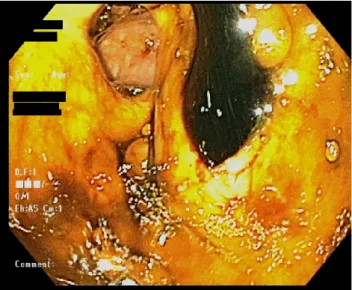
Fig. 2. Dark copious bile flowing through LAMS. Note that distal flange is not visible and is retracted into gastric wall. LAMS, lumen apposing metallic stent.
Fig. 3. A 0.035 inch jag guidewire being passed through the LAMS.
LAMS, lumen apposing metallic stent.
A
A BB
Fig. 4. Fluoroscopic image showing coiled guidewire in the gallbladder lumen with deployment of both stents (A). Distal flange of SEMS seen in gastric lumen after successful placement (B).
SEMS, self-expanding metallic stents.
with an electrocautery tip, followed by deployment of a 10×10 mm AXIOS (Boston Scientific, Marlborough, MA, USA) stent with the proximal end in the GB lumen and with the distal end in the gastric lumen. A copious amount of dark, black bile was observed flowing through the stent (Fig. 2). On further inspection, the distal flange of stent was noted to be retracted in the gastric wall due to GB contraction (Fig. 2). Attempts to reposition the stent with cold biopsy and raptor forceps failed. Due to anatomical position and difficult access, we were unable to advance a guidewire through the stent. A double chan- nel upper endoscope (GIF-2TH180, Olympus, Tokyo,
Japan) was then passed into the stomach. The tip of a 5 Fr biliary catheter was modified into a hockey stick shape and advanced through the biopsy channel of the endoscope. A 0.035-inch jag guidewire (Boston Scientific, Marlborough, MA, USA) was passed through the biliary catheter and stent under endoscopic and fluoroscopic guidance (Fig. 3). Multiple loops of the guidewire were coiled inside the GB lumen through LAMS. A 10×40 mm fully covered biliary SEMS (Boston Scientific, Marlborough, MA, USA) was then deployed over the guidewire with dis- tal flange inside the stomach (Fig. 4). Our patient tol- erated the procedure well, and follow-up acute abdomen series showed no evidence of free air with continuous im- provement of her liver enzymes. She was discharged in stable condition two days after the procedure. Due to in- volvement of the superior mesenteric artery and poor
Garg R and Barawi M. A Guidewire May Save the Day 85
Vol. 72 No. 2, August 2018
functional status, she then decided to undergo palliative measures and was discharged to home hospice care.
DISCUSSION
According to literature review, similar cases describing the placement of fully covered SEMS within the LAMS have been reported previously. A case series reported as many as 4 out of 13 patients needing a second stent placement during EGBD.3,4 However, such cases have been reported with the use of the ‘Cold LAMS’ technique, which already involves the placement of a guidewire be- fore stent deployment.3,4 Technical failure has been at- tributed to thickened GB wall, resulting in stent migration. There have been no significant differences in the outcomes and adverse events of patients with EGBD when compared with percutaneous GBD; however, favor- able trend towards endoscopic intervention has been observed.5-7 A study reported that about 10.7% (8/75) of patients experienced adverse events, including recurrent cholecystitis (3), stent migration (2), and Bouveret syn- drome (1), with all these complications managed conservatively.8 EGBD has also been shown to be better tolerated with low risk of adverse events as compared to percutaneous GBD.7 Catheter-related problems are minimal in EGBD; however, the risk of perforation, bile peritonitis, bleeding, and stent migration have been reported.7,9 In our patient, the proposed mechanism of displacement is sudden retraction of the GB wall after decompression with stomach distention as a potential contributor. Based on our literature review, cases of spontaneous migration of LAMS on repeat endoscopy have been reported, but cases of immediate migration have not been reported. We propose that there might be a component of selective reporting here since patients with immediate migration require urgent surgical treat- ment for the prevention of peritonitis and perforation, which might preclude their study inclusion. We propose that a guidewire should routinely be used for patients un- dergoing EGBD with ‘Hot LAMS’ technique, which can be passed through the delivery system before stent deployment. Placement of a guidewire in GB lumen be-
fore stent deployment would likely allow for a safe and reliable placement of another stent in case of displace- ment or migration of the first stent. A careful endoscopic examination should be performed to confirm the correct placement of LAMS. If the stent is deployed without any complications, the guidewire can then be removed through the stent, or if needed, it can be used to place another stent in case of malalignment or displacement of the first stent. Hence, we believe that routine use of a guidewire for the placement of “Hot LAMS” might as well “Save the day” for patients who otherwise might ex- perience complications.
REFERENCES
1. Patil R, Ona MA, Papafragkakis C, Anand S, Duddempudi S.
Endoscopic ultrasound-guided placement of the lumen-apposing self-expandable metallic stent for gallbladder drainage: a prom- ising technique. Ann Gastroenterol 2016;29:162-167.
2. Xu MM, Kahaleh M. EUS-guided transmural gallbladder drainage:
a new era has begun. Therap Adv Gastroenterol 2016;9:138-140.
3. de la Serna-Higuera C, Pérez-Miranda M, Gil-Simón P, et al.
EUS-guided transenteric gallbladder drainage with a new fistu- la-forming, lumen-apposing metal stent. Gastrointest Endosc 2013;77:303-308.
4. Law R, Grimm IS, Stavas JM, Baron TH. Conversion of percuta- neous cholecystostomy to internal transmural gallbladder drain- age using an endoscopic ultrasound-guided, lumen-apposing metal stent. Clin Gastroenterol Hepatol 2016;14:476-480.
5. Irani S, Ngamruengphong S, Teoh A, et al. Similar efficacies of en- doscopic ultrasound gallbladder drainage with a lumen-apposing metal stent versus percutaneous transhepatic gallbladder drainage for acute cholecystitis. Clin Gastroenterol Hepatol 2017;15:738-745.
6. Khan MA, Atiq O, Kubiliun N, et al. Efficacy and safety of endo- scopic gallbladder drainage in acute cholecystitis: is it better than percutaneous gallbladder drainage? Gastrointest Endosc 2017;85:76-87.e3.
7. Chan JHY, Teoh AYB. Current status of endoscopic gallbladder drainage. Clin Endosc 2018;51:150-155.
8. Dollhopf M, Larghi A, Will U, et al. EUS-guided gallbladder drain- age in patients with acute cholecystitis and high surgical risk us- ing an electrocautery-enhanced lumen-apposing metal stent device. Gastrointest Endosc 2017;86:636-643.
9. Teoh AYB, Serna C, Penas I, et al. Endoscopic ultrasound-guided gallbladder drainage reduces adverse events compared with percutaneous cholecystostomy in patients who are unfit for cholecystectomy. Endoscopy 2017;49:130-138.