Korean J Gastroenterol Vol. 69 No. 6, 359-362 https://doi.org/10.4166/kjg.2017.69.6.359 pISSN 1598-9992 eISSN 2233-6869
CASE REPORT
Korean J Gastroenterol, Vol. 69 No. 6, June 2017 www.kjg.or.kr
궤양성 대장염에서 위 유문부 폐쇄를 일으킨 거대세포바이러스 감염 1예
강성환, 이기명1, 신성재1, 임선교1, 황재철1, 김진홍1
성애병원 소화기내과, 아주대학교 의과대학 소화기내과1
Cytomegalovirus Gastric Ulcer Complicated with Pyloric Obstruction in a Patient with Ulcerative Colitis
Sung Hwan Kang, Kee Myung Lee1, Sung Jae Shin1, Sun Kyo Lim1, Jae Chul Hwang1 and Jin Hong Kim1
Division of Gastroenterology, Department of Internal Medicine, Sungae Hospital, Seoul, Division of Gastroenterology, Department of Internal Medicine, Ajou University School of Medicine1, Suwon, Korea
In patients with inflammatory bowel disease (IBD), cytomegalovirus (CMV) infections could aggravate the course of IBD but it is difficult to distinguish CMV infection from IBD exacerbation endoscopically. Usually, CMV tends to localize to the colon and other organic in- volvements were reported very rare in the IBD patients. Herein, we report a case that CMV gastric ulcer complicated with pyloric ob- struction in a patient with ulcerative colitis during ganciclovir therapy, which was resolved by surgical gastrojejunostomy with review of literature. (Korean J Gastroenterol 2017;69:359-362)
Key Words: Cytomegalovirus; Gastric ulcer; Gastric outlet obstruction; Ulcerative colitis
Received December 15, 2016. Revised June 3, 2017. Accepted June 5, 2017.
CC This is an open access article distributed under the terms of the Creative Commons Attribution Non-Commercial License (http://creativecommons.org/licenses/
by-nc/4.0) which permits unrestricted non-commercial use, distribution, and reproduction in any medium, provided the original work is properly cited.
Copyright © 2017. Korean Society of Gastroenterology.
교신저자: 신성재, 16499, 수원시 영통구 월드컵로 164, 아주대학교 의과대학 소화기내과
Correspondence to: Sung Jae Shin, Division of Gastroenterology, Department of Internal Medicine, Ajou University School of Medicine, 164 World cup-ro, Yeongtong-gu, Suwon 16499, Korea. Tel: +82-31-219-5149, Fax: +82-31-219-5999, E-mail: [email protected]
Financial support: None. Conflict of interest: None.
INTRODUCTION
Cytomegalovirus (CMV) infections are usually asympto- matic in most immunocompetent individuals. However, in the case of immunocompromised patients with malignant tu- mors, organ transplantations, and immunosuppressant drugs, it can cause severe opportunistic infections, such as hepatitis, gastrointestinal disease, retinitis, and encephalitis.1
The gastrointestinal involvement of CMV infection in im- munosuppressed patients can occur in any part of the gastro- intestinal tract, manifested with esophagitis, gastritis, in- testinal ulcers, and focal and diffuse colitis. In addition, CMV infections have been known to complicate the course of in-
flammatory bowel disease (IBD), especially ulcerative colitis (UC).2 It has been reported that the involvement of CMV infection simultaneously in the stomach and colon is extremely rare in UC.
Therefore, we report, with literature review, a case in which CMV gastric ulcer was complicated into complete gastric outlet ob- struction in the middle of antiviral therapy in a patient with ulcer- ative colitis, which was superinfected by CMV infection.
CASE REPORT
A 36-year-old female patient was admitted presenting wa- tery diarrhea and epigastric pain. Diarrhea developed re- motely 6 months ago, which exacerbated 10 days prior to ad-
360 강성환 등. 궤양성 위 유문부 폐쇄를 일으킨 거대세포바이러스 감염
The Korean Journal of Gastroenterology
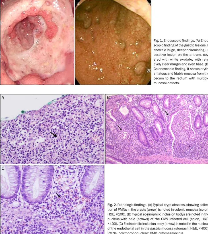
Fig. 1. Endoscopic findings. (A) Endo- scopic finding of the gastric lesions. It shows a huge, deepencirculating ul- cerative lesion on the antrum, cov- ered with white exudate, with rela- tively clear margin and even base. (B) Colonoscopic finding. It shows eryth- ematous and friable mucosa from the cecum to the rectum with multiple mucosal defects.
Fig. 2. Pathologic findings. (A) Typical crypt abscess, showing collec- tion of PMNs in the crypts (arrow) is noted in colonic mucosa (colon, H&E, ×100). (B) Typical eosinophilic inclusion bodys are noted in the nucleus with halo (arrows) of the CMV infected cell (colon, H&E,
×400). (C) Eosinophilic inclusion body (arrow) is noted in the nucleus of the endothelial cell in the gastric mucosa (stomach, H&E, ×400).
PMNs, polymorphonuclear; CMV, cytomegalovirus.
mission with epigastric pain. She had gotten Cesarean sec- tion one month ago, and no other medical history was reported. On physical examination, abdominal tenderness, without rebound tenderness, was observed in the epigastric area. Laboratory results revealed leukocytosis, mild anemia, and hypoalbuminemia: white blood cell 25,500/mm3 (neutrophil
70%, lymphocyte 20%, eosinophil 5%), hemoglobin 11.2 g/dL, hematocrit 33.4%, platelet 481,000/mm3, aspartate transaminase/alanine transaminase 7/12 IU/L, Total pro- tein/albumin 5.3/2.0 mg/dL, total bilirubin 0.2 mg/dL, alka- line phosphatase 102 IU/L. According to an upper endoso- copy, a huge ulcerative lesion invading the entire antrum cir- C
A B
A B
Kang SH, et al. Cytomegalovirus Gastric Ulcer with Pyloric Obstruction in a Patient with Ulcerative Colitis 361
Vol. 69 No. 6, June 2017 Fig. 3. Upper gastrointestinal series finding. It shows no passage of
contrast-material through the pylorus and marked distention of the stomach, suggesting total obstruction at the gastric pylorus.
cumferentially was found, with a positive result of rapid ure- ase test. The margin of ulcer was relatively clear, and the base was covered with exudates. Erythematous and friable muco- sa with partially multiple mucosal defects were discovered across the entire colon, according to a colonoscopy for the evaluation of diarreha (Fig. 1). She was diagnosed endo- scopically as active ulcerative colitis, accompanied by gastric ulcer. We started with steroid pulse therapy (intravenous me- thy-prednisolone 20 mg daily) for ulcerative colitis and proton pump inhibitor for gastric ulcer. In spite of steroid and proton pump inhibitor treatment, her symptoms did not improve. On the 8th day, according to her pathologic report, crypt abscess and inflammatory cell infiltration were found in the colonic mucosa, consistent with ulcerative colitis, and eosinophilic intranuclear inclusion body with acute inflammatory change was also noted, implying CMV infection (Fig. 2). To manage CMW infection, anti-viral agent, such as ganciclovir, was started and steroid treatment was tapered out. After the treatment using antiviral agent, her symptoms were greatly improved; however, on the 10th day of anti-viral therapy (the 44th day from admission), she complained of continuous vomiting. In a follow-up upper endoscopy, a large amount of fluid collections and pyloric stricture with healing ulcer were
seen in the stomach, and as a result, the scope was not able to pass through. In the upper gastrointestinal series on the 45th day since admission, the dye did not pass through the pylorus, suggesting a complete pyloric obstruction (Fig. 3).
Consequently, endoscopic balloon dilatation was attempted, but failed, and surgical gastrojejunostomy was performed on the 47th day post-hospitalization. After the operation, vomiting was relieved. Then, she was discharged and she did not re- lapse during the follow-up period.
DISCUSSION
The pathophysiology of gastrointestinal ulcer by CMV in- fection is believe to be associated with the invasion of the en- dothelium of the vessels in the submucosa and causes vasculitis. Fibrin clots are formed, blocking direct blood flow and causing ischemia, ultimately leading to the occurrence of mucosal ulcers.3 According to the endoscopic findings, er- ythema, erosion, ulcer, bleeding, mucosal hypertrophy, and pseudo-polyps can be shown solitarily or multiply. However, it is known that there is no typical endoscopic finding of CMV infection in the gastrointestinal tract.4 Therefore, if clinically suspected, it is necessary to perform a biopsy in the ulcer base, rather than the margin since CMV mainly invades the endothelium of the submucosal layer, fibroblasts, smooth muscle cells, and glandular cells of the gastrointestinal tract.5
In IBD patients, CMV infections could aggravate the course of IBD. The incidence of CMV infections in IBD patients receiv- ing immunosuppressive therapy has been reported to be be- tween 15.8 and 34%.6 Moreover, it is difficult to endoscopi- cally distinguish CMV infections from IBD exacerbation.
Hence, we should suspect CMV infection in patients with IBD who were refractory to immunosuppressive drugs, such as steroid. Additionally, CMV usually tends to localize around the colon, with a very rare occurrence of dissemination to other organs in patients with IBD.7 In our case, CMV simultaneously invaded the stomach and colon in a UC patient, as confirmed by the pathological report. Therefore, CMV infection can be suspected if there were no other accompanying symptoms except colon-related ones in IBD patients.
During the treatment of CMV infection with anti-viral agents, complications of complete gastric outlet obstruction occurred as a result of gastric ulcer healing process, which
362 강성환 등. 궤양성 위 유문부 폐쇄를 일으킨 거대세포바이러스 감염
The Korean Journal of Gastroenterology
was resolved by a surgical gastrojejunostomy. In general, gas- tric ulcer disease may be the underlying cause in less than 5 to 8 percent of patients with gastric outlet obstruction.8 Mostly gastric ulcer disease is associated with duodenal or pyloric channel ulceration. In our case, gastric ulcer was located on the pyloric channel and antrum, circumferentially and the pa- tient was a high risk group for gastric outlet obstruction.
Therefore, to prevent gastric outlet obstruction, it would be better to place a temporary stent in the pylorus before start- ing any anti-viral and anti-ulcer medications.9 In our case, temporary stent insertion was not possible due to complete obstruction.
In conclusion, it is important to suspect the possibility of CMV infection in IBD patients who were unresponsive to im- munosuppressant drugs with other accompanying symp- toms or signs that might suggest the involvement of other organs.
REFERENCES
1. Weller TH. The cytomegaloviruses: ubiquitous agents with pro- tean clinical manifestations. II. N Eng J Med 1971;285:267-274.
2. Hannant KL, Rotterdam HZ, Bell ET, Tapper ML.Cytomegalovirus infection of the alimentary tract: a clinicopathological correlation.
Am J Gastroenterol 1986;81:944-950.
3. Lee G, Kim NI, Gu JT, Suh JI, Yang CH, Lee CW. A case of cytomegalo- virus colitis in an immunocompetent adult. Korean J Gastroenterol 2000;35:649-653.
4. Chon SY, Lim YJ, Kim MY, et al. A case of cytomegalovirus (CMV) colitis in a patient after splenectomy. Korean J Gastrointest Endosc 2003;26:158-162.
5. Foucar E, Mukai K, Foucar K, Sutherland DE, Van Buren CT. Colon ulceration in lethal cytomegalovirus infection. Am J Clin Pathol 1981;76:788-801.
6. Papadakis KA, Tung JK, Binder SW, et al. Outcome of cytomegalo- virus infections in patients with inflammatory bowel disease. Am J Gastroenterol 2001;96:2137-2142.
7. Ye BD, Kim SG, Kim JS, et al. A case of cytomegalovirus-asso- ciated multiple gastric ulcers in ulcerative colitis. Int J Colorectal Dis 2007;22:1419-1420.
8. Gisbert JP, Pajares JM. Review article: helicobacter pylori in- fection and gastric outlet obstruction-prevalence of the infection and role of antimicrobial treatment. Aliment Pharmacol Ther 2002;16:1203-1208.
9. Dormann AJ, Deppe H, Wigginghaus B. Self-expanding metallic stents for continuous dilataion of benign stenoses in gastro- intestinal tract - first results of long-term follow-up in interim stent application in pyloric and colonic obstructions. Z Gastroenterol 2001;39:957-960.