J o u r n a l o f R h e u m a t i c D i s e a s e s V o l . 2 1 , N o . 1 , F e b r u a r y , 2 0 1 4
http://dx.doi.org/10.4078/jrd.2014.21.1.40 □ Case Report □
40
<Received:May 16, 2013, Revised (1st: May 23, 2013, 2nd: May 25, 2013), Accepted:May 27, 2013>
Corresponding to:Seong Wook Kang, Department of Internal Medicine, Chungnam National University School of Medicine, 282, Munhwa-ro, Jung-gu, Daejeon 301-721, Korea. E-mail:kangsw@cnuh.co.kr
pISSN: 2093-940X, eISSN: 2233-4718
Copyright ⓒ 2014 by The Korean College of Rheumatology
This is a Free Access article, which permits unrestricted non-commerical use, distribution, and reproduction in any medium, provided the original work is properly cited.
Osteonecrosis of the Humeral Head after Cerebral Angiography
In Seol Yoo, Chan-Keol Park, Young Kim, Seung Taek Song, Si Wan Choi, Jin Hyun Kim, Seong Wook Kang
Department of Internal Medicine, Chungnam National University School of Medicine, Daejeon, Korea
A 79-year-old woman was admitted to our hospital for shoulder pain. A physical examination revealed a tender right shoulder with limitation of active, and preservation of passive, motion. She had undergone a cerebral angiog- raphy with coil embolization two months prior to admission. After the procedure, she was presented with pain in the right upper arm and shoulder. Due to persis-
tent shoulder pain, an MRI of the shoulder was per- formed, and osteonecrosis of the humeral head was detected. We present a case of osteonecrosis of the hum- eral head after cerebral angiography.
Key Words. Osteonecrosis, Cerebral angiography, Humeral head
Introduction
Osteonecrosis, also known as avascular necrosis, is defined as the death of bone cells due to decreased blood flow (1).
The humeral head remains the second most common site of osteonecrosis following the femoral head (2). Most osteonec- rosis cases are related to traumatic interruption of the blood supply to the bone, which are most commonly caused by hu- merus fracture and surgical procedures (3,4). Cerebral angiog- raphy is an invasive procedure associated with a small, but definite risk of vessel injury which can block blood flow and cause a stroke (5). However, there have been no reported cas- es of osteonecrosis of a humeral head involving the axillary arteries. Here, we present the case of a 79-year-old woman who developed osteonecrosis of the head of the humerus after cerebral angiography.
Case Report
A 79-year-old Korean woman was admitted to the hospital because of a two-month history of pain in her right shoulder and upper arm. She was diagnosed with type 2 diabetes melli- tus and is being treated with hypoglycemic agents for seven years. Also, she has been taking amlodipine 5 mg, losartan
100 mg, aspirin 100 mg and clopidogrel 75 mg once daily for angina pectoris.
Two months before admission, she had undergone cerebral angiography with coil embolization for an aneurysm involving the right internal carotid artery and left anterior choroidal artery. The puncture site was the right brachial artery at the antecubital fossa. Soon after the procedure, she presented with a swelling in her right antecubital fossa and upper arm pain.
At that time, the swelling of the right antecubital fossa was thought to be iatrogenic hematoma caused by catheterization.
Therefore, she was discharged without further treatment.
However, the intensity of pain had gradually increased. There was no other significant medical history for two months.
At the time of admission, she complained of pain in the right upper arm and shoulder. The initial vital signs may be sum- marized as follows: body temperature 36.6oC, pulse rate 96/minute, respiration rate 20/minute, blood pressure 120/80 mm Hg. On physical examination, the right shoulder and upper arm were tender and swollen. Both active and passive ranges of motion of the shoulder provoked severe pain. There was no neurological deficit. The CBC results may be summarized as follows: WBC 20,450 cells/μL (neutrophil 87.0%, lympho-
Osteonecrosis of the Humeral Head 41
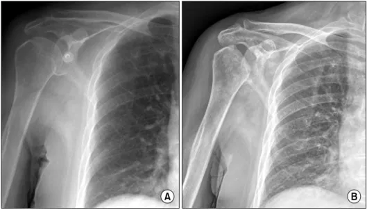
Figure 1. Radiograph of the right shoulder. (A) Radiograph of the right shoulder before cerebral an- giography shows normal humeral head. (B) Radiograph of the right shoulder reveals severe osteone- crosis of the humeral head.
Figure 2. MRI of the right sho- ulder. (A) Sagittal T1 weighted image show extensive low signal abnormalities of the humeral epi- physis, metaphysis and diaphysis that indicate avscular necrosis. (B) Coronal T2 weighted fat saturated image show severe osteonecrotic changes in the humeral head.
cyte 7.0%, monocyte 4.0%), platelet count 325,000 cells/μL, hemoglobin 9.4 g/dL, Hct 28.0%. The erythrocyte sed- imentation rate (ESR) and the C-reactive protein (CRP) level were elevated (ESR 49 mm/hr, CRP 1.3 mg/dL). Laboratory tests for antinuclear antibody and antiphospholipid antibodies were not performed. Radiographs showed necrosis of the hum- eral head which was stage V according to the classification of Cruess (6), while no abnormality was found in the radio- graph from the examination two months prior (Figure 1A, B).
MRI showed severe osteonecrotic changes in the humeral head and increased signal abnormalities and tenosynovitis of the long head tendon of biceps brachii (Figure 2A, B). Surgical
intervention by total joint replacement was planned.
Discussion
The conditions associated with osteonecrosis must com- promise the blood supply of the humeral head in one of four ways: (1) mechanical disruption of blood vessels, (2) injury to or compression of the arterial walls, (3) obstruction of arte- rial inflow such as thrombosis and embolism, and (4) ob- struction of venous outflow (7).
Main arterial supply to the humeral head is from a branch of the anterior humeral circumflex artery, the arcuate artery.
The arcuate artery ascends on the lateral border of the inter-
42 In Seol Yoo et al.
tubercular groove and enters the bone at the level of the great- er tuberosity (7-9).
The subchondral bone of the humeral head is especially vul- nerable to thrombotic and embolic phenomena, because the ar- terioles in this area become sinusoids that turn 180° to return to the intraosseous circulation (7). Also, the lack of collateral blood flow in the humeral head enhances this vulnerability.
In our patient, the cause of the osteonecrosis might be the injury or thrombosis of the arcuate artery. Although, we did not use MRA to confirm this conjecture, there are two reasons for it. First, the progression of osteonecrosis was very rapid.
Radiographic changes were detected within two months from onset of symptoms (Figure 1A and B). Darder et al. showed that it may take up to four years for radiological signs of os- teonecrosis to develop in patients with four-part fractures of the proximal humerus (10). In addition, the osteonecrosis was accompanied by seroma and tenosynovitis of the long head tendon of biceps brachii. In our patient, the catheter was in- serted into the right brachial artery and passed through the ax- illary artery, the origin of the anterior humeral circumflex artery. Thus, thrombosis or emboli of the ascending branch of the anterior humeral circumflex artery is likely responsible for the osteonecrosis in our patient. We found no other predis- posing factors for osteonecrosis (i.e., corticosteroid therapy, alcohol abuse, or connective tissue disorders) in our patient.
Such clinical complications following cerebral angiography are rare (5); nonetheless, arteriography is still associated with significant morbidity and mortality. In addition, the trans axil- lary route showed higher incidence of complication than the trans femoral route (11).
Therefore, it is important to maintain an appropriate degree of vigilance when patients who have undergone arteriography, present signs or symptoms suggestive of osteonecrosis of the humeral head.
Summary
We describe here the case of osteonecrosis of the humeral head complicated by cerebral angiography. Therefore, the
physicians should keep in mind that osteonecrosis can occur after angiographic procedures.
Acknowledgements
We certify that there is no conflict of interest with any finan- cial organization regarding the material discussed in the manuscript.
References
1. Mankin HJ. Nontraumatic necrosis of bone (osteonecro- sis). N Engl J Med 1992;326:1473-9.
2. Sarris I, Weiser R, Sotereanos DG. Pathogenesis and treatment of osteonecrosis of the shoulder. Orthop Clin North Am 2004;35:397-404.
3. Solomon DJ, Navaie M, Stedje-Larsen ET, Smith JC, Provencher MT. Glenohumeral chondrolysis after arthro- scopy: a systematic review of potential contributors and causal pathways. Arthroscopy 2009;25:1329-42.
4. Lee CK, Hansen HR. Post-traumatic avascular necrosis of the humeral head in displaced proximal humeral fractures.
J Trauma 1981;21:788-91.
5. Dawkins AA, Evans AL, Wattam J, Romanowski CA, Connolly DJ, Hodgson TJ, et al. Complications of cere- bral angiography: a prospective analysis of 2,924 consec- utive procedures. Neuroradiology 2007;49:753-9.
6. Cruess RL. Experience with steroid-induced avascular ne- crosis of the shoulder and etiologic considerations regard- ing osteonecrosis of the hip. Clin Orthop Relat Res 1978;(130):86-93.
7. Hasan SS, Romeo AA. Nontraumatic osteonecrosis of the humeral head. J Shoulder Elbow Surg 2002;11:281-98.
8. Laing PG. The arterial supply of the adult humerus. J Bone Joint Surg Am 1956;38-A:1105-16.
9. Gerber C, Schneeberger AG, Vinh TS. The arterial vascu- larization of the humeral head. An anatomical study. J Bone Joint Surg Am 1990;72:1486-94.
10. Darder A, Darder A Jr, Sanchis V, Gastaldi E, Gomar F.
Four-part displaced proximal humeral fractures: operative treatment using Kirschner wires and a tension band. J Orthop Trauma 1993;7:497-505.
11. AbuRahma AF, Robinson PA, Boland JP, Umstot RK, Clubb EA, Grandia RA, et al. Complications of arteriog- raphy in a recent series of 707 cases: factors affecting outcome. Ann Vasc Surg 1993;7:122-9.