중추신경계 생식세포종양의 최근 치료 경향
한정우
1,2
ㆍ고경남3
ㆍ김지윤4
ㆍ백희조5
ㆍ이지원6
ㆍ심규원7
ㆍ조재호8
ㆍ김동석7
1
연세대학교 의과대학 소아과학교실,
2연세암병원 소아청소년암센터 소아혈액종양과,
3울산대학교 의과대학 소아과학교실,
4
경북대학교 의과대학 경북대학교병원 소아과학교실,
5전남대학교 의과대학 소아과학교실,
6
성균관대학교 의과대학 소아과학교실,
7연세대학교 의과대학 신경외과학교실,
8연세대학교 의과대학 방사선종양학교실
Current Trends in Management for Central Nervous System Germ Cell Tumor
Jung Woo Han, M.D.
1,2, Kyung-Nam Koh, M.D.
3, Ji Yoon Kim, M.D.
4, Hee Jo Baek, M.D.
5, Ji Won Lee, M.D.
6, Kyu-Won Shim, M.D.
7, Jaeho Cho, M.D.
8and Dong-Seok Kim, M.D., Ph.D.
71
Division of Pediatric Hemato-Oncology, Department of Pediatrics, Yonsei University College of Medicine, Yonsei University Health System,
2
Department of Pediatric Hemato-Oncology, Yonsei Cancer Center, Yonsei University Health System,
3Division of Pediatric Hematology/Oncology, Department of Pediatrics, University of Ulsan College of Medicine & Asan Medical Center, Seoul,
4Department of Pediatrics, Kyungpook National University Hospital, Kyungpook National University School of Medicine, Daegu,
5Department of Pediatrics, Chonnam National University Medical School, Chonnam National University Hwasun Hospital, Gwangju,
6Department of Pediatrics, Samsung Medical Center, Sungkyunkwan University School of Medicine,
7Department of Pediatric Neurosurgery, Severance Hospital, Yonsei University
Health System,
8Department of Radiation Oncology, Yonsei Cancer Center, Yonsei University Health System, Seoul, Korea
Central nervous system germ cell tumor is a rare but important tumor in childhood brain tumors. It requires a multidisciplinary approach to increase survival and promote quality of life, and all three treatment modalities including surgery, radiotherapy and chemo- therapy has its own distinct role for germ cell tumor. For germinoma, radiotherapy alone can cure the disease but, the effort to limit the long term toxicity and the proper combi- nation of chemotherapy and radiotherapy are under investigation. Craniospinal irradi- ation is reserved only for the disseminated germinoma or nongerminomatous germ cell tumor (NGGCT). For germinoma, craniospinal irradiation of 20 to 24 Gy is sufficient to control microscopic disease in the spinal axis. Chemotherapy and radiotherapy com- posed of 30 to 40 Gy of local field radiotherapy and 20 to 24 Gy of whole ventricular irradiation are required for localized germinoma, but the proper combination of two modalities has yet to be defined. For NGGCT, both the chemotherapy and radiotherapy should be performed, and survival rate is substantially increasing with modern treatment protocols. The omission of craniospinal irradiation is being tried for the localized NGGCT in international cooperative group trials. Surgery has its role for the resection of residual disease after the treatment, and the extent of resection in NGGCT has the prognostic implication. Bifocal germ cell tumors and basal ganglia germ cell tumor have distinctive clinical course and mandate special attention. To advance clinical and biological per- spectives in central nervous germ cell tumor, the cooperation and communication of the multidisciplinary specialists are essential.
pISSN 2233-5250 / eISSN 2233-4580 http://dx.doi.org/10.15264/cpho.2016.23.1.17 Clin Pediatr Hematol Oncol 2016;23:17∼27
Received on March 30, 2016 Revised on April 11, 2016 Accepted on April 21, 2016
Corresponding Author: Dong-Seok Kim Department of Pediatric, Neurosurgery in Severance Children’s Hospital, Brain Korea 21 Project for Medical Science, Yonsei University College of Medicine, Yonsei University Health System, CPO Box 8044, 50-1 Yonsei-ro, Seodaemun-gu, Seoul 03722, Korea Tel: +82-2-2228-2160
Fax: +82-2-393-9979 E-mail: dskim33@yuhs.ac
Key Words: Neoplasms, Germ cell and embryonal, Brain neoplasms, Germinoma,
Neoplasms
Table 1. Classification of germ cell tumors
Germ cell tumors Morphology code
Germinomas 9064/3
Nongermionmatous germ cell tumors
Embryonal carcinoma 9070/3
Yolk sac tumor 9071/3
Choriocarcinoma 9100/3
Teratoma 9080/1
Immature teratoma 9080/3
Mature teratoma 9084/0
Teratoma with malignant transformation 9084/3
Mixed germ cell tumors 9085/3
서 론
중추신경계 생식세포종양은 전체 뇌종양 중 약 1-2%를 차 지하는 종양이다. 여타 중추신경계종양과 달리, 생식세포종양 에는 수술, 방사선, 항암화학요법의 다양한 역할이 공존하고, 이들의 적절한 조합으로 생존율의 향상과 삶의 질 제고에 기 여할 수 있다. 이에, 각 치료 방침의 이해와, 다학제간 협력이 무엇보다 중요한 종양이라고 할 수 있다[1,2]. 본 종설에서는 중추신경계 생식세포종양의 임상 양상과 다양한 치료 방법의 역할에 대해 고찰하고자 한다.
본 론
1) 중추신경계 생식세포종양(germ cell tumor)의 기원 중추신경계 생식세포종양(germ cell tumor, GCT)는 드문 신경계 종양으로, 원인에 대해 두 가지 학설이 있다. 첫째는 원시생식세포(primordial germ cell)가 잘못 이주(migration) 하여 생식기능선(genital ridge) 대신 배아의 중추신경계(em- bryonic central nervous system)에 정착하였다는 것이고, 둘 째는 다능성줄기세포(pluripotent stem cell)가 중추신경계 내 에 존재하다가 생식세포종으로 변화한다는 것이다[3].
2) 역학
빈도는 전통적으로 서양에서 0.4-3.4%, 일본과 대만에서 2.1-9.4%를 차지하는 것으로 알려져 있었다[4]. 미국의 등록사 업보고에 따르면 0-19세 신경종양 중 3.9%를 차지하고 10만 명당 0.1명이 발생한다. 남녀의 성비는 2.1:1로 남성에서 더 흔한 것으로 알려져 있다. 발생 중앙 연령은 16.0세이다. 또한 백인에서 1.5배 정도로 흑인보다 많다. 0-19세에 0.22명으로 가장 높고, 20-34세에 0.10명, 이후 연령대에서는 0.02-0.05명 으로 매우 드물게 발생한다. 매년 미국에서는 약 290명이 발 생하는 것으로 알려져 있다[5]. 일본에서는 구마모토(Kuma- moto)현에서 1989-1998년까지 10만명당 0.20명이 발생하고, 남:녀 2.58이라고 하여 서양보다 높다고 알려졌다. 특히 15세 미만에서 0.55명으로 높고, 10-19세에 빈발하였다. 조직 확인 비율은 65.8%였다[6]. 같은 지역에서 1989-2008년까지 최근 보고에 의하면 발생률은 0.22, 성비는 3.36이었고 전체 중추 신경계 종양 중 1.2%를 차지하였다. 특히 2005-2008년에는 발생률 0.10, 성비 1.62로 보고되어 서양과 차이가 없었다[7].
미국과 일본의 등록사업을 통합한 연구에서 일본에서는 송 과샘(pineal gland)에 40.2%, 안장위(suprasellar)에 41.8%가
발생하였고 미국에서는 송과샘 46.4%, 안장위 65.6%가 발생 하였다[8]. 여성에서는 75%가 안장위에 나타나고, 남성에서는 67%가 송과샘에 발생된다고 하는데, 일본 남성의 57.7%와 미 국 남성의 86.6%에서 송과샘에, 일본 여성의 82.3%와 미국 여성의 66.1%에서 안장위에 위치 하였다[4,8]. 송과샘 종양의 남녀비는 11.7-14.4였고, 안장위 종양의 남녀비는 1.8-1.1이었 다. 미국와 일본에서 전체 종양 중 77.5-82.0%가 뇌종자세포 종(뇌종자종, intracranial germinoma)였다. 전 연령대에서 비 슷한 뇌종자종 분포를 보였으나 0-9세에서는 42.9-51.1%만이 뇌종자종이었다. 뇌종자종과 전체 생식세포종양의 절대발생 수는 미국과 일본 모두 10-14세에 가장 많았다[8].
3) 분류
WHO (World Health Organization)분류에서 생식세포종양 은 뇌종자종과 비종자종성 생식세포종양(non germinomatous germ cell tumor, NGGCT)으로 분류된다(Table 1) [9].
뇌종자종은 일반적으로 종양표지자를 표현하지 않지만, 융
합영양세포(syncytiotrophoblast)가 포함되어 있는 경우 베타-
인간융모막성선자극호르몬(beta-human chorionic gonado-
trophin (hCG, hCG))가 상승할 수 있다. 상승범위에 대해서
는 이견이 있으나 50 mIU/mL까지는 뇌종자종의 범주로 간주
하는 것이 추세이고, 연구자에 따라서는 50-100 mIU/mL까지
도 뇌종자종으로 간주할 때도 역시 치료 성적이 유사하다고
하고 있다[10]. 미국에서도 100 mIU/mL까지는 뇌종자종으로
간주하여 치료하여도 예후에 차이가 없었으나 그 이상인 경우
는 주의가 필요하다고 의견이 있다[11]. 일부에서는 200 mIU/mL
또는 그 이상인 경우에도 예후에 차이가 없다고 주장하기도
한다[12]. 이를 종합하여 유럽에서는 hCG>50 mIU/mL 그리
고/또는 AFP (Alpha fetoprotein)>25 ng/mL 또는 기관의 참
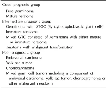
Table 2. Japanese classification of germ cell tumors
Good prognosis groupPure germinoma Mature teratoma
Intermediate prognosis group
Germinoma with STGC (Syncytiotrophoblastic giant cells) Immature teratoma
Mixed GTC consisted of germinoma with either mature or immature teratoma
Teratoma with malignant transformation Poor prognostic group
Embryonal carcinoma Yolk sac tumor Choriocarcinoma
Mixed germ cell tumors including a component of embyonal carcinoma, yolk sac tumor, choriocarcinoma or other malignant neoplasm
고치 이상을 분비성 생식세포종양(secreting germ cell tumor) 로 정의하고 있고[13,14], 미국에서는 조직학적 진단이 없는 경우 AFP≤10 ng/mL 또는 기관의 참고치 이하, hCG≤50 mIU/mL를 뇌종자종으로, 조직학적 진단이 있는 경우 hCG≤
100 mIU/mL까지로 정의하고 있다[15].
난황낭종양(york sac tumor, YST)이나 융모막암종(chorio- carcinoma)은 각각 AFP와 hCG를 특이적으로 분비하며, 예후 가 나쁘다[2]. 일본에서는 생식세포종양을 불량예후군(Poor prognosis group), 중간예후군(intermediate), 양호예후군(good) 으로 분류하고 있다(Table 2). 마츠타니 등은 도쿄대학교에서 153명의 조직학적으로 확인된 생식세포종양을 분석하였다 [16]. 비종자종성 생식세포종양에 대해 Japanese Intracranial Germ Cell Tumor Study Group의 항암화학요법과 방사선치 료를 하였고, 뇌종자종 50예에 대해 43명은 방사선치료를, 7 명은 항암치료와 방사선치료를 하였다[16]. 뇌종자종과 성숙 기형종은 10년 92.7-92.9%의 생존율을 보였으나, 순수악성생 식세포종양(pure malignant germ cell tumor (N=11); 융모망 암종, 배아암종 또는 난환낭종양) 또는 조직에 순수생식세포 종양이 포함된 혼합생식세포종양(mixed germ cell tumor, N=12)에서는 5년 생존율이 9.3-27.3%에 불과 하였다. 이를 바탕으로 3가지 단계의 분류가 제안 되었다[16]. 중간예후군 의 예후는 시스플라틴 기반의 항암화학요법과 방사선 치료로 5년 생존율 92-97% 정도를 보이며, 이는 방사선 치료 단독 군의 62%에 비해 높다[17]. 불량예후군의 예후는 과거 10-20%
에 머물렀으나 현재는 최소 60%를 상회 한다[16,18].
4) 임상증상
임상증상은 환자의 연령, 종양의 위치, 종류, 분비물질, 크 기에 따라 다양하게 나타난다[19]. 송과샘 위치 종괴는 폐쇄성 수두증을 발생하여 뇌압이 상승하는 경우가 많아, 뇌실복강단 락술(ventriculoperitoneal shunt)를 시행하거나 제3뇌실창냄 술(ventriculostomy)을 시행할 수 있으며, 이 중 현실적으로 가능하다면 제3뇌실창냄술이 추천된다[20]. 또한 파리노증후 군(Parinaud’s syndrome, dorsal midbrain syndrome; 상향주 시력 마비, pseudo-argyll robertson 동공, 눈꺼풀 뒤당김, 일 몰 징후(setting sun sign), 폭주후퇴 안진 등의 증상복합증후 군), 사시, 복시를 포함한 안구 증상 등이 나타날 수 있다. 내 분비계 이상이나 이차성징 발달 이상은 안장위 종양에 비해 적다[21].
안장위 종양에서는 내분비 이상 특히 중추성요붕증이 흔히 종양의 첫 증상으로 발생한다[22]. hCG 상승으로 LH (luteinizing hormone), FSH (follicular stimulating hormone) 상승과 성조 숙증이 발생한다[2,22]. 그 외에 뇌하수체기능저하증, 성선기 능저하, 사춘기 성숙지연, 갑상선기능저하, 성장 장애 등 내분 비 증상이 나타난다[3,19].
5) 진단
정확한 진단은 치료의 결정에 반드시 필요하다[2]. 임상 증 상, 종양표지자 및 영상검사, 뇌척수액검사 등으로 진단 및 병기를 결정한다. 가능한 한 생검으로 조직학적 확진이 필요 하다. 종양표지자로는 AFP, hCG와 PLAP (placental alkaline phosphatase)이 있으며, 혈액 및 뇌척수액에서 시행한다. 종 양 때문만 아니라 정상에서도, 혈청보다 뇌척수액에서 종양표 지자가 더 높게 측정되는 경향이 있어 주의를 요한다[3,20,23].
종양표지자는 진단 시뿐만 아니라 치료 반응을 관찰할 수 있 으므로 정기적으로 추적되어야 하며, 진단 시 음성이라 하여 도 추적 검사를 주기적으로 행해야 한다[20]. 뇌척수액 세포학 적 검사는 양성 결과를 치료 결정에 반영하는 국가나 기관에 서는 반드시 시행하는 것을 추천하며, 뇌실보다는 요추 천자 로 시행하여야 한다[20]. 영상 검사에는 뇌 및 척수 자기공명 영상이 추천된다[20]. 기저체(basal ganglia)에 발생한 종양은 초기에 조영증강이 잘 되지 않고 구분이 어려운 경우가 많으 므로 진단과 추적에 주의를 요한다[24].
병기는 생식세포종양에 특화된 체계는 없으며, 수모세포종
(medulloblastoma)의 병기 체계를 따라 M0-M4까지 분류하여
적용한다[2,25]. 뇌척수전이는 뇌종자종에서 약 7-12%에서 나
타나며 난황낭종양에서는 23%까지 보고된다[4,26]. 드물지만
3%에서 폐, 뼈 등으로 전신 전이를 하는 경우가 있고, 뇌실복 강단락술을 받은 경우 10%까지 복강 내 전이가 발생하기도 하며, 매우 드물지만 동시에 고환 또는 난소에 종괴가 있는 경우도 배제할 수 없으므로, 복부, 가슴, 뼈 및 고환 부위의 영상 검사도 필요 시 고려하여 시행하기도 한다[3,4].
6) 생식세포종양의 치료-뇌종자종(germinoma)
(1) 방사선치료 단독연구-두개척추방사선조사(craniospinal ir- radiation, CSRT)
두개척추방사선조사(craniospinal irradiation, CSRT)는 뇌 종자종 치료에 충분한 효과를 갖는다. MAKEI (the maligue keimzelltumoren) 83/86 연구(N=60)에서는, 두개척추방사선 조사 36 Gy 및 원발부위추가치료(primary tumor boost, PTB) 14 Gy를, MAKEI 89연구에서 두개척추방사선조사 30 Gy 및 원발부위추가치료 15 Gy를 조사하여 5년 무재발생존율(Re- lapse Free Survival, RFS)은 91%±3.9%, 5년 전체생존율 (overall survival, OS)는 93.7±3.6%으로, 두개척추방사선조 사 30-36 Gy, 국소부위 방사선치료(local field radiotherapy, LFRT) 45-50 Gy는 뇌종자종을 완치시킬 수 있었다[27].
두개척추방사선조사로 완치가 가능함이 알려지면서 용량 을 낮추는 시도가 이루어 졌다. Shibamato 등은 50 Gy 이상 을 시작으로 점차 감량하여 뇌척수전이 예방 용량으로 두개척 추방사선조사 20-24 Gy 정도가 충분한 것으로 생각되었다[28].
Children’s Hospital of Philiadelphia (CHOP, Pennsylvania) 에서 최소 30.6 Gy 이상의 두개척추방사선조사를 조사를 시 작으로 감량하여 23.4-27 Gy (89년까지) 및 그 이후 19-19.8 Gy까지 조사량을 낮추었으며 전이가 없는 환자에서는 18-19.8 Gy의 두개척추방사선조사를 받아도 높은 생존율을 보였다 [29]. 허 등은 국소부위방사선치료 54 (40-56.1) Gy, 전뇌방사 선치료(whole brain radiotherapy, WBRT) 36 (19.8-44) Gy, 척추축(spinal axis) 24 (13.1-36) Gy를 투여 하여 5년 및 10년 전체생존율 96.9%였다[30]. 따라서 뇌종자종에서 필요한 두개 척추방사선조사는 최소 20-24 Gy 정도로 생각된다. Cho 등은 최근에 이를수록 원발부위 용량 59에서 39.3 Gy, 두개척추방 사선조사 34.2에서 19.2 Gy로 낮추어 치료하였으며, 두개척 추방사선조사를 진행한 60예 중 51예(85%)가 두개척추방사선 조사 25 Gy 미만을, 22예(36.7%)가 20 Gy 미만(중앙값 19.5 Gy)를 받아, 두개척추방사선조사 19.5 Gy 정도로 완전한 조 절을 이룰 것으로 기대 되었다[31]. 따라서 뇌종자종에서 전이 가 있다 하더라도 일반적으로 두개척추방사선조사 20-24 Gy 가 충분하다고 보여진다[32].
(2) 방사선치료 단독연구-제한된 영역의 방사선조사 원발부위 40-50 Gy의 방사선치료는 재발을 일으키지 않으며 두개척추방사선조사는 뇌종자종 치료에 명확한 효과를 갖는다 [28,33]. 그러나 치료 효과를 얻으며 장기후유증을 예방하기 위 해, 방사선치료의 용량과 범위에 많은 연구가 진행되었다.
파종성 전이 여부는 예후에 관계가 있으나 두개척추방사선 조사 여부는 예후에 관계가 없어, 완전한 뇌척수 전이 평가가 이루어진다면, 비전이성 뇌종자종에서 두개척추방사선조사는 필요 없다[34]. 비전이성 뇌종자종에서 두개척추방사선치료 대신 국소부위방사선치료하는 경우 질환을 조절하는데 일정 부분 효과가 있다[35]. 그러나, 국소부위방사선치료만으로는 비전이성 뇌종자종 치료시 척수나 두개내, 뇌실 부위 재발이 많았으므로 최소한 뇌실을 방사선치료에 포함할 필요성이 있 다[36]. 특히 40 Gy미만의 조사를 한 경우 국소부위방사선치 료는 두개 내 재발 확률이 높아 불충분하였다[37]. 40 Gy 정도 의 치료는 조사부위 내부에서는 재발을 방지하였지만 뇌실이 포함되지 않은 국소부위방사선치료는 재발 위험성이 있으며, 범위를 넓힌 전뇌방사선치료는 뇌종자종 치료에 충분한 조절 효과를 발휘하였다[38].
결과적으로 전뇌실방사선치료(whole ventricle irradiation, WVI) 이상의 치료가 뇌종자종 치료에 필요하다[39]. 전뇌실방 사선치료 및 원발부위추가치료 포함 원발 부위 40 Gy 이상 정확한 CT 시뮬레이션으로 투여한 환자에서는 재발이 없었다 [40]. 전뇌실방사선치료를 점차 감소시켜 20-40 Gy 수준까지 낮추고, 원발부위추가치료도 총 30-50 Gy까지 낮추어 시행하 여도 재발 없이 잘 치료 되었다[41,42]. 또한 항암화학요법의 반응성에 따라, 반응이 좋은 환자에서는 방사선치료의 범위가 축소될 수 있는 가능성도 제시되었다[43]. 현재 전뇌실방사선 치료 20-24 Gy, 원발부위 최소 30-40 Gy 정도가 뇌종자종 치 료에 충분한 용량으로 생각된다.
(3) 항암화학요법 및 방사선치료
항암치료의 병합은 방사선치료의 용량과 범위를 낮출 것으
로 예상되어 연구되어 왔다[20]. Allen 등은 전보조(neoadju-
vant) 항암화학요법으로 방사선치료의 용량을 원발부위 55
Gy에서 33.1 (30-45) Gy, 두개척추방사선조사를 36 Gy에서
26.2 (20.0-30.0) Gy로 낮추었다고 보고하였다[44]. POG 9530
에서는 4회의 항암화학요법 후 국한성 뇌종자종인 경우 국소
부위방사선치료 30.6-50.4 Gy 시행하고 91.7% (11/12)가 중
앙추적기간 66개월에 무진행생존하여 항암화학요법 병합 시
국소부위방사선치료만으로 관해를 유지할 수 있음을 보여주
었다[45].
Table 3. Pre-radiotherapy chemotherapy regimen for KSPNO clinical trial (A/B/A/B, total 4 courses every 3 weeks, alternating)
Germinoma NGGCT
Course A Carboplatin 450 mg/m2 D1 450 mg/m2 D1-2
Etoposide 150 mg/m2 D1-3 150 mg/m2 D1-3
Bleomycin - 15 mg/m2 D3
Course B Cyclophosphamide 1,000 mg/m2 D1-2 2,000 mg/m2 D1-2
Etoposide 150 mg/m2 D1-3 150 mg/m2 D1-3
Bleomycin - 15 mg/m2 D3
NGGCT, nongerminomatous germ cell tumor.
하지만 국소부위방사선치료는 항암치료 병합에도 불충분 하다는 의견이 많다. SFOP에서 항암화학요법을 병합하였으 나 비전이성 뇌종자종에 대해 국소부위방사선치료 40 Gy를 조사하고, 두개 내 재발이 다수 발생하여 국소부위방사선치료 만으로는 질병 조절에 부족하였다[46]. SIOP CNS 96 뇌종자종 연구에서는 항암화학요법 후 비전이성 뇌종자종에 국소부위 방사선치료 40 Gy를 사용하여, 뇌실막하 재발을 경험하였다 [13]. 반면 항암화학요법 병용시에도 국소부위방사선치료보다 는 뇌실 이상에 방사선치료한 경우 재발이 없었다. Aoyama 등 은 그들의 경험에서 최종적으로 뇌종자종에서 WV 24 Gy+
원발부위추가치료 6-16 Gy로 치료 하는 것을 권고하였다[47].
Children’s Hospital LA (CHLA)에서도 역시 항암화학요법에 전뇌실방사선치료 21.6-25.5 Gy 및 원발부위추가치료 추가, 총 30.0-30.6 Gy가 사용되어 투여하여 3년 무사건생존율 89.5%, 전체생존율 100%로 질환이 조절됨을 보고하였다[11].
비전이성 뇌종자종의 치료 시 국소부위방사선치료는 뇌실막 하 국소 재발뿐만 아니라 척수재발 역시 높으며 대신, 전뇌실 방사선치료는 척수재발률에서 두개척추방사선치료와 비슷하 였다[48]. 따라서 항암화학요법 병합시에도 전뇌실방사선치료 이상이 추천된다.
(4) 전이성 뇌종자종
전이성 뇌종자종에는 두개척추방사선치료가 필요하며 잘 조절된다[31,45,49]. 항암화학요법을 병합할 때 두개척추방사 선치료를 줄일수 있는지에 대해서는 아직 명확하지 않지만, 대부분의 기관에서 항암화학요법을 병용한다[45,49]. 최근에 는 선량을 낮추어 19.5-25 Gy 미만의 저용량 두개척추방사선 조사로도 질환이 잘 조절됨이 알려져 있다[31,50].
(5) 항암화학요법 단독 치료
생식세포종양은 일반적으로 항암화학요법 반응성이 좋으 며, 따라서 중추신경계 생식세포종양에서도 항암화학요법 단
독 치료법이 연구되었다. 항암화학요법 단독으로는 질환을 조 절하기 어렵다[51]. 첫번째 국제임상시험으로서 진행된 연구 에서, 항암화학요법은 높은 완전관해율(77.4%)을 얻었지만, 2 년 전체생존율은 뇌종자종 84%, 비종자종성 생식세포종양 62%으로 기대보다 낮았다. 하지만, 54명의 생존 환자 중 22명 (40.7%)은 방사선치료 없이 생존하였으므로 항암치료 단독으 로 생존할 수 있는 환자들도 소수이지만 있었다는 것이 알려 졌다[52]. 3번째 국제 CNS GCT 연구에서는 항암화학요법 만 으로 11/25 (44%)에서 재발하였고, 7/11은 뇌종자종, 4/14명 은 비종자종성 생식세포종양이었으며 이와 같이 항암화학요 법 단독으로는 치료가 불가능하나, 완전관해율은 높았고 소수 의 환자들은 항암화학요법 단독으로 충분한 기간 생존하였다 [53].
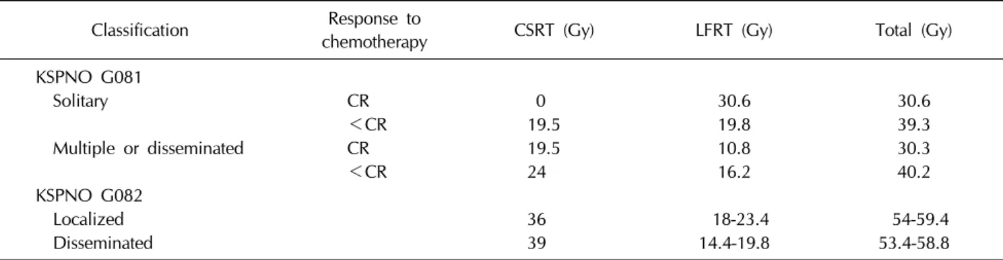
(6) KSPNO 뇌종자종 임상시험
대한소아뇌종양학회에서는 G051/081 임상 시험을 진행하
였다(Table 3, 4). KSPNO G051임상 시험에서 방사선단독치
료군과 항암화학요법/방사선치료 병용군으로 나뉘어 진행되
었으나, 2008년 이후 G081/G082로 통합되어 방사선단독치료
군은 제외되었다. G051 방사선치료 단독군 30명과 G081 치
료 결과 122명이 등록 되었다. 조직학적 진단은 115명(94.2%)
에서 이루어 졌고, 임상적으로 5예가 진단되었다. 항암화학요
법 후 75%에서 완전관해 또는 VGPR (very good partial re-
sponse)이 관찰되었고 1명의 질환 관련 사망이 있었다. 완전
관해환자 중 5명이 최종 재발하였고 PD는 1명에서 관찰되었
고, 알 수 없는 이유로 뇌출혈이 발생한 1명, 총 7명에서 질환
관련사건(event)이 발생하였다. 재발한 환자 1명과, 뇌출혈로
1명이 사망 하였으며, 항암화학요법 중에도 PD를 보였던 1명
이 최종적으로 사망하여 전체 사망 환자는 3명이었다. 5년 무
사건생존율 93%, 전체생존율 97%였다.
Table 4. Radiotherapy plan for KSPNO G081 and G082
Classification Response tochemotherapy CSRT (Gy) LFRT (Gy) Total (Gy)
KSPNO G081
Solitary CR 0 30.6 30.6
<CR 19.5 19.8 39.3
Multiple or disseminated CR 19.5 10.8 30.3
<CR 24 16.2 40.2
KSPNO G082
Localized 36 18-23.4 54-59.4
Disseminated 39 14.4-19.8 53.4-58.8
CR, complete remission; CSRT, craniospinal irradiation; LFRT, local field irradiation.
7) 비종자종성 생식세포종양(nongerminomatoud germ cell tumor, NGGCT)
(1) 비종자종성 생식세포종양의 치료
전통적으로 비종자종성 생식세포종양의 예후는 매우 좋지 않아 전통적으로 마츠타니 분류의 불량예후군은 5년 생존율 9.3-27.3%에 불과하였다[16]. 또한 1990년대 이전의 생식세포 종양 5년 생존율은 뇌종자종 약 45-64%, 비종자종성 생식세포 종양 25.7%였다[4,54].
현재 비종자종성 생식세포종양의 치료에는 항암화학요법 과 두개척추방사선조사가 모두 필요하다[20,47,55-57]. 항암화 학요법과 병용하여 방사선치료를 하는 경우에도, 두개척추방 사선조사를 하지 않으면 척수 재발이 흔하였다[58]. 두개척추 방사선조사를 한 환자들의 재발율은 낮았다[18,59]. MAKEI 89 분석에서는 cisplatin 400 mg/m
2이상 사용 되었을 때 생존 율이 높았고(18/22 vs. 2/8), 두개척추방사선조사 30 Gy, 원발 부위추가치료 15-20 Gy를 받은 환자에서 가장 생존율이 높았 다[60]. 항암화학요법으로는 cisplatin, carboplatin, etoposide, ifoafamide, bleomycin, vinblastine 등이 사용된다[20,45,47,57].
POG 9530 고위험군치료에서 항암화학요법(etoposide, cis- platin, vincristine, cyclophosphamide)과 방사선요법으로 78.6%
(11/14)가 58개월에 무진행생존하여 생존율 향상을 보여 주었 다[45]. COG-ACNS0122 연구에서는 비종자종성 생식세포종 양에 대해 CE (carboplatin 600 mg/m
2D1, etoposide 90 mg/m
2/d D1-3) 또는 IE (ifosfamide 1,800 mg/m
2D1-5, eto- poside 90 mg/m
2D1-5)를 총 6회 치료하고 완전관해인 경우 방사선치료, 완전관해 이하인 경우 잔존종양 수술을 진행하여 완전관해 또는 부분관해인 경우 방사선치료, SD 또는 부분관 해이면서 종양표지자 양성인 때 고용량항암화학요법 및 조혈 모세포 이식을 진행한 후 방사선를 하였다. 두개척추방사선조
사 36 Gy, 국소부위방사선치료 54 Gy를 투여 하였고 척수의 육안적 전이 종괴에 대해 45 Gy의 부스트를 하였다. 5년 무사 건생존율 84%, 전체생존율 93%를 나타내었다. 항암화학요법 후 잔존종양 수술은 15명에서 시행되었고, 단 2명(13%)만 비 종자종성 생식세포종양의 잔존세포가 있었다. 치료 중 진행한 5명 중 4명은 기형종이었다. 치료 중 단 2명만 이식의 대상이 되었고 두 명 다 생존하였다. 비종자종성 생식세포종양의 종 류에 따라 생존율 차이가 없었으며 항암화학요법 후 완전관해 또는 부분관해를 얻은 환자의 생존율이 3년 무사건생존율 92%, 전체생존율 98%에 이르렀다[61]. 대부분의 재발은 18개 월 이내에 일어나지만 5년이후에 발생되는 경우도 있어 장기 간의 추적을 요한다[4,16,55].
종합하면 비종자종성 생식세포종양의 방사선치료는 원발 부위 총 50 Gy, 두개척추방사선조사는 30-36 Gy 정도로 적용 된다. 또한 비종자종성 생식세포종양의 치료에는 수술, 방사 선치료, 항암화학요법 등이 모두 쓰인다. 최근 진행되는 임상 시험인 COG-ACNS 1123에서는 국한성 비종자종성 생식세포 종양에서 SIOP CNS 96의 예비 결과와 첫번째 국제임상시험 에서 완전관해를 이룬 15/21명이 살아 있고, 두번째 국제임상 시험에서 완전관해를 이룬 환자가 부분관해보다 오래 생존하 며, 완전관해를 이루지 못한 5명 모두 사망한 결과 등을 근거 로, 항암화학요법에 반응이 있는 환자에 대해서 두개척추방사 선조사 대신 침범부위 방사선치료(involved filed RT)를 시행 하고 있다[15].
(2) KSPNO 비종자종성 생식세포종양 임상시험
대한소아뇌종양학회에서는 G052/082 임상시험을 진행하
였다(Table 3, 4). 조직검사에서 비종자종성 생식세포종양으
로 확진되거나 혈청, 뇌척수액 종양표지자 검사에서 hCG가
50 mIU/mL 이상, AFP이 10 ng/dL 이상인 경우 비종자종성
생식세포종양으로 진단하였다. 조직학적으로 뇌종자종이었으 나 종양표지자 양성이었던 22예와, 임상적 또는 조직학적으로 확진된 비종자종성 생식세포종양 46예를 포함하여 총 99예가 등록되었다. 사망은 8예였고, 질환관련 6예, 독성 사망 2예 있었다. 항암화학요법에 의해 49.5%가 완전관해 또는 매우양 호한 부분관해(very good partial remission)을 보였고, PD는 11예(11.1%)에서 있었다. 5년 무사건생존율은 74%, 전체생존 율 89%였다.
8) 최근의 국제 임상시험 동향
COG-ACNS1123은 국소성 중추신경계 생식세포종양의 치 료를 연구하고 있다. AFP>10 ng/mL와 함께 조직검사로 확인 된 경우 hCG 100 mIU/mL까지를, 조직검사로 확인하지 않는 경우 hCG≤50 mIU/mL를 뇌종자종으로 한다. 항암화학요법 후 반응에 따라 방사선치료 또는 이차 수술을 계획한다. 항암 화학요법에 완전관해인 경우 전뇌실방사선치료를 18 Gy까지 낮추어 치료한다. 국한성 비종자종성 생식세포종양인 경우 두 개척추방사선조사를 제외하고 전뇌실방사선치료 30.6 Gy, 국 소부위방사선치료 23.4 Gy를 사용하였다. 전이성 비종자종성 생식세포종양인 경우 COG-ACNS0122 프로토콜에 따른다.
SIOP CNS GCT II 연구에서는 그동안 잘 알려지지 않았던 기형종(teratoma)에 대한 치료 방침을 설정하고자 노력하고 있다. 또한 AFP가 1,000 ng/mL으로 극히 높은 군을 고위험군 으로 지정하고 조혈모세포 구제술을 응용한 중등용량 항암화 학요법을 사용한다. 특히 뇌종자종에서도 항암화학요법 후 기 형종이 남아 있는 경우, 완전 절제가 되지 않으면 재발 위험이 있다는 SIOP CNS 96연구의 하위분석 결과에 따라, 기형종이 있는 경우 절제가 완전하지 않다면 54.4 Gy까지 국소 방사선 용량을 높일 것을 주장하고 있다. 비전이성 뇌종자종에는 전 뇌실방사선치료 24.0 Gy를 투여하고, 항암요법에 완전관해미 만인 경우 국소부위방사선치료 14.0 Gy를 추가한다. 비전이 성 비종자종성 생식세포종양에는 두개척추방사선조사없이 54 Gy의 국소부위방사선치료만을 사용하며, 전이성 비종자종성 생식세포종양에 두개척추방사선조사 30 Gy, 국소부위방사선 치료 24 Gy를 투여한다.
9) 기타 관련된 이슈들
(1) 중추신경계 생식세포종양에서의 수술의 역할
종양표지자가 정상인 경우, 조직검사 없이 치료 하여 높은 성적을 보고하기도 한다[62]. 그러나 대개, 정상일 경우라도, 영상소견에 상관 없이 진단을 위한 수술적 조직검사가 행해져 야 한다[20]. 근치적절제술(radical surgery)은 뇌종자종에서
무재발생존율이나 전체생존율에 영향을 주지 않으며 조직검 사 단독으로도 최종진단은 대개 정확하다[63]. 이와 같이 영상 검사가 발달하고, 정위 조직검사의 기술이 좋아져 진단에 어 려움이 없으며, 특히 시상하부, 뇌하수체 등에 종괴가 위치할 때는 합병증을 초래할 위험이 많으므로, 뇌종자종의 근치적절 제는 피하는 것이 좋다. 또한 SIOP CNS GCT 96 연구를 보면 잔존 종양이 남은 67명의 환자들은 다른 환자들에 비해 무사 건생존율과 무진행생존율이 차이가 없었다(0.95 vs. 0.93, P=0.54; 0.97 vs. 0.94 P=0.41) [13]. 다만 방사선치료 없이 항암화학치료만 단독으로 시행하는 경우에는, 근치적절제술 을 받은 환자들이 결과적으로 생존하였으므로, 수술의 역할이 있을 것으로 예상할 수 있다[52].
비종자종성 생식세포종양은 종양표지자가 상승되어 진단 에 적합한 경우 조직검사를 하지 않을 수 있다[20]. 다만, 항암 화학요법 후에도 잔존 종양이 남는 경우가 많고 종양 표지자 가 지속적으로 상승되어 있는 등 잔존 종양에 대해 수술하는 것은 추천된다[20]. 기형종, 괴사, 섬유화된 조직일 경우가 많 으며, 수술로써 치료가 종료된다[19]. 비종자종성 생식세포종 양인 경우 절제 범위에 따른 생존율 차이를 보인다[59,64]. 수 술은 합병증과 사망을 초래할 수 있으므로, 증상이 없다면 1 차적 절제는 피하고 잔존종양이 남을 때 수술하는 지연 접근 법을 추천한다[65,66]. SIOP CNS 96 연구에 따르면 수술적 절 제를 하지 않는 것보다는, 항암치료 후 지연 수술을 받거나, 부분 절제 후 지연 수술을 받는 경우 가장 결과가 좋았다고 하였다[67]. 결과적으로 비종자종성 생식세포종양에서 절제범 위는 생존에 영향을 줄 수 있으며, 북미에서는 지연수술을 권 고한다.
(2) 기형종증식증후군(growing teratoma syndrome, GTS) 종양표지자가 정상화 되면서 항암화학요법 중간 또는 그 후 크기가 커지며, 병리학적으로 비종자종성 생식세포종양의 잔존종양이 없을 때 기형종증식증후군이라고 부른다[68,69].
빈도는 2-7%로 알려져 있다[69,70]. 1997-2008년의 170명의 환자 분석 중 11명(6.5%)에서 GTS가 발생하였고 모두 비종자 종성 생식세포종양이었다. 9명은 전절제, 2명은 부분절제로 치료 되었다. 잔존 GTS의 재발과 진행이 2명의 환자에서 사 망을 초래 하였다[69]. 따라서 비종자종성 생식세포종양 치료 중 증상, 의식, 신경학적 징후의 악화 소견이 있다면 영상학적 검사 확인 및 수술이 필요하다.
(3) hCG의 상승과 뇌종자종
Shibamato 등은 방사선단독치료으로 뇌종자종과 hCG 분
비 종자종 사이에 10년 전체생존율및 무재발생존율은 각각 89%와 100%로 두 군간에 차이가 없었다고 하였다[12]. 반면 Sawamura는 hCG 분비 뇌종자종(5-200 mUI/mL)과 순수뇌종 자종 간에는 질환특이무진행생존율(cause specific progres- sion)에 차이가 있다고 하였다(P<0.001) [65]. Fujikami 등은 뇌종자종과 hCG 분비 뇌종자종 사이에 재발률 12.4%와 12.8%, 5년 전체생존율 98.3%와 100%로 차이가 없었다고 하 였으나, 방사선치료는 27.8 Gy 대 36.9 Gy (P<0.01)로, 항암 화학요법의 횟수도 3.1회 vs. 5.3회(P<0.01)로 hCG 분비 뇌 종자종에서 더 많이 사용되어 치료 강도가 달랐음을 유념할 필요가 있다[71]. Ogino 등도 생존율 차이가 없었다고 하였으 나, hCG 분비 뇌종자종 환자들은 두개척추방사선조사를 더 많이 받았다[72]. Ogawa 등은 뇌종자종과 hCG 분비 뇌종자종 사이에 hCG의 상승 여부가 예후에 차이가 없었다고(각각 94%, P=0.95) 하였지만, 이 연구 역시 방사선치료 단독 연구 이다[38]. CHLA의 연구에서도 hCG 100 mIU/mL을 넘은 환자 가 1명 재발하였음에 유의할 필요가 있다[11]. Lim 등도 그들 의 분석에서 hCG가 5명에서는 200 이상, 4명은 500 mIU/mL 이상을 나타내었으나 모두 생존하여, hCG의 수치는 예후에 크게 상관이 없었으나, 두개척추방사선조사가 모두 투여되었 기 때문에 hCG 효과가 상쇄 되었을 가능성이 있다[73]. 따라 서 항암치료 및 방사선치료 병용으로 방사선치료의 용량이 적 어질 경우 hCG 분비 종양의 예후는 나빠질 가능성을 고려해 야 한다.
현재 북미 임상시험 ACNS 1123에서는 뇌종자종의 진단 기 준으로서, 조직학적으로 확진되는 경우 혈청 및 뇌척수액 hCG 100 mIU/mL까지로 하였다[15].
(4) 양초점 생식세포종양(bifocal germ cell tumor)
송과샘과 안장위에 동시에 침범하나 연뇌막파종의 증거가 없을 때 중추신경계 생식세포종양을 양초점 생식세포종양 (bifocal germ cell tumor, BFG)이라고 부른다[2,4]. 약 6-10%
를 차지하며 진단 중간연령은 12.9세였다[2,4]. 예후에 관하여 이견이 존재하며, 반응이 좋아 국한성으로 볼 수 있다는 견해 와, 연수막전이를 하므로 전이성질환으로 봐야한다는 견해가 있다[74]. Phi 등은 전체 생식세포종양 환자 중 23예(12.7%)의 양초점 생식세포종양을 확인하였고 이 중 뇌종자종은 18예였 으며 동시 발생 보다는 전이성일 가능성이 많을 것이라고 주 장하였다. 또한 양초점생식세포종양은 방사선 치료범위가 더 넓은 경향이었음에도 무사건생존율 62.8%로 낮았다(P<
0.01) [75]. 두개척추방사선치료 대신 항암화학요법 후 전뇌실 방사선치료 2,400-4,000 cGy와 원발부위추가치료(1,600 cGy)
만으로도 질환이 잘 조절된다[76]. 또한 Weksberg 등도 항암 화학요법을 진행한다면 두개척추방사선조사를 제외한 방사선 요법이 가능할 것이라고 하였다. 반면 전이가 있는 경우에는 두개척추방사선조사의 여부가 생존율에 큰 영향(100% vs.
69%. P=0.013)을 나타내므로 양초점생식세포종양이 발견될 경우 철저한 전이여부확인이 반드시 필요하며, 전이가 없을 때 두개척추방사선조사를 제외한다면 항암화학요법을 추가하 는 것이 좋을 것으로 분석된다[77]. 임상 시험으로서 현재 북 미에서는 전이성 질환으로, 유럽에서는 국한성 질환으로 보고 치료한다[48,74].
(5) 기저체 생식세포종양(basal ganglia germ cell tumor) 기저체 생식세포종양은 약 10%에서 발생한다고 알려져 있 으며 종양의 경계가 불분명하고 기저체 침범 범위를 정확히 확인하기 어렵기 때문에 보다 확장된 국소(extended local)또 는 전뇌방사선치료가 필요하다[78,79]. 영상검사 상 발견이 어 려워 진단이 지연되거나, 침범 범위를 잘못 설정하여 국소부 위방사선치료만으로 치료를 종료하는 경우가 있어 뇌실막하 전파를 막지 못하여 생존율이 낮아지므로, 최소한 뇌실을 포 함한 방사선치료가 필요하다[24]. 일부에서는 두개척추방사선 조사를 하기도 한다[78]. 이렇듯 기저체 생식세포종양에서는 방사선치료 범위의 확장이 필요하다.
결 론
뇌종자종은 현재의 치료 방식으로 충분히 완치를 이룰 수
있으며, 방사선 용량과 범위도 많이 축소하여 왔다. 방사선
및 항암화학요법의 각각의 부작용과 장기 후유증, 항암화학요
법과의 병용의 필요성과 그 역할에 대한 많은 의견들이 있지
만, 뇌종자종의 치료 방침에 끊임없는 천착으로 각 치료 방식
의 가장 적절한 능력과 조합 방식, 그 한계에 대한 결론을 도
출하기 위해 다학제간의 소통과 연구가 필요하다. 비종자종성
생식세포종양은 과거와 달리 병리 및 영상학적 발달, 방사선
치료와 항암화학요법의 병용, 적절한 수술적 조치 등으로 생
존율의 지대한 향상을 보여 왔다. 그러나 생식세포종양은 태
생적으로 중추신경계 종양으로서의 특성, 통계적으로 충분치
않은 환자 수, 다양한 임상양상, 임상시험의 난점 등으로 연구
자에게는 깔끔한 결론을 도출하는데 걸림돌이 많다. 다양한,
그리고 알려져 있지 않은 생식세포종양의 모습과 이 요약에
다루지 않은 생물학적 특징, 재발성 중추신경계 생식세포종
양, 더욱 다양해질 암종의 분자병리학적 분류와 각 치료 방식
에 가장 잘 반응할 환자들을 찾는 예측 인자(predictive mark-
er) 등에 대해, 연구자들의 더 많은 관심이 필요하다.
References