• 검색 결과가 없습니다.

Postintubation Tracheal Ruptures−

N/A
N/A
Protected

Academic year: 2021

Share "Postintubation Tracheal Ruptures−"

Copied!
6
0
0

로드 중.... (전체 텍스트 보기)

전체 글

(1)

DOI:10.5090/kjtcs.2011.44.3.260 ISSN: 2233-601X (Print) ISSN: 2093-6516 (Online)

*Department of Thoracic and Cardiovascular Surgery, Chonbuk National University Hospital, Chonbuk National University Medical School Received: October 22, 2010, Revised: December 31, 2010, Accepted: May 10, 2011

Corresponding author: Min-Ho Kim, Department of Thoracic and Cardiovascular Surgery, Chonbuk National University Hospital, Chonbuk National University Medical School, San 2-20, Geumam-dong, Deokjin-gu, Jeonju 561-180, Korea

(Tel) 82-63-250-1489 (Fax) 82-63-250-1480 (E-mail) mhkim@chonbuk.ac.kr

C

The Korean Society for Thoracic and Cardiovascular Surgery. 2011. All right reserved.

CC
This is an open access article distributed under the terms of the Creative Commons Attribution Non-Commercial License (http://creative- commons.org/licenses/by-nc/3.0) which permits unrestricted non-commercial use, distribution, and reproduction in any medium, provided the original work is properly cited.

Postintubation Tracheal Ruptures

− A case report −

Kyung Hwa Kim, M.D.*, Min-Ho Kim, M.D.*, Jong-Bum Choi, M.D.*, Ja-Hong Kuh, M.D.*, Jung-Ku Jo, M.D.*, Hyun Kyu Park, M.D.*

Tracheobronchial ruptures (TBR) rarely complicate surgical procedures under general anesthesia. Seemingly un- eventful intubations can result in injury to the trachea, which often manifests as hemoptysis and subcutaneous emphysema. We present 2 patients with postintubation TBR who were treated surgically and discuss considerations in the management of this potentially lethal injury.

Key words: 1. Tracheal rupture 2. Intubation, intratracheal

CASE REPORT 1) Patient 1

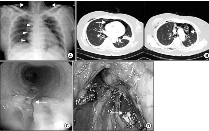
A 68-year-old woman who had a history of lumbar spine surgery under general anesthesia at another center was trans- ferred to our hospital with dyspnea and subcutaneous emphysema. One hour after extubation, she had hemotysis and dyspnea. During the following six hours, progressive sub- cutaneous emphysema and dyspnea developed. She was 145 cm tall and weighed 45 kg. A chest showed subcutaneous emphysema of the neck and pneumomediastinum (Fig. 1A).

Thoracic computed tomography (CT) scans showed subcuta- neous emphysema and the disappearance of tracheal posterior membranous wall continuity (Fig. 1B). We decided on an emergency operation. She reached the operating table 7 hours after lumbar spine surgery. During operation, a fiberoptic bronchoscope was used to verify double-lumen endobronchial tube placement, and showed a linear tracheal laceration of the posterior membranous wall at the distal part of the trachea

(Fig. 1C). She underwent a right thoracotomy and tracheal re- pair of about 7 cm of a membranous tracheal linear lacer- ation arising 3 cm from the carina (Fig. 1D). During the su- turing of the laceration, the endotracheal tube was withdrawn several times to allow good exposure of the tracheal tear.

The laceration was repaired with 4-0 Monosyn interrupted stitches (B. Braun Aesculap AG & Co KG, Tuttlingen, Germany), starting from the proximal end of the tear. Once the laceration was repaired, the anesthesiologist advanced the original orotracheal tube beyond the suture.

2) Patient 2

A 52-year-old woman who had a history of lumbar spine

surgery under general anesthesia at another center was trans-

ferred to our hospital with subcutaneous emphysema. She re-

ported having dyspnea and presented with subcutaneous

emphysema. She was 158 cm tall and weighed 60 kg. She

had a history of percutaneous coronary intervention due to

non-ST elevation myocardial infarction 3 years prior. Chest

(2)

Fig. 1. A chest film shows subcutaneous emphysema of the neck and pneumomediastinum (A), and computed tomography scans of the chest show subcutaneous emphysema and disappearance of tracheal posterior membranous wall continuity (B). A fiberoptic bronchoscope reveals a linear tracheal laceration of the posterior membranous wall at the distal part of the trachea (C). Intraoperative photographs show a membranous tracheal linear laceration of about 7 cm arising 3 cm from the carina (D).

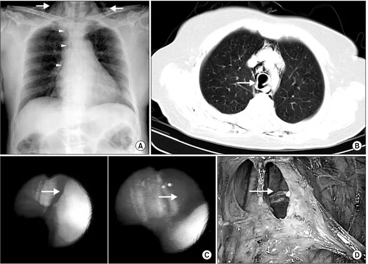
film showed a marked pneumomediastinum and subcutaneous emphysema of the neck (Fig. 2A). Thoracic computed tomog- raphy (CT) scans showed marked pneumomediastinum and pneumopericardium, but did not show lesions warranting sus- picion of tracheal or esophageal injury (Fig. 2B). The next day, progressive subcutanoues emphysema and dyspnea developed. A fiberoptic-bronchoscopy was performed. Bron- choscopy confirmed a 4-cm posterior membranous tracheal laceration approximately 5 cm from the carina (Fig. 2C). She was immediately brought to the operating table. She reached the operating table 23 hours after lumbar spine surgery. She also underwent a right thoracotomy and tracheal repair (Fig.

2D) in which the same technique as patient 1 was utilized

without the double-lumen endobronchial tube placement.

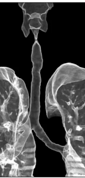
The postoperative course was uneventful, and the patients were discharged at the sixth and fifth postoperative days, respectively. Bronchoscopic follow-up showed a complete re- pair of the posterior tracheal wall tear (Fig. 3), and there were neither symptoms nor signs of tracheal stenosis (Fig. 4).

DISCUSSION

Despite the large number of intubations performed every

day, postintubation tracheal rupture (PiTR) is a very rare but

complication of general anesthesia. The first case series of

PiTR was not published until 1995 [1] and an estimate of the

(3)

Fig. 2. A chest film shows a marked pneumomediastinum and subcutaneous emphysema of neck (A) and computed tomography scans of chest show marked pneumomediastinum and pneumopericardium but does not show lesions indicating tracheal or esophageal injury (B).

Bronchoscopy reveals a 4-cm posterior membranous tracheal laceration approximately 5 cm from the carina (C). Intraoperative photographs show membranous tracheal linear laceration about 4 cm in length (D).

Fig. 3. Bronchoscopic follow-up shows a complete repair of the pos- terior tracheal wall tear (A=Patient 1; B=Patient 2).

incidence in the last decade ranged from 0.05% to 0.37% of all orotracheal intubations performed [2].

PiTR should be differentiated from tracheobronchial in- juries of traumatic origin since the different mechanisms lead

to different morphologic appearances and therapeutic options.

Traumatic TBR is usually the result of blunt chest trauma

and appears as horizontal or irregularly shaped disruptions in-

volving the main carina and often extending into the main

(4)

Fig. 4. Tracheobronchus 3D reconstruction of the first patient shows a well repaired trachea and no tracheal stenosis.

bronchi. PiTR, in contrast, usually presents as longitudinal lacerations of the posterior tracheal wall, either centrally or laterally, such that the membranous wall is avulsed from its cartilaginous insertion [2,3].

The exact mechanism underlying the lesion is uncertain.

There is a series of risk factors that contribute to PiTR; these factors may be divided into mechanical and anatomical.

Mechanical factors include multiple forced attempts at in- tubation, inexperience of the health professional, endotracheal tube introducers that protrude beyond the tip of the tube, overinflation of the cuff (diffusion of nitric oxide into the cuff), incorrect positioning of the tip of the tube, reposition- ing the tube without deflation of the cuff, inappropriate tube size, significant cough, and movements of the head and neck while the patient is intubated [3]. The anatomical factors in- clude congenital tracheal abnormalities, weakness of the pars membranosa of the trachea, chronic obstructive pulmonary disease and other inflammatory lesions of the tracheobron- chial tree, diseases that alter the position of the trachea (mediastinal collections, lymph nodes, or tumors), chronic use of steroids, advanced age, and female sex [3]. In the latter aspect, age also appears to play a role because, in the series published, there was not only evidence of female predom- inance, but the mean age of these women was over 50 years in the majority of the series published [3,4].

Some authors have suggested that PiTR may present more frequently in women because the pars membranosa is weaker in women than in men, the use of endotracheal tubes of a larger size than appropriate for women, or that women are not as tall and, as a consequence, the endotracheal tube is positioned significantly more distantly in a trachea that is al- ready smaller [5]. In the studies by Marty-Ane et al. [1], all patients had a height under 165 cm. Our experience is similar to those reported by other centers concerning the patients (short women) [5], the type of intubation (single-lumen or double-lumen tube) [6], and the site of the tear (the pars membranosa) [2,3].

The first signs of its presence usually appear within 12 hours of intubation [2,3,7]. Except for the case of intra- operative evidence, the appearance of symptoms such as head and neck emphysema, hemoptysis, and dyspnea should raise the suspicion of tracheobronchial ruptures.

Very often, the clinical manifestations of the lesion are not immediately obvious, and presentation can mimic that of oth- er clinical conditions. Early diagnosis or, if this is not possi- ble, high clinical suspicion may be associated with lower mortality, since either would likely lead to earlier therapeutic maneuvers. A delay in diagnosis could favor the onset of me- diastinitis, with deterioration in the clinical situation [2,8].

Clinical suspicion must be followed by diagnostic con- firmation, which is achieved by direct visualization of the tra- cheal rupture by bronchoscopy. This procedure provides data on the exact site and of the lesion, helps to plan the ther- apeutic approach, and can be used to reposition the tube or reintubate the patient if this is necessary [2].

Although bronchoscopic evaluation is the gold standard for diagnosis, CT scanning may provide valuable information.

Radiologic signs that suggest a TBR include subcutaneous emphysema without evidence of pneumothorax, irregularity in the posterior tracheal membrane, overdistention of the endo- tracheal balloon, and a displaced endotracheal tube in relation to the trachea [2,3]. Indeed, our initial suspicion that the first patient had a TBR was based on her CT scan findings.

Consensus has not yet been reached on the management of PiTR [2,3,7-9]. Early surgical repair has traditionally been considered the cornerstone of therapy for PiTR [1,2,4-8,10].

Recommendations regarding management of TBR complicat-

(5)

ing intubation have historically been based on experiences with blunt tracheobronchial ruptures in which surgical repair is usually required.

However, over the past 20 years, nonoperative management of TBR has been proposed for select patients in the following circumstances: stable vital signs, easy achievement of an ad- equate functional respiratory status under mechanical ven- tilation or in spontaneous ventilation, absence of esophageal injury, minimal mediastinal fluid collection, nonprogressive pneumomediastinum or subcutaneous emphysema, absence of sepsis, short ruptures, and delayed diagnosis [2,3,7-10]. If there is enough distance between the tear and the carina, the injury can often be excluded with the cuff of the endo- tracheal tube. In such circumstances, the use of an endo- tracheal tube with a subglottic suction port will avoid the pooling of secretions at the level of the injury. For TBR that involve the distal third of the trachea or extend into the car- ina, selective main stem bronchus intubation has had a high rate of success. In all cases, ventilator settings should attempt to minimize high inspiratory pressures and tidal volumes to avoid exacerbating the tear [2,3,7,9]. Because of the risk of mediastinitis, wide-spectrum antibiotics should be ad- ministered for at least 1 week after the injury has been identified. Complete healing usually occurs within 1 month, unless factors such as steroid use, immunosuppression, or se- vere malnutrition impede healing [7]. The use of expandable tracheal stents for the treatment of TBR has been described and recommended for patients who are poor surgical candi- dates with lesions not amenable to observation alone [11].

Operative management of TBR has been well described [1,2,4-8,10]. The surgical technique will depend on the type and of the lesion. In patients with lacerations of the proximal two-thirds of the trachea, a cervical approach is preferred [2,3,10]. A longitudinal anterior tracheotomy is made to ac- cess the injury, which is then closed primarily. Injuries in the distal one-third are best accessed through a right thoracotomy, with direct repair of the laceration without resection [2,3,10].

The length of the TBR as an indication for surgical manage- ment has been debated. Kaloud et al. recommended operating for any TBR lesion greater than 1 cm in length [5]. Gabor et al. [10], in cases of mixed iatrogenic and blunt tracheobron- chial injuries, recommended that ruptures greater than 2 cm

in length be repaired. Jougon et al., in contrast, proposed nonoperative management if the TBR was smaller than 4 cm [7]. In the future, minimally invasive techniques such as vid- eothoracoscopy may also be used.

Treatment is controversial, although it appears that con- servative management is associated with a better outcome.

The results of the recent larger case series published demon- strate that there is ever more evidence to support conservative management [2,3,7,9], allowing for the possible closure of the tracheal tear by adopting an expectant (conservative) approach. Moreover, some series have demonstrated that sur- gical repair in critically ill patients is a procedure with a mortality that can reach 71% [2].

The treatment guidelines until recently were based on a heterogeneous series of case reports involving different etiologies. Thus, goals when treating such patients should in- clude early recognition, appropriate antibiotic coverage, care- ful selection of operative candidates, and proper endotracheal tube and ventilator management.

We suggest that, except for selected cases in which non- surgical therapy is indicated, surgery is the treatment of choice in the vast majority of postintubation tracheal injuries.

When treated optimally, selected patients with TBR can fully and more quickly recover from surgical intervention than nonsurgical therapy.

REFERENCES

1. Marty-Ane CH, Picard E, Jonquet O, et al. Membranous tra- cheal rupture after endotracheal intubation. Ann Thorac Surg 1995;60:1367-71.

2. Minambres E, Buron J, Ballesteros MA, et al. Tracheal rup- ture after endotracheal intubation: a literature systematic review. Eur J Cardiothorac Surg 2009;35:1056-62.

3. Conti M, Marie P, Marquette CH, et al. Management of postintubation tracheobronchial ruptures. CHEST 2006;130:

412-8.

4. Chen EH, Logman ZM, Glass PS, et al. A case of tracheal injury after emergent endotracheal intubation: a review of the literature and causalities. Anesth Analg 2001;93:1270-1.

5. Kaloud H, Smolle-Juettner FM, Prause G, et al. Iatrogenic ruptures of the tracheobronchial tree. CHEST 1997;112:774-8.

6. Park SI, Won JH, Lee CK. Tracheal rupture following dou-

ble lumen endotracheal tube intubation. Korean J Thorac

Cardiovasc Surg 1999;32:765-7.

(6)

7. Jougon J, Ballester M, Choukroun E, et al. Conservative treatment for postintubation tracheobronchial rupture. Ann Thorac Surg 2000;69:216-20.

8. Sippel M, Putensen C, Hirner A, et al. Tracheal rupture af- ter endotracheal intubation: experience with management in 13 cases. Thorac Cardiovasc Surg 2006;54:51-6.

9. Fong PA, Seder CW, Chmielewski GW, et al. Nonoperative management of postintubation tracheal injuries. Ann Thorac

Surg 2010;89:1265-6.

10. Gabor S, Renner H, Pinter H, et al. Indications for surgery in tracheobronchial ruptures. Eur J Cardiothorac Surg 2001;

20:399-404.

11. Yopp AC, Eckstein JG, Savel RH, et al. Tracheal stenting of iatrogenic tracheal injury: a novel management approach.

Ann Thorac Surg 2007;83:1897-9.

수치

Fig. 1. A chest film shows subcutaneous emphysema of the neck and pneumomediastinum (A), and computed tomography scans of the  chest show subcutaneous emphysema and disappearance of tracheal posterior membranous wall continuity (B)
Fig. 2. A chest film shows a marked pneumomediastinum and subcutaneous emphysema of neck (A) and computed tomography scans of  chest show marked pneumomediastinum and pneumopericardium but does not show lesions indicating tracheal or esophageal injury (B)
Fig. 4. Tracheobronchus 3D reconstruction of the first patient  shows a well repaired trachea and no tracheal stenosis.

참조

관련 문서

He joyfully used techniques to enlarge the possibility of the instrumental representation with old master's works and his ability as a composer was

In this study, thermal efficiency of a flat-plate solar collector and a U-tube solar collector using a CuO nanofluid and water as a working fluid was investigated according to

As a result of performing a compound exercise of spinning and Zumba for 8 weeks, the change in α-amylase showed a significant difference in the exercise group (p<.01), and

Methods to overcome insufficient bone due to poor bone quality, the pneumatization of a maxillary sinus and other anatomical limitations of implant placement

Parents of the children completed a questionnaire, The questionnaire was consisted of the prevalence and types of alternative medicine used in the children,

In order to verify whether the museum exhibition of intangible cultural heritage has a significant influence on the visitor's behavioral intention, a

As a result of comparing the number of treatments per person by treatment behavior, the fluoride application during the preventive dental operation period was significantly

This work builds inversely a base performance model of a turboprop engine to be used for a high altitude operation UAV using measured performance data,