Clinical and Immunological Responses in Ocular Demodecosis
The purpose of this study was to investigate clinical and immunological responses to Demodex on the ocular surface. Thirteen eyes in 10 patients with Demodex blepharitis and chronic ocular surface disorders were included in this study and treated by lid scrubbing with tea tree oil for the eradication of Demodex. We evaluated ocular surface
manifestations and Demodex counts, and analyzed IL-1β, IL-5, IL-7, IL-12, IL-13, IL-17, granulocyte colony-stimulating factor, and macrophage inflammatory protein-1β in tear samples before and after the treatment. All patients exhibited ocular surface
manifestations including corneal nodular opacity, peripheral corneal vascularization, refractory corneal erosion and infiltration, or chronic conjunctival inflammatory signs before treatment. After treatment, Demodex was nearly eradicated, tear concentrations of IL-1β and IL-17 were significantly reduced and substantial clinical improvement was observed in all patients. In conclusion, we believe that Demodex plays an aggravating role in inflammatory ocular surface disorders.
Key Words: Blepharitis; Demodex; Immune Response; Ocular Surface; Tear Cytokine Jae Hoon Kim, Yeoun Sook Chun
and Jae Chan Kim
Department of Ophthalmology, College of Medicine, Chung-Ang University Hospital, Seoul, Korea Received: 27 February 2011
Accepted: 26 June 2011 Address for Correspondence:
Jae Chan Kim, MD
Department of Ophthalmology, College of Medicine, Chung-Ang University Hospital, 29 Heukseok-ro, Dongjak-gu, Seoul 156-755, Korea
Tel: +82.2-6299-1689, Fax: +82.2-825-1666 E-mail: jck50ey@kornet.net
This Research was supported by Chung-Ang University Research Grants in 2009.
The authors have no financial or proprietary interest in any of the materials or products mentioned in the manuscript.
http://dx.doi.org/10.3346/jkms.2011.26.9.1231 • J Korean Med Sci 2011; 26: 1231-1237
INTRODUCTION
Demodex (class Arachnida, superorder Acariformes) is an elon- gated ectoparasite found on the human body surface including the face, cheeks, forehead, nose, and eyelids (1). There are many species of Demodex, but only D. folliculorum and D. brevis are found on the human body (2). In the eye, Demodex can be found on the eyelashes, the lash follicles, and the meibomian glands, and is thought to be associated with blepharitis, allergic conjunc- tivitis, and pathological corneal features (2-5). Recently, a high prevalence of Demodex in eyelashes with cylindrical dandruff has been reported and a method of evaluating ocular demode- cosis by sampling and counting Demodex has been introduced (6, 7). In addition, Gao et al. (8, 9) reported the ocular Demodex- killing effects of tea tree oil (TTO) in vitro and in vivo, and intro- duced a new clinical treatment, lid scrubbing with TTO, that has proven effective for eradicating ocular demodecosis. Neverthe- less, the pathogenesis of Demodex on the ocular surface remains unclear up to recently. It has been merely presumed that inflam- matory or specific immune reactions may be associated with ocular demodecosis (4, 9).
Therefore, we evaluated the changes of the clinical manifesta- tions and the levels of tear cytokines following the eradication of Demodex to verify its pathogenicity, and to investigate the mechanisms of immunological response against Demodex on the ocular surface.
MATERIALS AND METHODS Patients
Thirteen eyes with ocular demodecosis and chronic ocular sur- face disorders of 10 patients were enrolled in this study. The 10 patients included six women and four men, with an average age of 48.3 ± 18.9 yr (range, 14 to 70 yr). Demographic and other clin- ical features are summarized in Table 1.
All patients reported ocular surface irritation and showed signs of ocular surface inflammation including conjunctival injections lasting over six months despite extensive patient-specific treat- ment including preservative-free artificial tears, corticosteroids, autologous serum, acyclovir (only for herpetic keratitis), antibi- otics, or lid scrubbing with baby shampoo. The medical records of all patients, including history of present illness and systemic diseases, were reviewed. All of the 10 patients underwent com- plete ophthalmologic examinations, including external photo- graphs, microscopic Demodex examination, and tear sampling.
The patients were treated with a four-week regimen of lid scrub- bing with TTO for ocular demodecosis and maintained prior topical treatments during that time. Four weeks after the initia- tion of treatment, we compared ocular surface manifestations, Demodex counts, and levels of inflammatory cytokines in tear samples measured before and after treatment.
Microscopic Demodex examination
Ocular demodecosis was confirmed by microscopic examina- tion of epilated lashes following the method described by Gao et al. (8) with some modifications. Briefly, two lashes with cylin- drical dandruff per lid were sampled and were placed separate- ly on a glass slide. Under a slit-lamp light microscope at a mag- nification of × 16, one drop of 20 μL saline was applied by pipette to the edge of the glass slide for lashes without retained dandruff.
For lashes with retained heavy dandruff, 20 μL of 100% alcohol was added. For the former, the number of Demodex was count- ed immediately and for the latter the counting time was delayed up to 20 min to allow the cylindrical dandruff to dissolve and to stimulate the migration of embedded Demodex. The Demodex count was recorded as the total number of mites found in a to- tal of four lashes per eye.
Treatments of ocular demodecosis
Weekly lid scrubs with 50% TTO were performed and daily lid scrubs with 10% TTO shampoo were advised for a minimum of four weeks, according to the method reported by Gao et al. (9).
In brief, at the clinic, after a drop of 0.5% proparacaine, a cotton tip wetted in 50% TTO was used to scrub the lid margin and lash roots for three sessions with a 10-min interval between each scrub. The patients were instructed to continue scrubbing daily at home, and advised to close their eyes and massage their lids with medium pressure for three to five minutes using a cotton tip wetted in 10% TTO shampoo. After treatment, the skin was rinsed with clean water and dried with a towel. We advised pa- tients to perform home lid scrubs twice daily.
Tear collection and multiplex bead immunoassay
Unstimulated tear fluid was collected from the inferior meniscus of each eye with the least possible irritation using a pre-weighed
polyester wick (Transorb rods; American Filtrona, Richmond, VA, USA) to obtain tear samples, as previously described (10).
Wicks were then placed into the end of a micropipette tip locat- ed within a 0.5 mL tube (Eppendorf, Fremont, CA, USA). The tear samples were immediately transported in an insulated cool- er to a -80°C freezer where they remained frozen until they were used for immunoassays. Tears were extracted from the saturat- ed wicks by adding a volume of buffer (50 mM Tris/HCl, 0.15 M NaCl, 10 mM CaCl2, 0.005% Brij35, 0.02% sodium azide [pH 7.5]) 10 times greater than the original volume of the tear sample to the pipette and then centrifuging at 12,000 rpm for five minutes.
The rods and pipette tips were carefully removed and the tear fluid aspirated. Cytokines and chemokines in these samples were analyzed using a Luminex® 200TM Total System (Invitro- genTM, Carlsbad, CA, USA). The cytokines and chemokines ana- lyzed included: IL-1β, IL-5, IL-7, IL-12, IL-13, IL-17, granulocyte- colony stimulating factor (G-CSF), and macrophage inflamma- tory protein-1β (MIP-1β). The concentrations of these factors in tears were calculated from standard curves of known concen- trations of recombinant human cytokines.
Statistical analysis
Summary data are expressed as means ± standard deviation (SD), analyzed by SPSS (version 13.0; SPSS, Inc., Chicago, IL, USA).
Pre-treatment and post-treatment parameters were evaluated by Wilcoxon signed-rank test, and P < 0.01 was considered sta- tistically significant.
Ethics statement
The institutional review board of Chung-Ang University Hospi- tal approved this study (2001-022-10) and all patients provided informed consent.
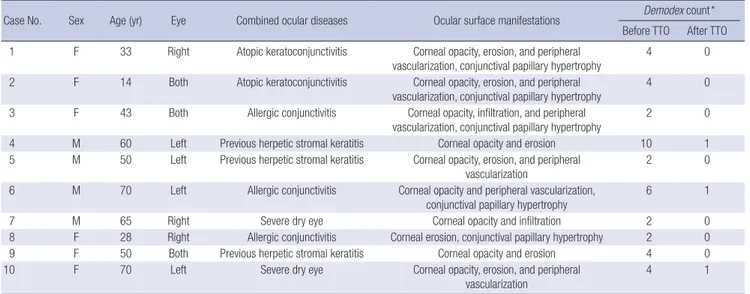
Table 1. Demographic and clinical features of patients with Demodex blepharitis and chronic ocular surface disorders
Case No. Sex Age (yr) Eye Combined ocular diseases Ocular surface manifestations Demodex count*
Before TTO After TTO 1 F 33 Right Atopic keratoconjunctivitis Corneal opacity, erosion, and peripheral
vascularization, conjunctival papillary hypertrophy
4 0
2 F 14 Both Atopic keratoconjunctivitis Corneal opacity, erosion, and peripheral
vascularization, conjunctival papillary hypertrophy 4 0 3 F 43 Both Allergic conjunctivitis Corneal opacity, infiltration, and peripheral
vascularization, conjunctival papillary hypertrophy 2 0
4 M 60 Left Previous herpetic stromal keratitis Corneal opacity and erosion 10 1
5 M 50 Left Previous herpetic stromal keratitis Corneal opacity, erosion, and peripheral
vascularization 2 0
6 M 70 Left Allergic conjunctivitis Corneal opacity and peripheral vascularization, conjunctival papillary hypertrophy
6 1
7 M 65 Right Severe dry eye Corneal opacity and infiltration 2 0
8 F 28 Right Allergic conjunctivitis Corneal erosion, conjunctival papillary hypertrophy 2 0
9 F 50 Both Previous herpetic stromal keratitis Corneal opacity and erosion 4 0
10 F 70 Left Severe dry eye Corneal opacity, erosion, and peripheral
vascularization 4 1
*The Demodex count was recorded as the total number of mites found in a total of four lashes per eye.
RESULTS
Ocular surface manifestations and Demodex count Demodex was found in all patients (Fig. 1). All patients reported ocular surface discomfort lasting over six months. They showed ocular surface manifestations including large corneal nodular opacity (four eyes; Fig. 2A-C), refractory corneal erosion and in- filtration (12 eyes; Fig. 2D-F), peripheral corneal vascularization (6 eyes; Fig. 2C, G, H), chronic palpebral conjunctival papillary hypertrophy (7 eyes; Fig. 2I), and chronic bulbar conjunctival injection (13 eyes). Three eyes had atopic keratoconjunctivitis, four eyes had allergic conjunctivitis, and two eyes had severe dry eye syndrome (dysfunctional tear syndrome level 3) (11). Four eyes had had herpes simplex stromal keratitis in the past.
After four weeks of weekly lid scrubs with 50% tea tree oil and daily lid scrubs with tea tree shampoo, the disappearance of dan- druff was noted in all patients’ eyelashes. Demodex was com- pletely eradicated in 10 of 13 eyes. The mean Demodex count per eye was reduced significantly, from 3.8 ± 2.2 to 0.2 ± 0.4 at four weeks after the initiation of treatment (Table 2; P = 0.001).
All patients showed improvement of bulbar conjunctival injec- tion (13 eyes), conjunctival papillary hypertrophy (7 eyes), and corneal erosions and infiltrations (12 eyes). Peripheral corneal vascularizations and nodular corneal opacities were markedly faded in three of six eyes and two of four eyes (Fig. 3).
Tear concentrations of inflammatory cytokines
The mean tear concentrations of all inflammatory cytokines be- fore and after treatment are presented in Table 2. Tear concen- trations of IL-1β (P = 0.001) and IL-17 (P = 0.001) were signifi- cantly reduced after treatment. Tear concentrations of IL-5, IL- 7, IL-12, IL-13, G-CSF, and MIP-1β were also reduced after treat- ment, but the reductions were not statistically significant (P > 0.01).
DISCUSSION
This study demonstrates that the clinical manifestations of ocu- lar demodecosis are considerably improved and the tear levels of IL-1β and IL-17 are significantly decreased after eradication of Demodex. Previously, several studies have reported that De- modex may cause ocular surface inflammation and pathologi- cal features and that lid scrubbing using tea tree oil was an effec- tive method for eradicating Demodex (4, 8, 9). Similarly, we ob- served chronic refractory pathological ocular surface appear- ances (such as large corneal nodular opacity, corneal vascular- ization, severe corneal erosion and infiltration, and conjuncti- val inflammatory reactions) in patients with ocular demodeco- sis, and we confirmed the clinical improvement of ocular mani- festation after the treatment of demodecosis in this study.
All patients in our study had not only intractable ocular sur- face manifestations but prior inflammatory ocular surface dis-
Table 2. The mean Demodex count and tear concentrations of cytokines (pg/mL) in eyes with Demodex blepharitis and chronic ocular surface disorders before and after lid scrub treatment with tea tree oil
Demodex
count* IL-1β IL-5 IL-7 IL-12 IL-13 IL-17 G-CSF MIP-1β
Before TTO 3.8 ± 2.2 1141.5 ± 440.3
244.6 ± 110.6
9,928.0 ± 4,684.5
487.6 ± 153.6
430.7 ± 256.5
1,907.8 ± 861.0
477.6 ± 149.2
3,178.2 ± 2,030.6
After TTO 0.2 ± 0.4 561.7 ±
261.0
209.0 ± 91.8
8,958.2 ± 3,999.2
442.8 ± 165.8
407.9 ± 250.8
1,124.2 ± 545.1
471.9 ± 166.1
3,062.7 ± 1,913.3
P value† 0.001 0.001 0.29 0.09 0.25 0.10 0.001 0.69 0.06
*The mean Demodex count per eye; †Wilcoxon signed-rank test. TTO, tea tree oil; G-CSF, granulocyte-colony stimulating factor; MIP-1β, macrophage inflammatory protein-1β.
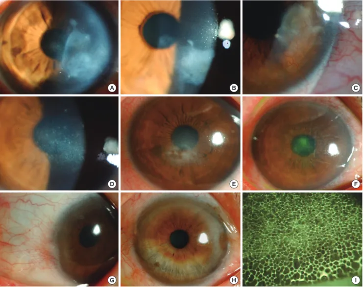
Fig. 1. Representative microscopic photographs of Demodex folliculorum in patients with ocular demodecosis. Two D. folliculorum with an eyelash (A, arrows) and one free D.
folliculorum are found in epilated eyelashes of patients.
A B
orders including herpetic stromal keratitis, atopic keratoconjunc- tivitis, allergic conjunctivitis, and severe dry eye. Recommend- ed conventional treatment was not successful but the eradica- tion of Demodex improved the pathological ocular surface man- ifestations. This suggests that Demodex plays a pathological role or at least an aggravating role in inflammatory ocular surface disorders, especially immunological disorders. Type III or type IV hypersensitivity immune response is an important patholog- ical mechanism in herpetic stromal keratitis and type I hyper- sensitivity immune responses is a primary mechanism in atop- ic keratoconjunctivitis and allergic conjunctivitis. So we believe that the pathogenesis of Demodex may be associated with hy- persensitive immune responses on the ocular surface.
The levels of tear cytokines can be used to indicate inflamma- tory or immunological responses on the ocular surface. In this study, we observed that concentrations of IL-1β and IL-17 were reduced after treatment with TTO lid scrubs, and that these re-
A B C
D E F
G H I
Fig. 2. Clinical features of patients with ocular demodecosis (A, case 4; B, case 5; C, case 9; D, case 1; E, case 3; F, case 7; G, case 2; H, case 6; I, case 8). Large corneal nodular opacity (A-C), refractory corneal erosion and infiltration (D-F), peripheral corneal vascularization (C, G, H), and palpebral conjunctival papillary hypertrophy (I) are ob- served in patients with ocular demodecosis.
ductions were statistically significant.
IL-1 is a potent inducer of other inflammatory cytokines, in- cluding IL-6, IL-8, TNF-α, and granulocyte-macrophage colony- stimulating factor (GMCSF), and stimulates production of colla- genase and matrix metalloproteinase (MMP) enzymes by epithe- lial cells, keratocytes, and inflammatory cells (12-14). IL-1 induces the destruction of extracellular matrix and renders inflammatory damage on the ocular surface. Clinically, there have been reports that IL-1β is present at increased levels in tears in patients suffer- ing inflammatory ocular surface disorders such as dry eye, Sjögren syndrome, and keratoconjunctivitis sicca (15). It is therefore rea- sonable to conclude that the improvements of chronic refracto- ry corneal epithelial erosions and ocular surface inflammatory signs observed after eradicating Demodex reflect reductions in IL- 1. However, it is difficult to specify the particular inflammatory process in ocular demodecosis because IL-1 is a cytokine involv- ed in various immune responses and inflammatory processes.
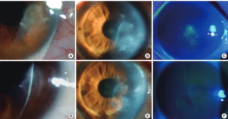
A B C
D E F
Fig. 3. Clinical appearance before and after lid scrubbing treatment with tea tree oil in patients with ocular demodecosis. (A and D, case 4; B and E, case 9; C and F, case 7).
There are corneal nodular opacities (A, B), bulbar conjunctival injections (A, B) and central corneal infiltration (C) before the treatment. However, four weeks after the treatment, corneal opacities are markedly faded and conjunctival injections had resolved (D, E). Central corneal infiltration had also resolved (F).
IL-17 is an important cytokine in inflammatory and autoim- mune conditions (16). It is secreted mainly by activated helper T cells and enhances the generation, activation, and migration of neutrophils through the induction of proinflammatory cytokines (17, 18). T cells secreting IL-17, named Th17 cells, are now con- sidered developmentally distinct from Th1 and Th2 cells and are thought to play a key role in autoimmune reactions (19, 20). IL- 17 activates T cells and other immune cells to produce a variety of cytokines and cell adhesion molecules not only in the early neutrophil-mediated inflammatory response, but also in the in- duction of both Th1-type and Th2-type immune responses (21, 22). Synergy with other inflammatory cytokines such as IL-1β, tumor necrosis factor (TNF)-α, and IFN-γ leads to up-regulation of gene expression and progression and amplification of local inflammation (23, 24). Levels of IL-17 are significantly increased in autoimmune diseases including rheumatoid arthritis synovi- um, asthmatic airways, during allograft rejection, and in other chronic inflammatory diseases including multiple sclerosis and psoriasis (25-28). In this study, the reduction in tear levels of IL- 17 after Demodex eradication suggests that ocular demodecosis is associated with elevated cell mediated immune conditions, especially the Th17 cell immune response. IL-17 is also a potent inducer of angiogenic chemokines such as vascular endothelial growth factor-A (VEGF-A) from a number of cells, including ke- ratinocytes, fibroblasts and epithelial cells (29, 30). It is reason- able to explain that decreased levels of tear IL-17 following De- modex eradication contribute to the improvement of abnormal
corneal vascularization.
There are some limitations in this study including the small number of subjects, the possible anti-inflammatory effect of tea tree oil, and the possibility of other inflammatory factors asso- ciated with Demodex, such as bacterial distribution. Future stud- ies should include normal control subjects or blepharitis control subjects without Demodex, a larger number of subjects, an as- sessment of the anti-inflammatory effects of tea tree oil and the bacterial distribution in ocular demodecosis, and a wider anal- ysis of cytokines associated with the Th17 immune response in- cluding IL-6, IL-21, IL-22, IL-23, and transforming growth factor beta (TGF-β). With further study, we may be able to confirm the pathogenic mechanism of Demodex on the ocular surface.
In summary, we examined chronic refractory inflammatory pathological ocular surface manifestations in patients with ocu- lar demodecosis and verified that lid scrubbing with tea tree oil was an effective method to eradicate Demodex leading to clini- cal improvement. We also noted that tear concentrations of IL- 1β and IL-17 were significantly decreased after the eradication of Demodex in the patients. Therefore, we believe that Demodex plays an aggravating role in inflammatory ocular surface disor- ders and that the treatment of Demodex induces the recovery of pathologic clinical manifestations.
REFERENCES
1. Baima B, Sticherling M. Demodicidosis revisited. Acta Derm Venereol
2002; 82: 3-6.
2. English FP, Nutting WB. Demodicosis of ophthalmic concern. Am J Oph- thalmol 1981; 91: 362-72.
3. Coston TO. Demodex folliculorum blepharitis. Trans Am Ophthalmol Soc 1967; 65: 361-92.
4. Kheirkhah A, Casas V, Li W, Raju VK, Tseng SC. Corneal manifestations of ocular demodex infestation. Am J Ophthalmol 2007; 143: 743-9.
5. Rodríguez AE, Ferrer C, Alió JL. Chronic blepharitis and Demodex. Arch Soc Esp Oftalmol 2005; 80: 635-42.
6. Kheirkhah A, Blanco G, Casas V, Tseng SC. Fluorescein dye improves mi- croscopic evaluation and counting of demodex in blepharitis with cylin- drical dandruff. Cornea 2007; 26: 697-700.
7. Gao YY, Di Pascuale MA, Li W, Liu DT, Baradaran-Rafii A, Elizondo A, Kawakita T, Raju VK, Tseng SC. High prevalence of Demodex in eyelashes with cylindrical dandruff. Invest Ophthalmol Vis Sci 2005; 46: 3089-94.
8. Gao YY, Di Pascuale MA, Li W, Baradaran-Rafii A, Elizondo A, Kuo CL, Raju VK, Tseng SC. In vitro and in vivo killing of ocular Demodex by tea tree oil. Br J Ophthalmol 2005; 89: 1468-73.
9. Gao YY, Di Pascuale MA, Elizondo A, Tseng SC. Clinical treatment of ocular demodecosis by lid scrub with tea tree oil. Cornea 2007; 26: 136-43.
10. Jones DT, Monroy D, Pflugfelder SC. A novel method of tear collection:
comparison of glass capillary micropipettes with porous polyester rods.
Cornea 1997; 16: 450-8.
11. Behrens A, Doyle JJ, Stern L, Chuck RS, McDonnell PJ, Azar DT, Dua HS, Hom M, Karpecki PM, Laibson PR, Lemp MA, Meisler DM, Del Castillo JM, O’Brien TP, Pflugfelder SC, Rolando M, Schein OD, Seitz B, Tseng SC, van Setten G, Wilson SE, Yiu SC. Dysfunctional tear syndrome: a Del- phi approach to treatment recommendations. Cornea 2006; 25: 900-7.
12. Woessner JF Jr. Matrix metalloproteinases and their inhibitors in connec- tive tissue remodeling. FASEB J 1991; 5: 2145-54.
13. Strissel KJ, Girard MT, West-Mays JA, Rinehart WB, Cook JR, Brincker- hoff CE, Fini ME. Role of serum amyloid A as an intermediate in the IL-1 and PMA-stimulated signaling pathways regulating expression of rabbit fibroblast collagenase. Exp Cell Res 1997; 237: 275-87.
14. Strissel KJ, Rinehart WB, Fini ME. Regulation of paracrine cytokine bal- ance controlling collagenase synthesis by corneal cells. Invest Ophthalmol Vis Sci 1997; 38: 546-52.
15. Pflugfelder SC, Jones D, Ji Z, Afonso A, Monroy D. Altered cytokine bal- ance in the tear fluid and conjunctiva of patients with Sjögren’s syndrome keratoconjunctivitis sicca. Curr Eye Res 1999; 19: 201-11.
16. McGeachy MJ, Anderton SM. Cytokines in the induction and resolution of experimental autoimmune encephalomyelitis. Cytokine 2005; 32: 81-4.
17. Fossiez F, Djossou O, Chomarat P, Flores-Romo L, Ait-Yahia S, Maat C, Pin JJ, Garrone P, Garcia E, Saeland S, Blanchard D, Gaillard C, Das Ma- hapatra B, Rouvier E, Golstein P, Banchereau J, Lebecque S. T cell inter- leukin-17 induces stromal cells to produce proinflammatory and hema- topoietic cytokines. J Exp Med 1996; 183: 2593-603.
18. Jovanovic DV, Di Battista JA, Martel-Pelletier J, Jolicoeur FC, He Y, Zhang
M, Mineau F, Pelletier JP. IL-17 stimulates the production and expression of proinflammatory cytokines, IL-beta and TNF-alpha, by human mac- rophages. J Immunol 1998; 160: 3513-21.
19. Harrington LE, Hatton RD, Mangan PR, Turner H, Murphy TL, Murphy KM, Weaver CT. Interleukin 17-producing CD4+ effector T cells develop via a lineage distinct from the T helper type 1 and 2 lineages. Nat Immu- nol 2005; 6: 1123-32.
20. Stockinger B, Veldhoen M. Differentiation and function of Th17 T cells.
Curr Opin Immunol 2007; 19: 281-6.
21. Umemura M, Yahagi A, Hamada S, Begum MD, Watanabe H, Kawaka- mi K, Suda T, Sudo K, Nakae S, Iwakura Y, Matsuzaki G. IL-17-mediated regulation of innate and acquired immune response against pulmonary Mycobacterium bovis bacille Calmette-Guérin infection. J Immunol 2007;
178: 3786-96.
22. Nakae S, Komiyama Y, Nambu A, Sudo K, Iwase M, Homma I, Sekikawa K, Asano M, Iwakura Y. Antigen-specific T cell sensitization is impaired in IL-17-deficient mice, causing suppression of allergic cellular and hu- moral responses. Immunity 2002; 17: 375-87.
23. Ruddy MJ, Wong GC, Liu XK, Yamamoto H, Kasayama S, Kirkwood KL, Gaffen SL. Functional cooperation between interleukin-17 and tumor necrosis factor-alpha is mediated by CCAAT/enhancer-binding protein family members. J Biol Chem 2004; 279: 2559-67.
24. Albanesi C, Cavani A, Girolomoni G. IL-17 is produced by nickel-specific T lymphocytes and regulates ICAM-1 expression and chemokine produc- tion in human keratinocytes: synergistic or antagonist effects with IFN- gamma and TNF-alpha. J Immunol 1999; 162: 494-502.
25. Chabaud M, Lubberts E, Joosten L, van Den Berg W, Miossec P. IL-17 derived from juxta-articular bone and synovium contributes to joint deg- radation in rheumatoid arthritis. Arthritis Res 2001; 3: 168-77.
26. Antonysamy MA, Fanslow WC, Fu F, Li W, Qian S, Troutt AB, Thomson AW. Evidence for a role of IL-17 in organ allograft rejection: IL-17 promotes the functional differentiation of dendritic cell progenitors. J Immunol 1999;
162: 577-84.
27. Teunissen MB, Koomen CW, de Waal Malefyt R, Wierenga EA, Bos JD.
Interleukin-17 and interferon-gamma synergize in the enhancement of proinflammatory cytokine production by human keratinocytes. J Invest Dermatol 1998; 111: 645-9.
28. Molet S, Hamid Q, Davoine F, Nutku E, Taha R, Pagé N, Olivenstein R, Elias J, Chakir J. IL-17 is increased in asthmatic airways and induces hu- man bronchial fibroblasts to produce cytokines. J Allergy Clin Immunol 2001; 108: 430-8.
29. Starnes T, Robertson MJ, Sledge G, Kelich S, Nakshatri H, Broxmeyer HE, Hromas R. Cutting edge: IL-17F, a novel cytokine selectively expressed in activated T cells and monocytes, regulates angiogenesis and endothelial cell cytokine production. J Immunol 2001; 167: 4137-40.
30. Numasaki M, Fukushi J, Ono M, Narula SK, Zavodny PJ, Kudo T, Rob- bins PD, Tahara H, Lotze MT. Interleukin-17 promotes angiogenesis and tumor growth. Blood 2003; 101: 2620-7.
AUTHOR SUMMARY
Clinical and Immunological Responses in Ocular Demodecosis
Jae Hoon Kim, Yeoun Sook Chun and Jae Chan Kim
Demodex is thought to be associated with blepharitis, allergic conjunctivitis, and pathological corneal features on the ocular surface. However, the pathogenesis of Demodex on the ocular surface remains unclear. We evaluated clinical manifestations and tear cytokine level changes following the eradication of Demodex on the inflammatory ocular surface disorders. After a course of treatment of lid scrubbing with tea tree oil in patients with ocular demodecosis, Demodex was nearly eradicated, and there was substantial clinical improvement and tear concentrations of IL-1β and IL-17 were significantly reduced in all patients. Therefore, we believe that Demodex plays an imporatant aggravating role in inflammatory ocular surface disorders.