INTRODUCTION
The autosomal dominant cerebellar ataxias (ADCA) are a heterogeneous group of neurodegenerative disorders with overlapping phenotypes that include progressive dysfunction of the cerebellum, basal ganglia, brainstem, cerebral cortex, spinal cord, and peripheral nerves. Based on the phenotypic characteristics, Harding (1) classified ADCAs into three types:
ADCA I, ADCA II, and ADCA III. Disorder in ADCA II show cerebellar ataxia accompanied by macular degeneration.
Advances in human molecular genetics have led to a reclas- sification of ADCA based on identification of the genotypes, and the term ADCA is being replaced by spinocerebellar ataxia (SCA) and episodic ataxia. SCA currently include SCA 1 through 8 and 10 through 14. SCA 1-3, 6, 7, and 12 are caused by an expanded CAG repeat within the encoding re- gion of the respective genes (2-7). SCA8 is caused by an un- translated CTG expansion (8), and SCA10 by ATTCT expan- sion (9). In other SCAs, the causative mutations remain un- known. SCA 1-4 have extracerebellar involvement, thus cor- respond to Harding's category of ADCA I (2-4, 10). SCA7 has retinal degeneration (ADCA II) (6). SCA 5, 6, 8, 11, and 14 appear to be purely cerebellar ataxia (ADCA III) (5, 11).
SCA7 is caused by the expansion of CAG trinucleotide re- peat in the chromosome 3p12-21.1. The affected gene region encodes for the ataxin-7. Ataxin-7 is an 892 amino acid nu- clear protein of unknown function. The N-terminal segment of ataxin-7 contains a polyglutamine stretch, and is believed to exert a toxic effect when the polyglutamine stretch is ex-
panded. Normal SCA7 alleles range from 6 to 17 CAG re- peats (12). Pathological alleles range from 37 and above (13- 15). As in other SCAs, SCA7 has a broad spectrum of phe- notypic abnormalities depending on the number of CAG repeats (16). Ataxia is a universal feature and constant in all age groups. On the other hand spasticity and ocular symp- toms are variable according to the onset age. Visual loss and electroretinographic changes may precede funduscopic abnor- malities (17). Retinal degeneration constitutes an important component in juvenile and early adult onset (<25 yr), but is variable in late onset patients (>40 yr) (18). In late onset pa- tients visual failure due to retinal degeneration is a late symp- tom, and in some patients, it is not found for periods varying between 17 and 27 yr after the onset of ataxia (18).
Herein, we describe a SCA7 patient without retinal degen- eration for 7 yr.
CASE REPORT
The patient was a 60-yr-old man who had had progressive gait disturbance and ataxia since the age of 52. Unsteady gait was the first symptom especially during running. Unsteady gait progressed until he became in a wheelchair-bound state.
Dysarthria began at the age of 56. He had no significant past medical history. Family history was absent for neurological disorders.
On admission, he was alert, oriented and dysarthric. Eye examination by ophthalmologist showed an uncorrected
Byeong-Chae Kim, Myeong-Kyu Kim, Ki-Hyun Cho, Beom S. Jeon*
Department of Neurology, Chonnam National University Medical School, Gwangju; Seoul National University*, College of Medicine, Seoul, Korea
Address for correspondence Ki-Hyun Cho, M.D.
Department of Neurology, Chonnam National University Medical School, 8 Hak-dong, Dong-gu, Gwangju 501-190, Korea
Tel : +82.62-220-6160, Fax : +82.62-228-3461 E-mail : [email protected]
577 J Korean Med Sci 2002; 17: 577-9
ISSN 1011-8934
Copyright � The Korean Academy of Medical Sciences
Spinocerebellar Ataxia Type 7 without Retinal Degeneration
: A Case Rreport
A 60-yr-old man developed progressive gait disturbance and limb ataxia at the age of 52. Family history was absent for neurological disorders. Examinations showed pure cerebellar syndrome. There was no retinal degeneration for 7 yr. A brain MRI done at the age of 56 showed atrophy of the cerebellar hemispheres and vermis. Genetic test confirmed the spinocerebellar ataxia type 7 with CAG repeat number of 42.
Key Words : Spinocerebellar Ataxias; Retinal Degeneration; Cerebellar Ataxia
Received : 3 May 2001 Accepted : 30 August 2001
578 B,-C. Kim, M.-K. Kim, K.-H. Cho, et al.
visual acuity of 20/40 in the right and 20/60 in the left with Snellen rating for distance vision, normal visual field, and normal color discrimination with Ishihara color test. There was neither ophthalmoparesis nor nystagmus. Funduscopic examination showed neither macular pigmentation nor disc pallor (Fig. 1). There was no muscular atrophy or weakness.
These ophthalmologic data were the same results as those at the age of 56. Pathologic reflexes were absent. Sensation and autonomic functions were normal. He was able to walk only with one-person assistance. Routine blood count, urinalysis, serum chemistries, and thyroid functions were normal.
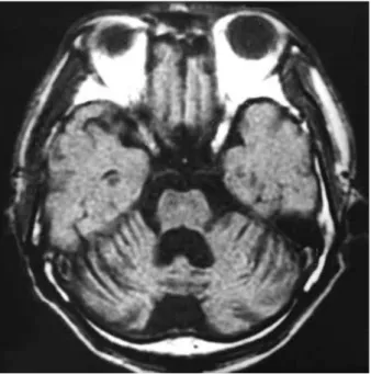
Visual evoked potential was normal. A brain MRI done at the age of 56 showed a global atrophy of the cerebellar hemispheres and vermis, whereas the pons, medulla, and upper spinal cord were spared (Fig. 2).
Genomic testing revealed a pathological expansion of CAG repeats in the SCA7 gene (repeat number=42/10).
DISCUSSION
We have previously reported that there are cases of SCA1, 2, 3, 6, and 7 in Korea with SCA2 being the most common (19). Genetically confirmed SCA7 with retinal degeneration was previously reported in Korea (20). Phenotypically, our patient had pure cerebellar syndrome without retinal degen- eration, therefore was not thought to be ADCAII or SCA7.
Our case again emphasizes the phenotypic variability of SCAs, and the need for genetic confirmation whenever possible. The degree of CAG expansion roughly determines the onset age
and phenotype (12). There was a strong negative correlation between the age at onset and the size of the CAG repeat ex- pansion in SCA7 patients. Larger expansions were associated with an earlier onset, a more severe and rapid clinical course, and higher frequencies of decreased vision, ophthalmoplegia, extensor plantar response, and scoliosis (14). CAG repeat number of 42 in the SCA7 gene is usually associated with the late adulthood onset and less frequent retinal involve- ment (14) as in our case.
REFERENCES
1. Harding AE. Clinical features and classification of inherited ataxias.
Adv Neurol 1993; 61: 1-14.
2. Orr HT, Chung M-Y, Banfi S, Kwiatkowski TJ Jr, Servadio A, Beaudet AL, McCall AE, Duvick LA, Ranum LP, Zoghbi HY. Expansion of an unstable trinucleotide CAG repeat in spinocerebellar ataxia type 1. Nat Genet 1993; 4: 221-6.
3. Pulst SM, Nechiporuk A, Nechiporuk T, Gispert S, Chen XN, Lopes- Cendes I, Pearlman S, Starkman S, Orozco-Diaz G, Lunkes A, Dejong P, Rouleau GA, Auburger G, Korenberg JR, Figueroa C, Sahba S.
Moderate expansion of a normally biallelic trinucleotide repeat in spinocerebellar ataxia type 2. Nat Genet 1996; 14: 269-76.
4. Kawaguchi Y, Okamoto T, Taniwaki M, Aizawa M, Inoue M, Kata- yama S, Kawakami H, Nakamura S, Nishimura M, Akiguchi I, Kimu- ra J, Narumiya S, Kakizuka A. CAG expansions in a novel gene for Machado-Joseph disease at chromosome 14q32.1. Nat Genet 1994;
8: 221-8.
5. Zhuchenko O, Bailey J, Bonnen P, Ashizawa T, Stockton DW, Amos Fig. 1.Photograph of the right fundus of the patient at the age of
59. Neither atrophy nor granular pigmentary changes are seen in the macula and optic disk.
Fig. 2.The axial view of T1-weighted brain MRI shows a marked atrophy of the cerebellar hemispheres and 4th ventricular dilata- tion.
SCA7 without Retinal Degeneration 579
C, Dobyns WB, Subramony SH, Zoghbi HY, Lee CC. Autosomal dominant cerebellar ataxia (SCA6) associated with small polyglu- tamine expansions in the alpha 1A-voltage-dependent calcium chan- nel. Nat Genet 1997; 15: 62-9.
6. David G, Abbas N, Stevanin G, Durr A, Yvert G, Cancel G, Weber C, Imbert G, Saudou F, Antoniou E, Drabkin H, Gemmill R, Giunti P, Benomar A, Wood N, Ruberg M, Agid Y, Mandel JL, Brice A.
Cloning of the SCA7 gene reveals a highly unstable CAG repeat ex- pansion. Nat Genet 1997; 17: 65-70.
7. Holmes SE, O'Hearn EE, McInnis MG, Gorelick-Feldman DA, Kleiderlein JJ, Callahan C, Kwak NG, Ingersoll-Ashworth RG, Sherr M, Sumner AJ, Sharp AH, Ananth U, Seltzer WK, Boss MA, Vieria-Saecker AM, Epplen JT, Riess O, Ross CA, Margolis RL.
Expansion of a novel CAG trinucleotide repeat in the 5′region of PPP2R2B is associated with SCA12. Nat Genet 1999; 23: 391-2.
8. Koob MD, Moseley ML, Schut LJ, Benzow KA, Bird TD, Day JW, Ranum LP. An untranslated CTG expansion causes a novel form of spinocerebellar ataxia (SCA8). Nat Genet 1999; 21: 379-84.
9. Matsuura T, Yamagata T, Burgess DL, Rasmussen A, Grewal RP, Watase K, Khajavi M, McCall AE, Davis CF, Zu L, Achari M, Pulst SM, Alonso E, Noebels JL, Nelson DL, Zoghbi HY, Ashizawa T.
Large expansion of ATTCT pentanucleotide repeat in spinocerebel- lar ataxia type 10. Nat Genet. 2000; 26: 191-4.
10. Flanigan K, Gardner K, Alderson K, Galster B, Otterud B, Leppert MF, Kaplan C, Ptuek LJ. Autosomal dominant spinocerebellar ataxia with sensory axonal neuropathy (SCA4): clinical description and genetic localization to chromosome 16q22.1. Am J Hum Genet 1996;
59: 392-9.
11. Ranum LP, Schut LJ, Lundgren JK, Orr HT, Livingston DM. Spi- nocerebellar ataxia type 5 in a family descended from the grand- parents of President Lincoln maps to chromosome 11. Nat Genet 1994; 8: 280-4.
12. Gouw LG, Castaneda MA, McKenna CK, Digre KB, Pulst SM, Perlman S, Lee MS, Gomez C, Fischbeck K, Gagnon D, Storey E, Bird T, Jeri FR, Ptacek LJ. Analysis of the dynamic mutation in the SCA7 gene shows marked parental effects on CAG repeat transmis-
sion. Hum Mol Genet 1998; 7: 525-32.
13. Del-Favero J, Krols L, Michalik A, Theuns J, Lofgren A, Goossens D, Wehnert A, Van den Bossche D, Van Jand K, Backhovens H, Van Regenmorter N, Martin JJ, Van Broeckhoven C. Molecular genetic analysis of autosomal dominant cerebellar ataxia with retinal degeneration (ADCA type II) caused by CAG triplet repeat expan- sion. Hum Mol Genet 1998; 7: 177-86.
14. David G, Durr A, Stevanin G, Cancel G, Abbas N, Benomar A, Belal S, Lebre AS, Abada-Bendib M, Grid D, Holmberg M, Yahyaoui M, Hentati F, Chkili T, Agid Y, Brice A. Molecular and clinical correlations in autosomal dominant cerebellar ataxia with progres- sive macular dystrophy (SCA7). Hum Mol Genet 1998; 7: 165-70.
15. Johansson J, Forsgren L, Sandgren O, Brice A, Holmgren G, Holm- berg M. Expanded CAG repeats in Swedish spinocerebellar ataxia type 7 (SCA7) patients: effect of CAG repeat length on the clinical manifestation. Hum Mol Genet 1998; 7: 171-6.
16. Benton CS, MRCP RS, Rutledge SL, Bohlega S, Ashizawa T, Zoghbi HY. Molecular and clinical studies in SCA-7 define a broad clinical spectrum and the infantile phenotype. Neurology 1998; 51: 1081-6.
17. Enevoldson TP, Sanders MD, Harding AE. Autosomal dominant cerebellar ataxia with pigmentary macular dystrophy. A clinical and genetic study of eight families. Brain 1994; 117: 445-60.
18. Jobsis GJ, Weber JW, Barth PG, Keizers H, Baas F, van Schooneveld MJ, van Hilten JJ, Troost D, Geesink HH, Bolhuis PA. Autosomal dominant cerebellar ataxia with retinal degeneration (ADCA II):
clinical and neuropathological findings in two pedigrees and genetic linkage to 3p12-p21.1. J Neurol Neurosurg Psychiatry 1997; 62:
367-71.
19. Kim JM, Shin S, Kim JY, Joo SI, Park SS, Kim JW, Jeon BS. Spino- cerebellar ataxia type 2 in seven Korean families: CAG trinucleotide expansion and clinical characteristics. J Korean Med Sci 1999; 14:
659-64.
20. Lyoo CH, Hur K, Choi YC, Lee SC, Stevanin G, David G, Brice A, Lee MS. CAG repeat expansion in the SCA7 in Korean families pre- senting clinical features compatible with ADCA Type II. J Korean Neurol Assoc 1998; 16: 341-52.