INTRODUCTION
Vasospastic angina (VA) is characterized by chest pain at rest with circadian variation (1-3). It is caused by coronary vasospasm and can be diagnosed by the provocation of epicar- dial coronary constriction after the infusion of acetylcholine or ergonovine (4-6). VA is relatively common in East Asians and is frequently observed in normal coronary arteries (7, 8).
Moreover, it has been reported that the prevalence of Raynaud’s phenomena or migraine in VA patients is relatively high (9- 11). These findings suggest that VA may be a manifestation of generalized smooth muscle contractile disorder, and that an abnormal contractile response in the coronary microcircu- lation may also be present. An abnormal endothelial depen- dent vascular tone is regarded to be a main pathogenetic me- chanism in VA (12-14). One of physiologic stimuli used to evaluate endothelial function is the cold pressor test (15). The response of coronary blood flow to cold stimulation in a patient with endothelial dysfunction was reported to be impaired (16). However, changes in coronary blood flow and microvas- cular function under cold stimulation in VA have not been assessed.
Accordingly, the aims of this study were to evaluate micro- vascular function in VA at room temperature, and to assess
whether microvascular dysfunction can be induced by cold stimulation by measuring coronary flow velocities with trans- thoracic Doppler echocardiography (TTDE).
MATERIALS AND METHODS Study population
We studied 14 consecutive patients with chest pain at rest.
All of patients had angiographically normal coronary arteries and showed documented coronary spasm associated with chest pain or electrocardiographic (ECG) changes after intracoronary infusion of acetylcholine, as reported previously (17). Fifteen healthy subjects were included as a control group. Six patients in the control group received coronary angiography and did not have significant coronary artery stenosis. There were no symptoms or signs of heart disease in the remaining 9 control subjects. ECG and echocardiography were normal in these patients. No subject had valvular heart disease, idiopathic dilated or hypertropic cardiomyopathy, significant arrhyth- mia, decreased left ventricular function (ejection fraction <50
%) or chronic renal failure.
Seong Mi Park, Wan Joo Shim, Jung Cheon Ahn, Do Sun Lim, Young Hoon Kim, Young Moo Ro
Division of Cardiology, Department of Internal Medicine, Korea University Medical College, Seoul, Korea
Address for correspondence Wan Joo Shim, M.D.
Division of Cardiology, Department of Internal Medicine, Korea University Medical College, 126-1 Anam-dong, Seongbuk-gu, Seoul 136-705, Korea Tel : +82.2-920-5445, Fax : +82.2-920-1478 E-mail : [email protected]
204
Changes of Coronary Blood Flow in Vasospastic Angina under Cold Stimulation by Transthoracic Doppler Echocardiography
This study was done to evaluate changes of microvascular function under cold stim- ulation by measuring coronary flow velocities (CFVs) in vasospastic angina (VA) patients using transthoracic Doppler echocardiography (TTDE). 14 patients with VA and 15 healthy controls were included. CFVs were measured at the distal left anterior descending coronary artery by TTDE at baseline and under cold stimulation. Hyper- emia was induced by intravenous adenosine infusion (140 g/kg/min). At baseline, CFVs and coronary flow reserve (CFR) were not different between controls and VA patients. Under cold stimulation, the degree of increment of CFV with adenosine was lower in VA patients than in controls. Comparing baseline with cold stimulation, coro- nary flow reserve (CFR) increased (3.1±0.7 to 3.8±1.0, p=0.06) in controls. In con- trast, in VA patients, CFR was decreased (2.8±0.9 to 2.6±0.7, p=0.05) and coro- nary vascular resistance index markedly increased (0.35 to 0.43, p=0.01). Through- out the study, no patient experienced chest pain or ECG changes. In VA patients, CFR was preserved at baseline, but coronary blood flow increase in response to cold stimulation was blunted and CFR was decreased. These findings suggest that endothelial dependent vasodilation is impaired at the coronary microvascular and the epicardial artery level in VA under cold stimulation.
Key Words :Angina Pectoris, Variant; Coronary Vasospasm; Cold Stimulation; Regional Blood Flow; Microvas- cular Function; Echocardiography, Doppler
Received : 2 August 2004 Accepted : 9 December 2004
Study protocol
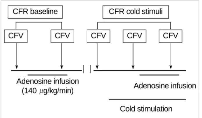
All subjects refrained from caffeine-containing beverages, alcohol and smoking for least 24 hr before the study. All vasoactive medications were also discontinued for 24 hr prior to the study. Coronary flow velocity (CFV) was measured using a high resolution ultrasound system (Sequoir C256 Acuson, Mountain view, CA, U.S.A.), with a 5-MHz transducer, via a transthoracic approach, in the distal left anterior descending coronary artery. Hyperemia was induced by intravenous adeno- sine infusion (140 g/kg/min) for 2 min and cold stimulation was performed by immersing both hands into ice water for 2 min. A diagram of the study protocol is shown in Fig. 1.
Briefly, CFV was recorded at rest and intravenous adenosine infusion before cold stimulation as baseline. After performing baseline measurements, ten minutes was allowed to allow the hemodynamic condition to fully recover to the resting state, cold stimulation by immersing both hand into ice water was started. CFV before and after adenosine infusion were recorded again under cold stimulation. Throughout the study, ECG was monitored continuously. Blood pressure and heart rate were checked simultaneously with CFV measurements.
Coronary flow reserve (CFR) was calculated as the ratio of hyperemic to resting mean diastolic coronary flow velocity.
The rate-pressure product (RPP) was defined as the product of heart rate and systolic blood pressure.
Coronary vascular resistance index (CVRI) was calculated as follows; [mean arterial pressure at peak blood flow veloci- ty/hyperemic diastolic velocity]÷[mean arterial pressure at basal flow velocity/basal peak diastolic velocity] (18).
Statistical analysis
For descriptive purposes, all data are presented as mean±
SD and relative frequencies, as indicated. Absolute changes of CFV induced by adenosine and cold stimulation were analyzed using Wilcoxon sign rank test and Mann-Whit- ney test. A p-value of <0.05 was considered statistically sig- nificant.
RESULTS
Table 1 summarizes the baseline characteristics of the study population. VA patients were older than the controls (57± 10 yr vs. 38±16 yr; p=0.001). The incidences of associated conditions (diabetes mellitus, smoking and dyslipidemia) were higher in VA patients than in controls, except hypertension (7% vs. 20%; p<0.05).
In all subjects, the intravenous infusion of adenosine induced significant increases in heart rate (HR) and RPP and decreases in blood pressure. Hemodynamic responses to adenosine were similar in both groups (Table 2). As shown in Table 3, base-
CFR baseline
Adenosine infusion
(140 g/kg/min) Adenosine infusion
Cold stimulation
CFV CFV CFV CFV CFV
CFR cold stimuli
Fig. 1.Study protocol. CFR, coronary flow reserve; CFV, coronary blood flow velocity.
*p<0.05, Vasospastic angina vs. Controls. �included 9-healthy young vol- unteers.
Vasospastic angina Controls
No. of patients 14 15
Men:Women 9:5 11:4
Age (yr) 37-67 23-70�
Mean age (yr)* 57±10 38±16
Hypertension* 1 (7%) 3 (20%)
Diabetes* 2 (14%) 0 (0%)
Smoking* 8 (57%) 0 (0%)
Dyslipidemia* 3 (21%) 0 (0%)
Table 1.Clinical characteristics of study populations
*p=not significant, Vasospastic angina vs. Controls, �p<0.05, rest vs. adeno- sine in each group. HR, heart rate; bpm, beats po2minute; SBP, systolic blood pressure; DBP, diastolic blood pressure; MBP, mean blood pres- sure; RPP, rate pressure product (mmHg/min/100).
Vasospastic angina rest adenosine�
Controls*
rest adenosine�
HR, bpm 62±6 80±7 60±5 83±7
SBP, mmHg 122±19 115±13 121±12 114±10
DBP, mmHg 72±15 67±12 72±8 65±9
MBP, mmHg 89±15 83±12 89±8 81±9
RPP 76±16 95±15 72±8 95±10
Table 2.The hemodynamic changes by adenosine infusion at baseline
*p=not significant, �p=0.08, Vasospastic angina vs. Controls. PSV, peak systolic velocity; PDV, peak diastolic velocity; MDV, mean diastolic velocity;
CFR, coronary flow reserve; CVRI, coronarv vascular resistance index.
Vasospastic angina rest adenosine*
Controls rest adenosine*
PSV, cm/sec 12±5 36±17 10±3 34±15 PDV, cm/sec 25±7 66±16 24±7 76±25 MDV, cm/sec 19±5 51±11 18±5 54±14
CFR 2.8±0.9 3.1±0.7*
CVRI 0.35 0.31�
Table 3.Coronary blood flow by adenosine infusion at baseline
line and hyperemic mean diastolic velocities of coronary flow were similar in VA patients and controls. As a result, CFR and CVRI were also similar in the two groups (CFR, 2.8± 0.9 vs. 3.1±0.7, p=0.21; CVRI, 0.35 vs. 0.31, p=0.08).
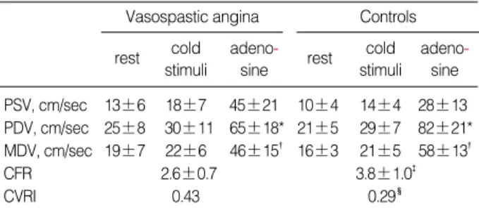
Under cold stimulation, heart rate, blood pressure, and RPP increased in both groups. In particular, increases in blood pres- sure and RPP were prominent in the VA group (Table 4). Co- ronary flow velocities were increased by cold stimulation in both groups, increases were lower in the VA group (p=0.03, Table 5).
Hemodynamic responses to adenosine during cold stimu- lation were similar in the two groups (Table 4). Unlike base- line response, hyperemic response to adenosine was blunted in the VA group under cold stimulation. In VA, the increase in diastolic coronary velocity was lower than in the controls, resulting in a lower CFR and a higher CVRI than for the con- trols (Table 5).
In summary, at room temperature, coronary flow velocity and CFR were similar in VA and control groups. Under cold stimulation, compared with baseline, CFR was marginally increased in controls (3.1±0.7 to 3.8±1.0, p=0.06). In con- trast, in the VA group, CFR decreased (2.8±0.9 to 2.6±0.7, p=0.05) and CVRI increased (0.35 to 0.43, p=0.01).
DISCUSSION
The present study shows the characteristics of coronary
blood flow in response to cold stimulation in VA patients by direct measurements of coronary blood flow velocities using TTDE.
Patients with angina caused by epicardial coronary artery spasm are diagnosed as either having ‘‘vasospastic angina’’ or
‘‘variant angina’’ (6). Coronary artery spasm may produce dif- fuse or focal arterial narrowing and be a manifestation of a generalized smooth muscle contractile disorder (9, 10, 19).
However, whether functional abnormality of the coronary microvascular circulation is present in vasosapstic angina is controversial (20, 21). In our study, as the coronary flow reserve was similar to that of controls at baseline, the microvascular function of VA appeared normal. This finding is agreement with a recent study by Sueda et al. (20). They reported no dif- ference in CFR in patients with acetylcholine induced spasm between the spasm positive and spasm negative vessels, and maintained normal in vessel with spasm as control.
There is also some controversy on the status of coronary vaso- motor tone. Kasai et al. (22) reported that the basal coro- nary artery tone of controls and VA patients does not differ significantly. However, Hoshio et al. (23) reported that coro- nary artery tone is significantly higher in VA patients than in control. In our study, the coronary vascular resistance index of VA patients at room temperature did not differ from that of the controls (0.35 vs. 0.31, p=0.08), but, as the p-value was 0.08, it is possible that CVRI might have been higher VA, if the study population had been larger.
The cold pressor test is recognized as a means of evaluating endothelial function and the functional integrity of the vas- cular wall (24, 25). Normally, cold stimulation induces the sympathetic release of norepinephrine and epinephrine and increases the heart rate, arterial blood pressure, and myocar- dial oxygen demand. This increase in myocardial metabolic demand has been shown to dilate epicardial arteries and in- crease coronary blood flow. Moreover, this was found to be mediated through the -adrenoreceptor by direct nitric oxide synthesis stimulation and through the flow-dependent release of endothelial-derived nitric oxide, despite -adrenergic coro- nary artery constriction by the direct stimulation of smooth
Vasospastic angina rest cold
stimuli
adeno- sine
Controls rest cold
stimuli adeno-
sine HR, bpm 62±6 70±10 84±10 61±7 68±8 85±9 SBP, mmHg 120±21 154±27* 127±13 119±12 138±11 123±11 DBP, mmHg 72±13 86±16 75±9 72±7 89±10 77±11 MBP, mmHg 89±15 114±18* 91±13 88±8 107±9 91±8 RPP 75±16 109±26 106±17 72±10 95±12 104±13 Table 4.The hemodynamic changes to cold stimulation and adenosine
*p<0.05, Vasospastic angina vs. Controls. HR, heart rate; SBP, systolic blood pressure; DBP, diastolic blood pressure; MBP, mean blood pres- sure; RPP, rate pressure product (mmHg/min/100).
Vasospastic angina rest cold
stimuli
adeno- sine
Controls rest cold
stimuli adeno-
sine PSV, cm/sec 13±6 18±7 45±21 10±4 14±4 28±13 PDV, cm/sec 25±8 30±11 65±18* 21±5 29±7 82±21*
MDV, cm/sec 19±7 22±6 46±15� 16±3 21±5 58±13�
CFR 2.6±0.7 3.8±1.0�
CVRI 0.43 0.29�
Table 5.Coronary blood flow by adenosine infusion under cold stimulation
*p, �p, �p, �p<0.05, Vasospastic angina vs. Controls. PSV, peak systolic velocity; PDV, peak diastolic velocity; MDV, mean diastolic velocity (cm/
sec); CFR, coronary flow reserve; CVRI, coronary vascular resistance index.
Vasospastic angina at baseline under cold
stimuli
Controls at baseline under cold
stimuli CFR 2.8±0.9 2.6±0.7� 3.1±0.7 3.8±1.0*
CVRI 0.35 0.43� 0.31 0.29
Table 6.Comparison of CFR and CVRI between baseline and cold stimulation
*p<0.05, at baseline vs. under cold stimuli in controls. �p,�p<0.05, at base- line vs. under cold stimulation in vasospastic angina; CFR, coronary flow reserve; CVRI, coronary vascular resistance index.
muscle cells (24, 26, 27). However, it is known that coronary dilatation by cold stimulation is impaired in patients with endothelial dysfunction such as hypertension or diabetes mel- litus (15, 28). Infrequently, cold stimulation may cause coro- nary vasospasm in VA, but the reported sensitivity is about 2 to 10%, which is too low to be clinically significant in the diagnosis of vasosapstic angina (29, 30). Our VA patients did not have chest pain, and did not show ECG changes, left ven- tricular wall motion abnormality on TTDE, or epicardial coronary artery vasospasm on coronary angiography during cold stimulation.
In this study, coronary blood flow incrementation by cold stimulation was lower in VA patients than in controls, and CFR decreased and CVRI increased in VA under cold stim- ulation. These findings suggest that sympathetic responses and microvascular integrity are different between VA patients and controls under cold stimulation. Constitutive nitric oxide synthase in the arterial endothelium continuously generates nitric oxide, which has been shown (31) to maintain basal vascular tone in animals and humans. Kugiyama et al. (32) recently showed that there is a deficiency in endothelial nitric oxide bioactivity at the sites of coronary artery spasm, and that this deficiency may play an important role in the patho- genesis of coronary spasm. Our data suggests that flow medi- ated endothelial dependent vasodilation was impaired at the coronary microvascular level in VA by cold stimulation.
There are several limitations in our study. First, the study population was small to derive definite conclusions. Second, the VA patients were older than the controls. Third, changes in epicardial coronary artery diameters under cold stimulation were measured on coronary angiography only in 8 patients (3 VA patients and 5 controls). However, none of these pati- ents showed coronary artery diameter changes by cold stim- ulation alone. In remaining study population, no patient expe- rienced chest pain or ECG changes under cold stimuation.
In conclusion, CFR is preserved at room temperature in vasospastic angina, but, compared to controls, increases in coronary blood flow in response to cold stimulation are blunt- ed and CFR is decreased. These findings suggest that endothe- lial dependent vasodilation is impaired at the coronary micro- vascular and epicardial artery levels in vasospastic angina un- der cold stimulation.
REFERENCES
1. Yasue H, Kugiyama K. Coronary spasm:clinical features and patho- genesis. Intern Med 1997; 36:760-5.
2. Yasue H, Touyama M, Shimamoto M, Kato H, Tanaka S. Role of autonomic nervous system in the pathogenesis of Prinzmetal variant form of angina. Circulation 1974; 50: 534-9.
3. Yasue H, Omore S, Takizawa A, Nagao M, Miwa K, Tanaka S. Cir- cardian variation of exercise capacity in patients with Prinzmetal’s variant angina; role of exercise induced coronary arterial spasm. Cir-
culation 1979; 59: 938-48.
4. Okumura K, Yasue H, Matsuyama K, Goto K, Miyagi H, Ogawa H, Matsuyama K. Sensitivity and specificity of intracoronary injection of acetylcholine for the induction of coronary artery spasm. J Am Coll Cardiol 1988; 12: 883-8.
5. Suzuki Y, Tokunaga S, Ikeguchi S, Miki S, Iwase T, Tomita T, Mura- kami T, Kawai C. Induction of coronary artery spasm by intracoro- nary acetylcholine: comparison with intracoronary ergonovine. Am Heart J 1992; 124: 39-47.
6. Maseri A, Chierchia S. Coronary artery spasm: demonstration, def- inition, diagnosis, and consequences. Prog Cardiovasc Dis 1982;
25: 169-92.
7. Nobuyoshi M, Abe M, Nosaka H, Kimura T, Yokoi H, Hamasaki N, Shindo T, Kimura K, Nakamura T, Nakagawa Y, Shiode N, Sakamo- to A, Kakura H, Iwasaki Y, Kim K, Kitaguchi S. Statistical analysis of clinical risk factors for coronary artery spasm: identification of the most important determinant. Am Heart J 1992; 124: 32-8.
8. Sugiishi M, Takatsu F. Cigarrette smoking is a major risk factor for coronary spasm. Circulation 1993; 87: 76-9.
9. Nakamura Y, Shinozaki N, Hirasawa M, Kato R, Shiraishi K, Kida H, Usuda K, Ishikawa T. Prevalence of migraine and Raynaud’s phe- nomenon in Japanese patients with vasospastic angina. Jpn Circ J 2000; 64: 239-42.
10. Miller D, Waters DD, Warnica W, Szlachcic J, Kreeft J, Theroux P.
Is variant angina the coronary manifestation of a generalized vaso- spastic disorder? N Eng J Med 1981; 304: 763-6.
11. Cannon RO 3rd, Cattau EL Jr, Yakshe PN, Maher K, Schenke WH, Benjamin SB, Epstein SE. Coronary flow reserve, esophageal motility and chest pain in patients with angiographically normal coronary arteries. Am J Med 1990; 88: 217-22.
12. Kiyotaka K, Masamichi O, Takeshi M, Seigo S, Hisao O, Michihiro Y, Yoshito I, Osamu H, Hiriaki K, Hirofumi S, Hirofumi Y. Nitric oxide-mediated flow-dependent dilation is impaired in coronary arteries in patients with coronary spastic angina. J Am Coll Cadiol 1997; 30:
920-6.
13. Motoyama T, Kawano H, Kugiyama K, Hirashima O, Ohgushi M, Tsunoda R, Moriyama Y, Miyao Y, Yoshimura M, Ogawa H, Yasue H. Vitamin E administration improves impairment of endothelium- dependent vasodilation in patients with coronary spastic angina. J Am Coll Cadiol 1998; 32: 1672-9.
14. Nabel EG, Ganz P, Gordon JB, Alexander RW, Selwyn AP. Dilation of normal and constriction of atherosclerotic coronary arteries caused by the cold pressor test. Circulation 1988; 77: 43-52.
15. Nitenberg A, Valensi P, Sachs R, Cosson E, Attali JR, Antony I. Prog- nostic value of epicardial coronary artery constriction to the cold pressor test in type 2 diabetic patients with angiographically normal coronary arteries and no other major coronary risk factors. Diabetes Care 2004; 27: 208-15.
16. Kang SH, Park HK, Lee CW, Kim JJ, Hong MK, Park SW, Park SJ.
Impaired flow-mediated vasodilation of epicardial coronary artery in vasospastic angina. J Korean Med Sci 1998; 13: 591-6.
17. Yasue H, Horio Y, Nakamura N, Fujii H, Imoto N, Sonoda R, Kugiya- ma K, Obata K, Morikami Y, Kimura T. Induction of coronary artery spasm by acetylcholine in patients with variant angina: possible role
of the parasympathetic nervous system in the pathogenesis of coronary artery spasm. Circulation 1986; 74: 955-63.
18. Antony I, Nitenberg A. Coronary vascular reserve is similarly reduced in hypertensive patients without any other coronary risk factors and in normotensive smokers and hypercholesterolemic patients with angiographically normal coronary arteries. Am J Hypertens 1997;
10: 181-8.
19. Rasmussen K, Ravnsbaek J, Funch-Jensen P, Bagger JP. Oesophageal spasm in patients with coronary artery spasm. Lancet 1986; 1: 174-6.
20. Sueda S, Kohno H, Fukuda H, Uraoka T. Coronary flow reserve in patients with vasospastic angina: correlation between coronary flow reserve and age or duration of angina. Coron Artery Dis 2003; 14:
423-9.
21. Anzai H, Saijo T, Nakajima R, Tezuka N, Takagi T, Tsunoda T, Ko- bayashi N, Nakamura S, Yamaguchi T. Evaluation of coronary flow reserve in patients with vasospastic angina. J Cardiol 2000; 36: 17-27.
22. Kasai JC, Tousoulis D, Gavrielides S, McFadden E, Galassi AR, Crea F, Maseri A. Comparison of epicardial coronary artery tone and reac- tivity in Prinzmetal’s variant angina and chronic stable angina pec- toris. J Am Coll Cardiol 1991; 17: 1058-62.
23. Hoshio A, Kotake H, Mashiba H. Significance of coronary artery tone in patients with vasospastic angina. J Am Coll Cardiol 1989; 14:
604-9.
24. Zeiher AM, Drexler H, Wollschlager H, Saurbier B, Just H. Coro- nary vasomotion in response to sympathetic stimulation in humans:
importance of the functional integrity of the endothelium. J Am Coll Cardiol 1989; 14:1181-90.
25. Zeiher AM, Drexler H, Wollschlager H, Just H. Modulation of coro-
nary vasomotor tone in humans: progressive endothelial dysfunction with different early stages of coronary atherosclerosis. Circulation 1991; 83: 391-401.
26. Nabel EG, Ganz P, Gordon JB, Alexander RW, Selwyn AP. Dilation of normal and constriction of atherosclerotic coronary arteries caused by cold pressor test. Circulation 1988; 77: 43-52.
27. Dimmeler S, Fleming I, Fisslthaler B, Hermann C, Busse R, Zeiher AM. Activation of nitric oxide synthase in endothelial cells by Akt- dependent phosphorylation. Nature 1999; 399: 601-5.
28. Antony I, Lerebours G, Nitenberg A. Angiotensin-converting enzyme inhibition restores flow-dependent and cold pressor test-induced dilations in coronary arteries of hypertensive patients. Circulation 1996; 94: 3115-22.
29. Dubois-Rande JL, Dupouy P, Aptecar E, Bhatia A, Teiger E, Hittinger L, Berdeaux A, Castaigne A, Geschwind H. Comparison of the effects of exercise and cold pressor test on the vasomotor response of nor- mal and atherosclerotic coronary arteries and their relation to the flow-mediated mechanism. Am J Cardiol 1995; 76: 467-73.
30. Waters DD, Szlachcic J, Bonan R, Miller DD, Dauwe F, Theroux P.
Comparative sensitivity of exercise, cold pressor and ergonovine test- ing in provoking attacks of variant angina in patients with active dis- ease. Circulation 1983; 67: 310-5.
31. Vallance P, Collier J, Moncada S. Effects of endothelium-derived nitric oxide on peripheral arteriolar tone in man. Lancet 1989; 2: 997-1000.
32. Kugiyama K, Yasue H, Okumura K, Ogawa H, Fujimoto K, Nakao K, Yoshimura M, Motoyama T, Inobe Y, Kawano H. Nitric oxide activity is deficient in spasm arteries of patients with coronary spas- tic angina. Circulation 1996; 94: 266-71.