로 나타나는 질환이다[1, 2]. 이러한 요로감염은 의료 영역에서 중 요한 부분을 차지하고 있는데, 이는 침습적인 의료시술의 사용 확 대와 노령 인구의 증가, 항생제남용과 이로 인한 내성균의 증가, 당 뇨 등 만성성인병의 증가, 면역저하 환자 및 만성질환자의 증가 등 으로 인해 감염의 발생률이 감소하고 있지 않기 때문이다[3-5]. 그 중에서 병원 내 중환자실이 요로감염의 유병률이 가장 높은 장소 이며, 병원 내 요로감염의 8-21%를 차지하고 있다[6, 7]. 요로감염 의 거의 모든 경우에서, 치료 원칙은 증상을 완화시키면서 병원성 세균을 제거하고 치료의 부작용 및 항균제 내성균의 발현을 줄이 는 것이다[1]. 이러한 요로감염의 원인균 및 항균제 내성 양상은 지 역에 따라 차이가 있을 수 있으므로 지역에 따른 원인균 및 항균제 내성률을 감시하는 것이 중요하다. 이에 이번 연구에서는 2007년 부터 2011년까지 경기 북부 지역 병원의 요 배양에서 분리된 미생 물의 균 종 및 항균제 내성을 연도별로 분석하고 그 변화 추이에 대해 고찰하고자 한다.
서 론
요로감염은 외래환자가 가장 많이 감염되는 질환 중의 하나로 무증상 세균뇨, 신우신염, 방광염, 요로패혈증 등 다양한 양상으
2007-2011년 경기 북부 지역 병원에서의 요 배양에 대한 고찰
Epidemiology and Resistance Patterns of Bacterial Pathogens in Urinary Tract Infections in the Northern Gyeonggi-do Area during 2007-2011
오은영·이혁민·임환섭·박윤희
Eunyoung Oh, M.D., Hyukmin Lee, M.D., Hwan Sub Lim, M.D., Younhee Park, M.D.
관동대학교 의과대학 진단검사의학교실
Department of Laboratory Medicine, Kwandong University College of Medicine, Goyang, Korea http://dx.doi.org/10.3343/lmo.2013.3.1.34
Corresponding author: Hyukmin Lee, M.D.
Department of Laboratory Medicine, Kwandong University, Myongji Hospital, 55 Hwasun-ro 14beon-gil, Deokyang-gu, Goyang 412-826, Korea
Tel: +82-31-810-7083, Fax: +82-31-962-1352, E-mail: [email protected]
Received: September 4, 2012 Revision received: September 27, 2012 Accepted: September 28, 2012
This article is available from http://www.labmedonline.org 2013, Laboratory Medicine Online
This is an Open Access article distributed under the terms of the Creative Commons Attribution Non-Commercial License (http://creativecommons.org/licenses/by-nc/3.0/) which permits unrestricted non-commercial use, distribution, and reproduction in any medium, provided the original work is properly cited.
Background: Bacteria that cause urinary tract infections (UTIs) are found with different frequencies in different regions; moreover, antibiotic susceptibility can also vary by region. We retrospectively studied and compared the species and antimicrobial susceptibility of bacterial pathogens isolated from patients with UTIs in the northern Gyeonggi-do area.
Methods: We analyzed urine specimens collected from patients who visited the Myongji Hospital between 2007 and 2011. The urine specimens were cultured, and bacteria were identified by biochemical examination with an API kit (bioMerieux Inc., USA). Antimicrobial susceptibility was de- termined by the disc diffusion method and the Vitek II system (bioMerieux Inc., USA).
Results: A total of 11,818 (31.4%) urine specimens were culture positive. The most common species identified were Escherichia coli (37.1%), Klebsiella pneumoniae (7.4%), Enterococcus faecium (6.1%), and Candida spp. (5.5%). The proportion of isolates producing extended-spectrum β-lactamases significantly increased during the study period.
Conclusions: E. coli, K. pneumoniae, and E. faecium were the 3 most common organisms identified. Of note, however, was the increasing fre- quency of Pseudomonas spp. and Proteus spp. isolated during the more recent years. Further studies are required from other centers in the northern Gyeonggi-do area.
Key Words: Urinary tract infection, Urine culture, Antibiotic susceptibility testing
대상 및 방법
2007년 1월부터 2011년 12월까지 경기도 고양시 덕양구에 위치 한 명지병원의 임상미생물검사실에 의뢰된 요 배양 검체 37,683건 을 대상으로 후향적으로 조사하였다. 채뇨는 카테터의 경우 카테 터 끝을 알코올로 닦은 후 멸균주사기를 이용하여 채뇨하고, 자가 배뇨가 가능한 경우 가능하면 아침 첫 요를 채취하는 것을 권장하 고, 배뇨한 첫 부분을 버리고, 중간부분을 무균용기에 채취하도록 하였다[8, 9]. 혈액한천 평판배지와 MacConkey 평판배지에 0.001 mL 직경의 백금이를 사용하여 접종한 후, 35℃에서 18-24시간 배 양하고, 균종 동정은 통상적인 생화학적 검사와 Vitek II system (bioMerieux Vitek Inc., Durham, NC, USA)를 이용하였고, 항균제 감수성검사는 디스크 확산법과 API kit (bioMerieux Inc., Dur- ham, NC, USA)를 이용하였다.
요로감염의 흔한 원인균에 대한 연도별 균 종 분리 비율과 중복 분리주를 제외된 환자에서 중요 항균제에 대한 감수성검사 결과 를 조사하였다. 각 원인균에 대한 연도별 균 분리 비율과 내원 및 입원 형태에 따른 원인균에 대해서는 카이제곱 검정을 이용하여
통계학적 유의성을 밝혔다.
결 과
2007년부터 2011년까지 총 37,683건의 요 배양이 의뢰되었고, 2007년 1,950건, 2008년 2,564건, 2009년 2508건, 2010년 2,337건, 2011년 2,459건으로 총 11,818검체(31.4%)에서 양성결과를 얻었다 (Table 1). Escherichia coli가 가장 흔하였고(37.1%), Klebsiella
pneumoniae (7.4%), Enterococcus faecium (6.1%), Candida al- bicans (5.5%), Candida trophicalis (5.5%), coagulase-negative
staphylococci (CoNS, 4.6%) 순이었다. 2007년부터 2011년 사이에 그람양성균은 2007년의 24.6%에서 2011년 16.1%로 점차 낮아졌 고, 효모형 진균은 2007년 14.7%에서 2011년 19.0%로 상승하였고 이는 통계학적으로 유의한 차이(P<0.000)가 있었다.검체가 채취된 환자의 병원 내원 및 입원 형태에 따라 외래, 응 급실, 일반 병실 및 중환자실 별로 분리된 원인균을 분석한 결과, 외래환자의 경우 E. coli (57.3%), CoNS (7.1%), K. pneumoniae (5.1%) 순이었고, 일반병실환자의 경우 E. coli (28.2%), E. faecium Table 1. Distribution of causative organisms of urinary tract infections from 2007 to 2011
Organisms No. (%) of isolates
2007 2008 2009 2010 2011 Total
Gram-positive 480 (24.6) 640 (25.0) 528 (21.1) 372 (15.9) 397 (16.1) 2,417 (20.5)
S. aureus 31 (1.6) 64 (2.5) 66 (2.6) 64 (2.7) 82 (3.3) 307 (2.6)
S. saprophyticus 3 (0.2) 5 (0.2) 9 (0.4) 3 (0.1) 4 (0.2) 24 (0.2)
Other CoNS 104 (5.3) 139 (5.4) 128 (5.1) 85 (3.6) 87 (3.5) 543 (4.6)
E. faecalis 136 (7.0) 138 (5.4) 92 (3.7) 85 (3.6) 94 (3.8) 545 (4.6)
E. faecium 138 (7.1) 221 (8.6) 188 (7.5) 96 (4.1) 82 (3.3) 725 (6.1)
Others 68 (3.5) 73 (2.8) 45 (1.8) 39 (1.7) 48 (2.0) 273 (2.3)
Gram-negative 1,184 (60.7) 1,475 (57.5) 1,544 (61.6) 1,566 (67.0) 1,594 (64.8) 7,363 (62.3)
E. coli 707 (36.3) 852 (33.2) 898 (35.8) 1,000 (42.8) 923 (37.5) 4,380 (37.1)
K. pneumoniae 138 (7.1) 209 (8.2) 201 (8.0) 150 (6.4) 173 (7.0) 871 (7.4)
Citrobacter spp. 27 (1.4) 31 (1.2) 36 (1.4) 46 (2.0) 40 (1.6) 180 (1.5)
Enterobacter spp. 60 (3.1) 79 (3.1) 77 (3.1) 58 (2.5) 67 (2.7) 341 (2.9)
Serratia spp. 17 (0.9) 14 (0.5) 14 (0.6) 19 (0.8) 16 (0.7) 80 (0.7)
Proteus spp. 50 (2.6) 82 (3.2) 65 (2.6) 69 (3.0) 88 (3.6) 354 (3.0)
P. aeruginosa 100 (5.1) 105 (4.1) 163 (6.5) 106 (4.5) 150 (6.1) 624 (5.3)
A. baumannii 16 (0.8) 25 (1.0) 20 (0.8) 27 (1.2) 32 (1.3) 120 (1.0)
Others 69 (3.5) 78 (3.0) 70 (2.8) 91 (3.9) 105 (4.3) 413 (3.5)
Yeast 286 (14.7) 449 (17.5) 436 (17.4) 399 (17.1) 468 (19.0) 2,038 (17.2)
C. albicans 103 (5.3) 135 (5.3) 121 (4.8) 133 (5.7) 160 (6.5) 652 (5.5)
C. glabrata 57 (2.9) 108 (4.2) 105 (4.2) 88 (3.8) 112 (4.6) 470 (4.0)
C. tropicalis 99 (5.1) 143 (5.6) 140 (5.6) 128 (5.5) 135 (5.5) 645 (5.5)
C. famata 3 (0.2) 10 (0.4) 21 (0.8) 9 (0.4) 17 (0.7) 60 (0.5)
Others 24 (1.2) 53 (2.1) 49 (2.0) 41 (1.8) 44 (1.8) 211 (1.8)
Total 1,950 (100.0) 2,564 (100.0) 2,508 (100.0) 2,337 (100.0) 2,459 (100.0) 11,818 (100.0)
Abbreviation: CoNS, coagulase-negative staphylococci.
(9.6%), K. pneumoniae (8.4%), 응급실 환자에서는 E. coli (61.9%),
Enterococcus faecalis (3.6.%), CoNS (3.6%), 중환자실 환자에서는 Candida glabrata (18.1%), C. albicans (13.8%), E. coli (12.5%) 순
으로 분리되었다(Table 2). 내원 및 입원 형태에 따라 분리된 원인 균의 차이는 통계학적 의의(P<0.05)가 있었다. 가장 흔한 균종은E. coli로 외래환자의 57.3%, 응급실 내원 환자 및 일반 병실 환자
에서는 각각 61.9%와 28.2%로 분리된 반면, 중환자실 환자에서는 효모형 진균이 46.9%로 가장 흔하였다. E. faecium의 경우 외래와 응급실의 경우 1.1-1.3%의 낮은 양성률을 보였으나 일반 병실과 응 급실의 경우 9.6%와 7.6%로 높은 양성률을 보였다. K. pneu-moniae의 경우 외래와 병실 별 차이가 크게 나타나지 않았다.
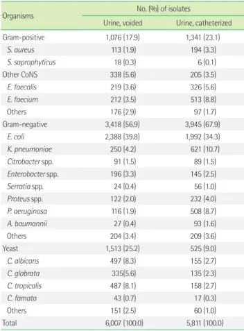
검체 양상을 중간뇨와 카테터를 이용한 경우로 나누어 분석한 결과, 중간뇨의 경우 E. coli (39.8%)와 효모형 진균이 가장 흔하게 분리되었고, 카테터 요에서는 E. coli (34.3%)와 더불어 그람 음성 균이 전반적으로 높게 분리되었다(Table 3).
그람음성균과 양성균에서 많은 비중을 차지하는 병원균에 대하 여 요로감염의 1차 치료제에 대한 항균제감수성 결과를 살펴보면
먼저 E. coli의 경우 levofloxcin에 70.5%의 감수성을 보이고, cotri- moxazole에 대하여 63.4%의 감수성을 보이는 것을 볼 수 있다 (Table 4). K. pneumoniae는 levofloxcin cotrimoxazole 모두에 61.6-66.5%의 감수성을 보이며 Pseudomonas aeruginosa는 levo- floxcin에 대해 50.7%의 감수성을 나타냈다. 그람양성균의 경우
Staphylococcus aureus의 경우 levofloxcin에는 56.1%의 감수성을
나타내지만 cotrimoxazole에 대해서는 95.3%의 높은 감수성을 나 타내고 있으며, CoNS의 경우 levofloxacin, cotrimoxazole에 대해 70.4%, 83.6%의 높은 감수성을 나타내고 있다. 특정 병원균에 대한 내성률을 살펴보면 S. aureus는 oxacillin에 대한 내성률이 2008 이 후 감소 추세에 있다가, 2009년 이후 비슷한 수치를 유지하고 있으 며, CoNS의 경우 oxacillin에 내성률이 평균 60%의 비율을 차지하 고 있었다. E. faecalis는 vancomycin에 대해 2008년까지 32%로 증 가추세를 보이다가 2009년 이후 약 22.7%로 감소하는 추세를 보이 고 있다(Fig. 1). E. coli 중 extended-spectrumβ
-lactamase의 경우 2007년 이후 꾸준하게 증가하는 추세로 평균 13.5%를 나타내었고,K. pneumoniae 중 extended-spectrum β
-lactamase의 경우 2007 년 24.3%에서 2011년 43.3%로 높은 증가율을 보이고 있었다.Table 2. Distribution of causative organisms of urinary tract infections according to wards and visit
Organisms No. (%) of isolates
OPD ER General ward ICU
Gram-positive 414 (19.4) 284 (12.7) 1,285 (23.8) 438 (21.2)
S. aureus 40 (1.9) 40 (1.8) 146 (2.7) 81 (3.9)
S. saprophyticus 11 (0.5) 7 (0.3) 6 (0.1) 0 (0.0)
Other CoNS 152 (7.1) 79 (3.6) 229 (4.2) 83 (4.0)
E. faecalis 79 (3.7) 79 (3.6) 295 (5.5) 92 (4.5)
E. faecium 24 (1.1) 28 (1.3) 517 (9.6) 156 (7.6)
Others 108 (5.1) 49 (2.2) 91 (1.7) 25 (1.2)
Gram-negative 1,686 (78.9) 1,896 (85.3) 3,128 (57.9) 661 (32.0) E. coli 1,226 (57.3) 1,374 (61.9) 1,522 (28.2) 258 (12.5) K. pneumoniae 109 (5.1) 170 (7.7) 453 (8.4) 139 (6.7) Citrobacter spp. 47 (2.2) 37 (1.7) 85 (1.6) 11 (0.5) Enterobacter spp. 81 (3.8) 54 (2.4) 161 (3.0) 45 (2.2)
Serratia spp. 10 (0.5) 13 (0.6) 48 (0.9) 9 (0.4)
Proteus spp. 54 (2.5) 82 (3.7) 197 (3.7) 21 (1.0) P. aeruginosa 66 (3.1) 76 (3.4) 373 (6.9) 109 (5.3)
A. baumannii 5 (0.2) 9 (0.4) 71 (1.3) 35 (1.7)
Others 88 (4.1) 78 (3.5) 214 (4.0) 33 (1.6)
Yeast 38 (1.8) 44 (2.0) 979 (18.3) 963 (46.9)
C. albicans 19 (0.9) 10 (0.4) 339 (6.3) 284 (13.8)
C. glabrata 11 (0.5) 17 (0.8) 248 (4.6) 194 (9.4) C. tropicalis 4 (0.2) 13 (0.6) 254 (4.7) 374 (18.1)
C. famata 2 (0.1) 1 (0.0) 26 (0.5) 31 (1.5)
Others 2 (0.1) 3 (0.1) 121 (2.2) 85 (4.1)
Total 2,138 (100.0) 2,219 (100.0) 5,396 (100.0) 2,065 (100.0) Abbreviations: OPD, outpatient department; ER, emergency room; ICU, intensive care unit; CoNS, coagulase-negative staphylococci.
Table 3. Distribution of causative organisms of urinary tract infections according to the specimen sampling technique
Organisms No. (%) of isolates
Urine, voided Urine, catheterized
Gram-positive 1,076 (17.9) 1,341 (23.1)
S. aureus 113 (1.9) 194 (3.3)
S. saprophyticus 18 (0.3) 6 (0.1)
Other CoNS 338 (5.6) 205 (3.5)
E. faecalis 219 (3.6) 326 (5.6)
E. faecium 212 (3.5) 513 (8.8)
Others 176 (2.9) 97 (1.7)
Gram-negative 3,418 (56.9) 3,945 (67.9)
E. coli 2,388 (39.8) 1,992 (34.3)
K. pneumoniae 250 (4.2) 621 (10.7)
Citrobacter spp. 91 (1.5) 89 (1.5)
Enterobacter spp. 196 (3.3) 145 (2.5)
Serratia spp. 24 (0.4) 56 (1.0)
Proteus spp. 122 (2.0) 232 (4.0)
P. aeruginosa 116 (1.9) 508 (8.7)
A. baumannii 27 (0.4) 93 (1.6)
Others 204 (3.4) 209 (3.6)
Yeast 1,513 (25.2) 525 (9.0)
C. albicans 497 (8.3) 155 (2.7)
C. glabrata 335(5.6) 135 (2.3)
C. tropicalis 487 (8.1) 158 (2.7)
C. famata 43 (0.7) 17 (0.3)
Others 151 (2.5) 60 (1.0)
Total 6,007 (100.0) 5,811 (100.0)
Abbreviation: CoNS, coagulase-negative staphylococci.
고 찰
요로감염은 원인이 되는 미생물 및 항균제에 대한 내성은 그와 관련된 지역의 역학과 관련이 있으며, 최근 몇 년 동안 크게 변화 해 왔다[2, 10]. 하지만, 한국에서 요로감염에 대한 지역에 따른 역 학 정보가 적은 것이 사실이다. 이에 이 연구에서는 2007년부터 2011년까지 경기 북부 지역 병원의 요 배양에서 분리된 미생물의 균 종 및 항균제내성을 연도별로 분석하고 그 변화 추이에 대해 고 찰하고자 하였다.
요로감염의 주요 원인균은 장내세균이며 특히 E. coli가 가장 많 은 빈도를 차지하는 것으로 알려져 있다. 이 연구에서도 총 양성 건수에서 그람음성세균이 적게는 57.5%에서 많게는 67.0%를 차지 하였으며 그 중 E. coli가 37.1%로 가장 흔하게 검출되었으며, K.
pneumoniae, E. faecium, 효모형 진균이 뒤를 따랐다. 전반적으
로 2007년 이후 E. coli, K. pneumoniae는 큰 차이를 보이지 않았 으며, 그 외 E. faecium은 2008년까지는 증가하다가 그 이후는 감 소를 하는 양상이었다. 특히 Proteus spp.가 2.6%에서 3.6%로 증가 하는 양상을 보였다. 이는 최근 국내 요로감염의 주요 원인균에 대한 연구에서 그람음성균에서 E. coli에 의한 감염은 감소하였으며,
P. aeruginosa, K. pneumoniae, Enterobacter spp., Proteus spp. 등
다른 그람음성균에 의한 감염이 증가하였다는 보고와 비슷한 양 상을 보이고 있다[11]. 검체가 채취된 환자의 내원 및 입원 형태별 로 나누어 봤을 때 외래, 응급실, 일반 병실 모두 E. coli가 각각 57.3%, 61.9%, 28.2%의 비율로 높게 나온 반면, 중환자실의 경우는 효모형 진균이 전체의 31.9%를 차지하였다. 중환자실의 경우 다른 문헌에서도 Candida spp.들이 중환자실 감염의 많게는 1/3까지 보고되고 있다고 한다[12].또한 중환자실의 경우 요로감염의 가장 중요한 위험인자는 카 테터의 유치 여부이며, 연구에 따라서는 중환자실에서 발생한 요 로감염의 95% 이상이 카테터와 연관이 있고 보고하였다[4, 13, 14].
이에 본 연구자에서 검체 양상에 따른 감염양상의 비교에서 카테 터를 유치한 그룹에서 E. faecalis, E. faecium, K. pneumoniae,
Proteus spp., P. areuginosa의 비율이 높게 나온 것을 볼 수 있었
다. P. aruginosa의 경우 단순 요로감염보다는 복잡성 요로감염에 서 더 많이 분리되는 결과를 보이며, 다른 연구에 의하면 외래환자 보다 입원환자에서 더 많이 분리되었다고 보고하고 있다[15].Table 4. Distribution of antimicrobial resistance of major uropathogens
Anti-microbial agent E. Coli (%) K. pneumoniae (%) P. aeruginosa (%) Proteus spp. (%) S. aureus (%) E. faecalis (%) E. faecium (%) CoNS (%)
Levofloxacin 26.3 35.7 41.4 12.2 42.2 26.9 95.4 24.9
Cotrimoxazole 35.3 25.1 88.0 43.2 5.5 17.4
Cefepime 13.6 36.8 27.3 31.1
Cefotaxime 13.9 37.2 53.2 31.1
Ceftazidime 13.7 38.1 31.1 31.1
Cephalothin 27.1 46.8 39.2
Gentamicin 24.6 31.9 36.5 36.5 44.7 25.1
ESBL(+) 13.6 38.3 41.0
ICR(+) 16.4 19.7
Abbreviations: CoNS, coagulase-negative staphylococci; ESBL, extended-spectrum β-lactamase; ICR, inducible clindamycin resistance.
Fig. 1. Antimicrobial resistance trend of major antibiotic-resistant bacteria isolated from the northern Gyeonggi area hospital.
Abbreviations: MRCoNS, methicillin-resistant coagulase-negative staphylococci; VAN-R-EFM, vancomycin-resistant E. faecium; ESBL-ECO, extend- ed-spectrum β-lactamase producing E. coli; ESBL-KPN, extended-spectrum β-lactamase producing K. pneumoniae; MRSA, methicillin-resistant Staphylococcus aureus.
MRCoNS VAN-R-EFM ESBL-ECO MRSA ESBL-KPN 80
70 60 50 40 30 20 10 0
%
2007 2008 2009 2010 2011
Year
한편, 요로감염의 치료는 감염의 각 유형에 따라 다른데 미국의 경우 주로 장내세균에 대한 1차 치료로 cotrimoxazole을 우선적으 로 권고하고 이에 대한 내성률이 20% 이상인 경우 fluoroquino- lone을 고려하는 지침을 사용하고 있다[16], 우리나라의 경우 단순 방광염 치료에는 경구용 fluoroquinolone을, 중증환자를 제외한 단순 급성신우신염환자에서는 일차로 정주용 ceftriaxone 혹은 gentamicin을 사용하고 이후 경구용 fluoroquinolone을 사용하고 있다. 가장 많은 빈도를 차지한 E. coli의 경우 fluoroquinolone과 cotrimoxazole에 대한 감수성은 각각 77-86%와 61-71%로서 미국 과 유럽에 비해 낮은 결과를 보인다[17]. 본 연구에서도 각각의 항균 제에 대하여 70.5%와 63.4%의 감수성을 나타내었다. 각 균주별 감 수성에 대해 분석한 결과, 2007년 extended-spectrum
β
-lactamase 를 생성하는 E. coli가 7.6%에서 23.1%로 증가하였고, K. pneu-moniae 또한 24.3%에서 43.3%로 높은 비율로 증가하는 결과를 보
였다. 이에 반해 Methicillin-resistant S. aureus (MRSA)나 MRCoNS 의 경우는 2007년 이후 소폭으로 감소하는 소견을 보였다.항균제감수성 양상은 지역이나 병원에 따라 차이를 보일 수 있 으므로 요로감염에 대한 원인균의 항균제감수성 정보를 알기 위 해 지속적인 조사를 통한 감시가 시행되어야 할 것이다. 이번 연구 는 단일 병원의 자료를 토대로 조사를 시행하였기 때문에 항균제 감수성 양상이 다른 연구에서 보인 결과와 다르게 나타날 수 있다.
좀 더 정확한 지역 내 항균제감수성 양상을 알기 위해서는 경기도 북부 지역내의 다기관 연구가 필요할 것으로 생각된다. 이를 통해 요로감염의 원인균에 대한 경험적 항균요법의 약제 선택에 도움이 될 수 있을 것이다.
요 약
배경: 요로감염의 원인균은 지역적 특성에 따라 빈도가 다양하고 항균제감수성 결과가 다르게 나타날 수 있다. 이에 저자들은 요로 감염의 원인균과 항균제감수성 결과를 후향적으로 연구하였다.
방법: 경기도 북부에 위치한 명지병원에서 2007부터 2011년까지 의뢰된 요 배양을 대상으로 하였다. 요검체를 접종 후 배양하였으 며, 균동정은 생화학검사와 API 키트로 시행하였고, 항균제감수성 검사는 디스크 확산법 및 Vitek II 시스템(bioMerieux Inc., USA)을 이용하였다.
결과: 총 11,818 (31.4%)검체에서 양성이 확인되었으며 가장 빈도 가 높은 것은 Escherichia coli 37.1%, Klebsiella pneumoniae 7.4%,
Enterococcus faecium 6.1% 그리고 효모형 진균 5.5% 순이었다.
Extended-spectrum
β
-lactamase 생성 세균은 연구기간 동안 증가 하였다.결론: E. coli, K. pneumoniae와 E. faecium이 본 연구에서는 가
장 흔한 요로감염의 원인균이었다. Pseudomonas aeruginosa와
Proteus spp.는 연구 기간 동안 꾸준히 증가되는 추세였다. 경기도
북부의 다른 기관과의 연관 연구가 더 필요하다.참고문헌
1. Kim YR, Huh JS, Kang SH. Patterns of antimicrobial susceptibility of the causative bacteria of urinary tract Infections in recent years in an island region. Korean J Clin Microbiol 2007;10:19-24.
2. Shin JH, Kim HR, Lee HR, Chung JI, Min K, Moon CS, et al. Etiology and antimicrobial susceptibility of bacterial pathogens causing com- munity-acquired urinary tract infection at a tertiary-care hospital. Ko- rean J Clin Microbiol 2005;8:142-7.
3. Kim HY, Yim SH, Cho HJ, Kim JS, Ha US, Kim DB, et al. Changes in causative organisms and antibiotic sensitivity in intensive care unit-ac- quired urinary tract infection. Korean J Urol 2009;50:1108-13.
4. Yu SM and Park KY. Risk factors for nosocomial urinary tract infection in the intensive care unit with a positive urine culture and foley cathe- terization. J Korean Acad Nurs 2007;37: 1149-58.
5. Foxman B. Epidemiology of urinary tract infections: incidence, mor- bidity, and economic costs. Am J Med 2002;113(S1):S5-13.
6. Lizioli A, Privitera G, Alliata E, Antonietta Banfi EM, Boselli L, Panceri ML, et al. Prevalence of nosocomial infections in Italy: result from the Lombardy survey in 2000. J Hosp Infect 2003;54:141-8.
7. Eriksen HM, Iversen BG, Aavitsland P. Prevalence of nosocomial infec- tions in hospitals in Norway, 2002 and 2003. J Hosp Infect 2005;60:40-5.
8. Patrick R, Murray, et al. eds. Manual of clinical microbiology. 9th ed.
Washington: ASM Press, 2007:323-4.
9. Jeong YS, Lee KW, et al. Diagnostic microbiology. 4th ed. Seoul: Seo- heung Publish, 2009:102-5.
10. Ferry S, Burman LG, Mattsson B. Urinary tract infection in primary health care in northern Sweden. I. Epidemiology. Scand J Prim Health Care 1987;5:123-8.
11. Ko HS, Choi DY, Han YT. A study of the changes of anitibiotic sensitiv- ity to the causative organisms of urinary tract infection for recent 5 years. Korean J Urol 1999;40:809-16.
12. Alvarez-Lerma F, Nolla-Salas J, León C, Palomar M, Jordá R, Carrasco N, et al. Candiduria in critically ill patients admitted to intensive care medical units. Intensive Care Med 2003;29:1069-76.
13. Richards MJ, Edwards JR, Culver DH, Gaynes RP. Nosocomial infec- tions in combined medical-surgical intensive care units in the United States. Infect Control Hosp Epidemiol 2000;21:510-5.
14. Mathaia D, Jones RN, Pfallera MA. Epidemiology and frequency of re- sistance among pathogens causing urinary tract infections in 1,510 hospitalized patients: A report from the SENTRY Antimicrobial Aur- veillance Program (North America). Diagn Microbiol Infect Dis 2001;
40:129-36.
15. Ryu KH, Kim MK, Jeong YB. A recent study on the antimicrobial sen- sitivity of the organisms that cause urinary tract infection. Korean J Urol 2007;48:638-45.
16. Warren JW, Abrutyn E, Hebel JR, Johnson JR, Schaeffer AJ, Stamm
WE. Guidelines for antimicrobial treatment of uncomplicated acute bacterial cystitis and acute pyelonephritis in women. Infectious Dis- eases Society of America (IDSA). Clin Infect Dis 1999;29:745-58.
17. The Korean Society of Infectious Diseases, The Korean Society for Chemotherapy; Korean Association of Urogenital Tract Infection and Inflammation; The Korean Society of Clinical Microbiology. Clinical guideline for the diagnosis and treatment of urinary tract infections:
asymptomatic bacteriuria, uncomplicated & complicated urinary tract infections, bacterial prostatitis. Infect Chemother 2011;43:1-25.