42
서 론
갑상선 유두암은 갑상선에서 발생하는 악성 종양의 70
∼80%를 차지하는 흔한 종양이지만 원발성 갑상선 림프 종은 전체 갑상선암의 약 0.1∼0.7%에 불과하다. 갑상선 MALT림프종은 B-cell origin의 비호지킨성 림프종으로 low grade에 속하고 대부분의 경우 Hashimoto 갑상선염의 경과 중 후천적으로 발생하며 최근 그 발생 비율이 증가 하고 있으며 급속히 커지는 경부 종물과 이로 인한 이차 적 압박, 폐쇄증상이 주증상이다. 갑상선 유두암 또한 Hashimoto 갑상선염에서 발생률이 유의하게 높고 임상적 으로 호르몬 억제 요법에 반응하지 않는 dominant nodule 이 주소이다. 두 질환 모두 갑상선에 국한된 경우 방사선 치료, 수술적 치료, 항암제 치료, 방사성동위원소 치료에 잘 반응하여 예후는 좋은 것으로 알려져 있다.
저자들은 Hashimoto 갑상선염 진단하에 수 년간 갑상선 호르몬 치료를 받아오던 53세 여성에서 갑상선 유두암과 동반된 갑상선 MALT림프종으로 진단된 1례를 경험하였 기에 문헌고찰과 함께 보고하는 바이다.
증 례
환 자: 신○, 53세 여자
주 소: 수 년간 계속된 우측 갑상선 종물 과거력 및 가족력: 특이사항 없음
병 력: 약 11년간이나 상기 주소로 갑상선 초음파 및 수 차례 미세침흡인세포검사에서 Hashimoto 갑상선염 진단 하에 갑상선 호르몬을 투여받았으나 갑상선 종물은 여전 하고, 갑상선기능검사에서 기능 저하증 소견을 보였으며 이 종물로 인한 기도 및 식도 폐색 증상은 호소하지 않았 다. 최근 개인 의원에서 마지막 시행한 미세침흡인세포검 책임저자:최영현, 전북 전주시 완산구 중화산동 1가 300번지
ꂕ 560-750, 전주예수병원 외과 Tel: 063-230-8229, Fax: 063-230-8228 E-mail: memi@Godpeople.com 게재승인일:2002년 6월 3일
본 증례는 2002년 춘계대한외과학회에서 포스터전시된 증례임.
중심 단어: 갑상선 MALT 림프종
ꠏꠏꠏꠏꠏꠏꠏꠏꠏꠏꠏꠏꠏꠏꠏꠏꠏꠏꠏꠏꠏꠏꠏꠏꠏꠏꠏꠏꠏꠏꠏꠏꠏꠏꠏꠏꠏꠏꠏꠏꠏꠏꠏꠏꠏꠏꠏꠏꠏꠏꠏꠏꠏ Departments of Surgery and 1Anatomical Pathology, Presby- terian Medical Center, Jeonju, Korea
갑상선 유두암과 동반된 갑상선 MALT 림프종 1예
전주예수병원 외과, 1해부병리과
최영현․오성수․이광민1․주명진1
Thyroid MALT Lymphoma Associated with Thy- roid Papillary Cancer
Young-Hyun Choi, M.D., Sung-Soo Oh, M.D., Kwang-Min Lee, M.D.
1 and Myung-Jin Joo, M.D.1There are thyroid lymphoma and thyroid papillary cancer in thyroid disease which can happen being associated with Hashimoto's thyroiditis. Thyroid lymphoma is a rare disease consisting of less than 1% of lymphoma and of 5% of thyroid cancer. It occurs with Hashimoto’s thyroiditis in 75%, and the cause is the immune reaction in which autoantibodies originated from thyroid are exposed B-cell continually. Also, the incidence of thyroid cancer, especially thyroid papillary cancer, increases in Hashimoto’s thyroiditis. The reason is that the genetic change-RET/PTC mutation- of Hashimoto’s thyroiditis is specific to thyroid papillary cancer than to other cancer. Patients usually complain neck nodule or sudden neck mass growing, hoarseness and respiratory difficulty.
FNA, USG, Neck CT, MRI, and RI scan can be used for diagnosis. We can choose radiation, operation, and che- motherapy in single form or combined form according to the stage and the location of disease. If a patient who has neck mass, the pathologic finding of it is similar to that of Hashimoto’s thyroiditis, and it is resistant to thyroid hormonal therapy, we should consider that it can be thyroid lymphoma or thyroid papillary cancer associated with Hashimoto’s thy- roiditis. We report a case of thyroid MALT lymphoma com- bined with occult papillary cancer which was resistant to thyroid hormonal therapy and which was successfully treated by operation and radiation therapy. (Korean J Endocrine
Surg 2002;2:42-46)
Key Words: Thyroid MALT lymphoma, Thyroid paillary can-
cer, Hashimoto’s thyroiditis최영현 외:갑상선 유두암과 동반된 갑상선 MALT 림프종 1예
43
ꠏꠏꠏꠏꠏꠏꠏꠏꠏꠏꠏꠏꠏꠏꠏꠏꠏꠏꠏꠏꠏꠏꠏꠏꠏꠏꠏꠏꠏꠏꠏꠏꠏꠏꠏꠏꠏꠏꠏꠏꠏꠏꠏꠏꠏꠏꠏꠏꠏꠏꠏꠏꠏꠏꠏꠏꠏꠏꠏꠏꠏꠏꠏꠏꠏꠏꠏꠏꠏꠏꠏꠏꠏꠏꠏꠏꠏꠏꠏꠏꠏꠏꠏꠏꠏꠏꠏꠏꠏꠏꠏꠏꠏꠏꠏꠏꠏꠏꠏꠏꠏꠏꠏꠏꠏꠏꠏꠏꠏꠏꠏꠏꠏꠏꠏ 사에서 갑상선 림프종이 의심되어 정확한 검사 및 치료위해 전원되었다.
이학적검사 소견: 내원당시 혈압, 맥박, 체온, 호흡수는 정상이었으며, 우측 갑성선엽 부위에 약 5×4 cm 크기의 고정된 부드러운 느낌의 압통이 없는 종괴가 만져졌고 국 소 발열이나 잡음은 없었다. 주위의 경부 임파절은 촉지 되지 않았고 심폐음은 정상이었으며 복부 진찰에서 특이 소견은 없었다.
검사실 소견: 혈액 검사 결과 백혈구 6,300/mm3, 혈색소 12.2 g/dl, 헤마토크리트 35.6%, 혈소판 260,000/mm3이었으 며, 생화학검사 결과 cholesterol 291 mg/dl, triglyceride 206 mg/dl으로 높은 것 외에는 정상이었고, 전해질 검사도 정 상소견이었다. 갑상선 기능검사에서 T3 0.93 ng/dl (0.8∼
Fig. 2. The area in Hashimoto's thyroiditis shows a dense lym- phoplasmacytic infiltrate with germinal center formation.
The follicular epithelium often shows Hṻrthle cell change (H & E, ×400).
Fig. 1. Gross specimen of Extranodal marginal zone B-cell lym- phoma of MALT type. The cut surface of thyroid lobe shows multinodularity with yellowish-tan color.
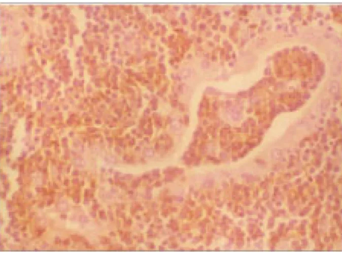
Fig. 3. A lymphoepithelial lesion is seen in the upper. It is a thyroid follicle infiltrated and expanded by lymphoid cells (H&E, ×400).
Fig. 5. Papillary carcinoma. The arborizing papillae have fibro- vascular cores (H&E, ×400).
Fig. 4. Immunostain for CD20 reveals that these cells are B-cell in nature (LSAB kit CD 20, ×400).
2.2), T4 8.09μg/dl (5∼13), free T4 1.03 ng/dl (0.8∼2.2) 그 리고 TSH 10.05μIu/ml (0.34∼3.5)이었다.
방사선학적 소견: 단순흉부촬영에서 이상소견은 없었 고, Tc-99m thyroid scan에서 갑상선엽부위의 minimal up- take소견을 보였다.
조직학적 소견: 갑상선 전 절제술 및 전경부 림프 곽청 술 시행 후 나온 조직 소견에서 우측엽은 5.5×4.5×3.5 cm 크기에 무게는 37 gm이었고 좌측엽은 4×3×2.5 cm 크기에 18 gm이었다. 절단면은 경계가 불분명한 다양한 크기의 yellowish-tan color의 견고한 결절성 모양이었다 (Fig. 1). 우측엽안에 약 6 mm 크기의 작고 단단한 결절이 발견되었다. 현미경 소견에서는 Hashimoto 갑상선염의 소 견(Fig. 2)과 함께 lymphoepithelial lesion (Fig. 3) 및 CD20 immunostain에서 양성 소견을 보였고(Fig. 4) 갑상선 유두 암의 조직소견도 관찰되었다(Fig. 5).
치료 및 경과: 경부 종물에 대한 검사 위해 시행된 수술 중 동결결절조직소견에서 MALT 림프종 및 occult 갑상선 유두암 소견을 보여 갑상선 전 절제술 및 전경부 림프 곽 청술 시행하였고 갑상선 유두암과 동반된 MALT 림프종 stage IE, REAL classification에 따라 “Extranodal marginal zone B-cell lymphoma of MALT type”으로 진단되었다. 수 술 후 약 1달 뒤 I-131 radioiodine ablation therapy (30 mCi) 받았고 이로부터 약 2주 후부터 4주 동안 전후 경부에 external radiation therapy (4,500 cGy) 받았으며 chemother- apy는 받지 않았다. 현재까지 특별한 이상 증상이나 소견 없이 갑상선 호르몬 복용하면서 추적 관찰 중이다.
고 찰
갑상선 림프종은 모든 림프종의 1% 이하, extranodal 비 호지킨성 림프종의 2%를 차지할 정도로 그 수가 적고 대 부분의 경우 B-cell에서 기원한 비호지킨성 림프종이며 (2,4,6) 아주 드문 경우 일차성 T-cell 비호지킨성 림프종도 보고되는데 이는 B-cell 림프종보다 생존율 및 예후가 불 량하다.(14) 대부분 항상 Hashimoto 갑상선염과 연관되어 발생하는데(1-7) Hashimoto 갑상선염 환자에서 갑상선 림 프종 발생의 상대적 위험도가 67∼80배라는 보고도 있 다.(10) 또한 Hashimoto 갑상선염에서는 갑상선암의 빈도 가 증가하는데 특히 갑상선 유두암의 빈도가 특이하게 높 다.(8) 갑상선 림프종의 대다수는(60∼75%) diffuse large cell type-intermediate grade이며 low grade malignant 림프종 -MALT type이 두 번째로 흔하다.(2-4,6) MALT 림프종의 경우 위장관계와 같은 다른 extranodal 부위에 유사한 병변 을 동반하기도 한다.(2,6)
MALT 림프종에서 보이는 세포유전학적 변화로는 t(11;18) (q21;q21)-이로 인한 API2/MLT fusion protein, trisomy 3, t(1;14)(p22;q32)-이로 인한 BCL-10 mutant, 그리고 BCL-6
alterations과 p53 mutation 등이 있다. 가장 흔한 것이 t(11;18)(q21;q21) translocation이고 약 50%에서 보이며 이 로 인해 API2 on 11q21와 MLT on 18q21 genes이 생겨나는 데 이 API2/MLT fusion protein이 림프종의 apoptosis에 억 제 작용을 하여 MALT 림프종이 생존하는데 기여할 것으 로 추정되고 있다. 또한 BCL-10 mutant은 wild type의 pro- apoptotic ability를 잃어버려 이 또한 MALT B-cell의 sur- vival에 기여한다.(2,10-12) 또한 Epstein-Barr virus (EBV)는 일부에서 Hashimoto 갑상선염이 악성림프종으로 변환되는 데 기여할 뿐 아니라(4∼9%) low grade 림프종이 high grade 로 변형되는 데도 기여한다.(3,4,13)
한편, Hashimoto 갑상선염에서 갑상선 유두암의 빈도가 증가되는 이유로는 분자생물학적 검사 결과 Hashimoto 갑 상선염에서 보이는 RET/PTC mutation products가 갑상선 유두암에 상대적으로 높은 특이도를 보이기 때문이다.(8) 갑상선 림프종은 여성에서 더 많이 발병하고(3:1∼8:
1), 평균연령은 60대(50∼80세)이며 약 1∼3개월 동안 급 속히 자라는 갑상선 종물 및 약 10∼30%에서 애성, 호흡 곤란, 연하곤란 등 국소 침범과 연관된 증상을 호소한 다.(1-3,6) 갑상선 림프종은 갑상선 기능검사 결과 주로 euthyroid 또는 hypothyroid 소견을 보이고(2,3,6) Grave's disease가 연관되는 경우가 아주 드물지만 Grave's disease 에서 갑상선암의 빈도는 일반인이나 중독성 갑상선 선종 환자보다 높다.(8) Hashimoto 갑상선염을 동반한 갑상선 유두암은 호르몬 억제 요법에 반응하지 않는 dominant nodule이 있는 경우 의심해볼 수 있으며 일부에서는 림프 종에서와 같이 결절이 갑자기 커지면서 동통이나 국소침 범, 압박증상을 호소할 수도 있다.(8)
진단에 필요한 방사선학적 검사로는 ultrasound, scan, CT, MRI 등이 있으며 특히 MRI는 인접조직, 림프절, 내경 정맥 그리고 경동맥 침범여부를 정확히 진단 가능케 하 며, 흉부 및 복부 CT는 폐문, 종격동 및 후복막 림프절 침 범 여부 판단을 위해 사용될 수 있다.(8) FNA는 Hashimoto 갑상선염과 연관된 갑상선 결절의 갑상선 유두암 여부를 검사하는 일차적 진단 방법으로 민감도가 90% 이상이지 만 이것만으로는 갑상선 림프종과 갑상선염을 정확히 감 별하기가 힘들어(2,4,6,8) 갑상선 림프종 대부분의 경우 open biopsy나 surgical resection으로 진단된다.(1,4,6) 그러 나 최근에는 FNA로 얻은 소량의 조직으로 PCR이나 Southern blot analysis등 분자생물학적 방법을 이용한 진단 이 증가하는 추세이다.(4,6,10,11)
갑상선 MALT 림프종에서 보이는 가장 흔한 조직소견 은 lymphoepithelial lesion (LEL)인데 이는 갑상선 follicles 이 neoplastic lymphoid cell로 꽉 들어찬 모습이며 종종 follicular epithelium이 부분적 또는 전부 이 병변으로 대치 된다.(3,10) 그 외 marginal zone cells, monocytoid B cells, small lymphocytes, 그리고 plasma cells이 다 나타나는 cel-
최영현 외:갑상선 유두암과 동반된 갑상선 MALT 림프종 1예
45
ꠏꠏꠏꠏꠏꠏꠏꠏꠏꠏꠏꠏꠏꠏꠏꠏꠏꠏꠏꠏꠏꠏꠏꠏꠏꠏꠏꠏꠏꠏꠏꠏꠏꠏꠏꠏꠏꠏꠏꠏꠏꠏꠏꠏꠏꠏꠏꠏꠏꠏꠏꠏꠏꠏꠏꠏꠏꠏꠏꠏꠏꠏꠏꠏꠏꠏꠏꠏꠏꠏꠏꠏꠏꠏꠏꠏꠏꠏꠏꠏꠏꠏꠏꠏꠏꠏꠏꠏꠏꠏꠏꠏꠏꠏꠏꠏꠏꠏꠏꠏꠏꠏꠏꠏꠏꠏꠏꠏꠏꠏꠏꠏꠏꠏꠏ lular hetrogenicity, 그리고 marginal zone이나 interfollicularregion에서 보이는 reactive follicles 등도 갑상선 림프종의 조직소견이다.(9)
갑상선 림프종의 치료는 림프종의 histologic subtype, tumor bulk, stage, 그리고 동반질환 등에 결정된다. Stage IE 및 IIE의 early stage 갑상선 림프종에서는 방사선 치료 가 단독으로 또는 수술적 치료와 같이 일차적 치료로 사 용되나, 최근 combination chemotherapy를 stage IE, IIE 환 자들에서 일차적 치료나 방사선 치료에 병합하여 사용하 여 원격 및 국소 재발을 감소시킨 좋은 결과를 보고한 경 우도 있다.(3,6,13)
저자에 따라 수술의 역할에 대해 의견이 다른데 어떤 이는 충분한 조직을 얻기 위한 biopsy 이상의 역할은 없고 갑상선 주위 nodal invovement가 있거나 extrathyroid exten- sion 있는 경우 resection이나 tumor debulking을 시행해서 는 안 된다고 한 반면에(2,6,10) 다른 이는 intrathyroid와 extrathyroid 수술적 감별이 치료결정과 예후 판단에 필수 적이고 갑상선 림프종이 early stage인 경우 thyroidectomy 가 림프 조직 증식을 자극하는 항원 제거 역할의 potential benefit의 효과가 있으며 bulky residual disease가 방사선 치 료 후 재발하는 데 전구 역할을 하므로 수술적 치료 후 방사선 치료를 함으로써 재발이 없었다는 보고도 있다.
(3,13,15) 또한 어떤 이는 MALT origin의 갑상선 림프종의 경우 antigenic stimulation 부위에 국소화되는 경향이 있어 국소 치료나 antigenic stimulus를 제거하는 치료만으로도 충분하다 하고 방사선 치료 후 5년 생존율이 MALT histo- logy인 경우 약 90%, 아닌 경우 55%라고 발표하였다.(17) Aggressive high grade large B-cell 림프종에서는 방사선, 항암제 병용 치료가 원칙이다. Vancouver group에서 발표 한 stage I 및 II 림프종에서 요구되는 최소한의 치료로는 CHOP 3 cycles과 침범된 부위의 3,000∼4,400 cGy의 방사 선 치료이고 2년 생존율은 약 85%라고 하였다.(2-4,6) 또 한 식도 및 기도 폐색증상이나 성대마미 증상을 보이는 환자에서도 combined mordality treatment가 적응증이 된 다.(1)
한편, Hashimoto 갑상선염을 동반한 갑상선 유두암에서 그렇지 않은 경우와 비교할 때 진단 및 치료방법의 변화 가 요구되지는 않고 수술적 합병증의 차이도 없으나 갑상 선 유두암이 Hashimoto 갑상선염을 동반한 경우 환자의 생존율이 증가한다는 보고가 있는데 명확한 이유는 아직 밝혀지지 않았다.(8)
일반적으로 갑상선 유두암은 자체로 예후가 양호하며 더 욱이 종양의 크기가 1.5 cm 이하, 한 쪽 엽만 침범한 경우, 국소 침입이나 원격전이가 없는 경우에서 예후가 좋다.
갑상선 림프종에서 MALT histology가 생존율에 유일한 중요 예후 인자라는 보고가 있고 그 외 좋은 예후 인자로 는 lack of bulk, stage IE disease, abscence of mediastinal or
retrosternal extension이 있고, 좋지 않은 예후 인자로는 수 술 후 residual tumor, extracapsular extension, retrosternal involvement, 그리고 severe local compression symptoms등 이 있으며 이런 인자들을 가진 환자에서는 chemotherapy 가 고려되어야 한다. 또한 불량한 예후와 연관되는 임상 적인 소견들로는 60세 이상, LDH 증가, Eastern Coopera- tive Oncology Group (ECOG) performance 2∼4 group, stage III/IV 그리고 한 곳 이상의 extranodal site involvement 등 이 있다.(2-4,6,17)
결 론
최근 저자들은 약 11년간이나 우측 갑상선 종물을 동반 한 갑상선 기능 저하증으로 수차례의 FNA 및 갑상선 호 르몬 치료를 받았으나 호전이 없었던 53세 여성에게서 수 술적 치료를 시행, 동결결절조직소견에서 occult 갑상선 유두암 및 MALT 림프종 소견을 보여 갑상선 전 절제술 및 전경부 림프 곽청술을 시행하였고 국소 방사선 조사의 병용 치료를 하여 현재까지 재발 및 합병증 없이 추적 관 찰 중인 1예를 경험하였기에 문헌고찰과 함께 보고하는 바이다.
REFERENCES
1) Haskell C, Berek J. Cancer treatment. 5th ed. Pennsylvania:
W.B. Saunders; 2001. p.1056-1058, 1428.
2) Devita V, Hellman S, Rosenberg S. Cancer: principles & prac- tice of oncology. 6th ed. philadelphia: LIPPINCOTT WIL- LIAMS & WILKINS; 2001. p.1759, 2223, 2301-2302.
3) Falk S. Thyroid disease. 2nd ed, New York: Lippincott-Raven;
1997. p.86-87, 403-409, 651-653.
4) Knowles D. Neoplastic hematopathlogy. 2nd ed, New York:
LIPPINCOTT WILLIAMS & WILKINS; 2001. p.1365-1370.
5) Cotran R, kumar V, Robbins S. Pathologic basis of disease.
5th ed. Pennsylvania: W.B. Saunders; 1994. p.1126-1127.
6) Ansell S, Grant C, Habermann T. Primary thyroid lymphoma.
Semin Oncol 1999;26(3):316-323.
7) Fenton JE, Kelly P, Stalk J, Phil D, O'Dwyer T. Lymphoma and Hashimoto's thyroiditis. J Laryngol Otol 1995;109:781- 783.
8) Singh B, Shaha A, Trivedi H. Coexistent hashimoto's thyroi- ditis with papillary thyroid carcinoma: impact on presentation, management, and outcome. Surgery 1999;126(6):1070-1076.
9) Harris N, Jaffe E, Stein H Banks P, Chan J, Cleary M, et al.
A revised european-american classification of lymphoid neo- plasm: a proposal from the international lymphoma study group. Blood 1994;84(5):1361-1392.
10) Kossev P, Livolsi V. Lymphoid lesions of the thyroid: review in light of the revised european-american lymphoma classifi- cation and upcoming world health organization classification.
Thyroid 1999;9(12):1273-1280.
11) Akagi T, Motegi M, Tamura A, Suzuki R, Hosokawa Y, Suzuki H, et al. A novel gene, MALT1 at 18q21, is involved in t(11;18) (q21;q21) found in low-grade B-cell lymphoma of mucosa associated lymphoid tissue. Oncogene 1999;18:5785- 5794.
12) Galdano G, Volpe G, Pastore C, Chlarle R, Capello D, Gloghl- ni A, et al. Detection of BCL-6 rearrangements and p53 muta- tions in Malt-lymphomas. Am J Hematol 1997;58:206-213.
13) Lam K, LO C, kwong L, Lee J, Srivastava G. Malignant lymphoma of thyroid: a 30-year clinicopathologic experience and on evaluation of the presence of epstein-barr virus. Am J Clin Pathol 1999;112:263-270.
14) Haciyanli M, Erkan N, Yorukoglu K, Sagol O, Harmancioglu
O. Primary non-Hodgkin's T-cell lymphoma of the thyroid gland complicating hashimoto's thyroiditis: case report. Thy- roid 2000;10(8):717-720.
15) Friedberg M, Coburn M, Monchik J. Role of surgery in stage IE non-Hodgkin's lymphoma of the thyroid. Surgery 1994;
16(6):1061-1067.
16) Leedman P, Sheridan W, Downey W, Fox R, Martin F. Com- bination chemotherapy as single modality therapy for stage IE and IIE thyroid lymphoma. Med J Aust 1990;152:40-43.
17) Laing R, Hoskin P, Hudson B, Hudson G, Harmer C, Bennett M, et al. The significance of malt histology in thyroid lym- phoma: a review of patients from the BNLI and regal marsden hospital. Clin Oncol 1994;6:300-304.