INTRODUCTION
Breast cancer is the most frequently diagnosed cancer in women, with a 12% lifetime risk of diagnosis, and the leading cause of cancer death among women, accounting for 14% of cancer deaths worldwide [1,2]. Trends in the incidence of breast cancer and death rates due to the disease differ among countries. Korea has one of the lowest breast cancer incidence rates (50.7 per 100,000 women-years in 2012) [3-5]; however, this incidence is rising rapidly, and breast cancer is now the second-most common cancer among women in Korea [3,6,7].
However, like many other developed countries, the age-ad- justed death rate in Korean patients with breast cancer has been decreasing [8]. Many reports have provided possible rel- evant explanations for the recent improvement in survival in patients with breast cancer. These explanations include na- tionwide screening programs with improved early detection of breast cancer [9], increases in the proportion of less aggres- sive cancers [8], and advances in adjuvant treatment, such as aromatase inhibitors for hormone receptor-positive tumors and trastuzumab for human epidermal growth factor receptor 2 (HER2)-positive tumors [10-12]. However, almost all of these studies have been from Western countries, and there are limited data to explain the improved breast cancer survival outcomes in Korea. To discriminate the influences on survival between tumor stage and time period, we analyzed survival outcome according to time at a single institution using our database of >10,000 patients.
Although improvement in the treatment outcomes, such as overall survival (OS) and disease-free survival (DFS), has been achieved on the strength of developed adjuvant treat-
Chronological Improvement in Survival of Patients with Breast Cancer:
A Large-Scale, Single-Center Study
Sae Byul Lee, Guiyun Sohn, Jisun Kim, Il Yong Chung, Hee Jeong Kim, Beom Seok Ko, Jong Won Lee, Byung Ho Son, Sung-Bae Kim1, Sei-Hyun Ahn
Division of Breast Surgery, Department of Surgery, and 1Department of Oncology, Asan Medical Center, University of Ulsan College of Medicine, Seoul, Korea
ORIGINAL ARTICLE
Purpose: This study aimed to chronologically evaluate survival of patients with breast cancer in Korea and investigate the ob- served changes during the last 20 years. We also sought to de- termine factors that may influence outcomes and changes in the duration of survival over time. Methods: We retrospectively ana- lyzed a total of 10,988 patients with breast cancer who were treated at our institution between January 1993 and December 2008. We divided the study period into three periods (P1, 1993–
1997; P2, 1998–2002; and P3, 2003–2008). We retrospectively reviewed the collected data from the Asan database, including age at diagnosis, clinical manifestations, pathology report, surgi- cal methods, types of adjuvant treatment modalities, type of re- currence, and follow-up period. Results: At a median follow-up of 8.2 years, we observed that survival outcomes have improved
recently. The 5-year breast cancer-specific survival (BCSS) rate also increased from 82.8% in P1 to 92.6% in P3 (p<0.001). The survival rate in patients with tumors at each stage increased in similar patterns in all patients, and, remarkably, there was a sig- nificant survival improvement in patients with stage III breast cancer (P1 vs. P3: 5-year BCSS, 57.4% vs. 80.0%, p<0.001).
The time period was a significant prognostic factor in multivari- ate analysis (P1 vs. P2: hazard ratio [HR], 0.83, p=0.035; P1 vs.
P3: HR, 0.75, p=0.015). Conclusion: The study results suggest an improvement in breast cancer survival in Korea, which is con- sistent with the development of treatments and early detection.
Key Words: Breast neoplasms, Prognosis, Recurrence, Survival
Correspondence to: Sei-Hyun Ahn
Division of Breast Surgery, Department of Surgery, Asan Medical Center, University of Ulsan College of Medicine, 88 Olympic-ro 43-gil, Songpa-gu, Seoul 05505, Korea
Tel: +82-2-3010-3490, Fax: +82-2-3010-6710 E-mail: ahnsh@amc.seoul.kr
Presented in part at the Global Breast Cancer Conference, Jeju, Korea, April 23–25, 2015.
Received: November 6, 2017 Accepted: January 4, 2018
Cancer
ments, patients with breast cancer can still experience any type of recurrence. Patients with breast cancer follow a variety of clinical courses depending on tumor characteristics such as size, lymph node metastasis status, and biological subtype.
Some relapse a few months after their initial operation, while others may relapse many years later. Therefore, identification of prognostic factors for relapse and death and predictive fac- tors for recurrence is very important to predict patient out- comes and determine the optimal form of adjuvant treatment.
The primary endpoint of this study was to analyze the changing patterns of survival and recurrence in Korean pa- tients diagnosed with breast cancer over the course of 16 years (1993–2008). We also analyzed the data to determine the fac- tors possibly influencing outcomes and changes in the dura- tion of survival over time.
METHODS
Patients and clinical data
We reviewed 11,119 patients with breast cancer who were treated at the Asan Medical Center, Seoul, Korea, between January 1993 and December 2008. We excluded 83 patients with a malignant phyllodes tumor, lymphoma, or sarcoma and 48 patients who underwent neoadjuvant chemotherapy, ultimately enrolling 10,988 patients. We divided the study pe- riod into three phases, according to significant changes in an- ti-hormonal therapy and chemotherapy, as follows: P1, 1993–
1997; P2, 1998–2002; and P3, 2003–2008. We analyzed the database of patients with primary breast cancer in each peri- od. All patient information and tumor characteristics were re- trieved from our prospectively collected database, including age, clinical manifestations, clinical and pathologic data, sur- gical methods, type of adjuvant treatment modality, type of recurrence, and follow-up period. We performed stage migra- tion according to the American Joint Committee on Cancer 7th classification. Nodal stage was re-classified according to the number of metastatic lymph nodes for patients diagnosed before 2002. Cases with 1–3, 4–9, and ≥10 metastatic lymph nodes were designated as N1, N2, and N3, respectively. Pa- tients who had a metastatic supraclavicular lymph node and no distant metastasis, who were previously regarded as having M1 disease, were designated as having N3 disease (Supple- mentary Tables 1-4, available online). This study was reviewed and approved by the Institutional Review Board of Asan Medical Center (20150185). Informed consent was waived because the study was based on retrospective clinical data.
Pathological data
Pathological data were evaluated in the Department of Pa-
thology at the Asan Medical Center. Estrogen receptor (ER) status, progesterone receptor (PR) status, and HER2 status were determined immunohistochemically. ER and PR were considered to be positive if >10% of cells showed positivity.
For HER2 overexpression analysis, cases graded 0, 1+ or 2+
were considered to be negative. Cases graded 2+ were evalu- ated by fluorescence in situ hybridization, and cases graded 3+
were regarded as positive.
Adjuvant treatment
Treatment varied for each patient. Considering each pa- tient’s general condition, treatments were administered based on the phenotype of the tumor. Endocrine therapy, such as aromatase inhibitors, tamoxifen, or a luteinizing hormone-re- leasing hormone (LHRH) analog, was administered to hor- mone receptor-positive patients. For triple-negative tumors, chemotherapy was administered. Adjuvant chemotherapy in- cluded an anthracycline or taxane. In the present study, che- motherapy was divided into cyclophosphamide, methotrex- ate, and 5-fluorouracil (CMF) or anthracycline-based; anthra- cycline- and taxane-based regimens were the most commonly used chemotherapeutic agents. After 2007, the use of trastu- zumab in the adjuvant setting for advanced breast cancer was covered by the Korean National Health Insurance. After adju- vant therapy, all patients had routine follow-up, including clinical examinations, laboratory tests, chest radiography, and mammography every 6 months during the first 5 years and annually thereafter until the first recurrence of their disease.
Statistical analysis
Data analysis was performed with SPSS version 18.0 (SPSS Inc., Chicago, USA). Linear regression analysis and a chi- square test were used to determine the trends in each parame- ter over time. OS was defined as the time from the initial sur- gery to the time of death, and breast cancer-specific survival (BCSS) was defined as the time from the initial surgery to the time of breast cancer-specific death, based on the Korean reg- istry cause-of-death code. DFS was defined as the time from the date of the initial surgery to the date of the first appear- ance of an initial relapse (locoregional or systemic) or cancer- specific death without any type of relapse. When more than one site was involved, patients were classified according to the dominant site of the metastasis. Survival curves were generat- ed using the Kaplan-Meier method, and the significance of survival differences among selected variables was verified us- ing the log-rank test. A multivariate Cox regression analysis with a backward elimination method was used to estimate hazard ratios and identify independent prognostic factors. All reported p-values are two-sided, and a value <0.05 was con-
Table 1. Clinicopathologic characteristics according to period at diagnosis of the 10,988 enrolled patients Factor
1993–1997 (n=1,051)
No. (%)
1998–2002 (n=2,703)
No. (%)
2003–2008 (n=7,234)
No. (%)
Total (n=10,988)
No. (%) p-value Linear association
Age at diagnosis (yr) <0.001 0.001
<31 41 (3.9) 82 (3.0) 203 (2.9) 326 (3.0)
31–40 279 (26.5) 616 (22.8) 1,395 (19.3) 2,290 (20.8)
41–50 378 (36.0) 1,156 (42.8) 3,245 (44.9) 4,779 (43.5)
51–60 234 (22.3) 557 (20.6) 1,560 (21.6) 2,352 (21.4)
61–70 84 (8.0) 217 (8.0) 633 (8.8) 934 (8.5)
71–80 31 (2.9) 68 (2.5) 178 (2.5) 277 (2.5)
>80 4 (0.4) 7 (0.3) 20 (0.3) 30 (0.3)
BMI (kg/m2) <0.001 <0.001
<18.5 48 (4.8) 98 (3.7) 260 (3.6) 406 (3.7)
18.5–22.9 433 (43.0) 1,215 (45.3) 3,290 (45.9) 4,938 (45.5)
23–24.9 235 (23.3) 663 (24.7) 1,700 (23.7) 2,598 (23.9)
≥25.0 291 (28.9) 705 (26.3) 1,918 (26.8) 2,914 (26.8)
Unknown 44 22 66 132
Operation method <0.001 <0.001
BCS 168 (16.0) 682 (25.2) 3,721 (51.4) 4,571 (41.6)
Mastectomy 834 (79.4) 1,935 (71.6) 3,361 (46.5) 6,130 (55.8)
Biopsy 49 (4.7) 86 (3.2) 152 (2.1) 287 (2.6)
Stage <0.001 <0.001
0 73 (7.0) 198 (7.3) 726 (10.0) 997 (9.1)
I 279 (26.8) 841 (31.3) 2,888 (40.0) 4,008 (36.6)
II 407 (38.9) 1,005 (37.4) 2,414 (33.5) 3,826 (34.9)
III 236 (22.7) 563 (20.9) 1,049 (14.5) 1,848 (16.9)
IV 48 (4.6) 84 (3.1) 148 (2.0) 280 (2.5)
Unknown 8 12 9 29
T stage <0.001 <0.001
Tis 74 (7.2) 198 (7.4) 727 (10.1) 999 (9.2)
T1 372 (36.2) 1,119 (42.1) 3,641 (50.6) 5,132 (47.2)
T2 451 (43.8) 1,121 (42.1) 2,388 (33.2) 3,960 (36.4)
T3 90 (8.7) 140 (5.3) 280 (3.9) 510 (4.7)
T4 42 (4.1) 83 (3.1) 158 (2.2) 283 (2.6)
Unknown 22 42 40 104
Node metastasis <0.001 <0.001
Negative 577 (57.2) 1,551 (58.8) 4,558 (63.4) 6,686 (61.7)
Positive 432 (42.8) 1,087 (41.2) 2,635 (36.6) 4,154 (38.3)
Unknown 42 65 41 148
Histologic grade <0.001 <0.001
G1 78 (11.3) 147 (7.6) 432 (7.1) 657 (7.6)
G2 308 (44.4) 897 (46.1) 3,444 (56.8) 4,649 (53.4)
G3 307 (44.3) 900 (46.3) 2,186 (36.1) 3,393 (39.0)
Unknown 358 759 1,172 2,289
Nuclear grade <0.001 <0.001
G1 22 (7.5) 73 (6.5) 463 (6.9) 558 (6.9)
G2 174 (59.6) 495 (43.8) 3,807 (57.0) 4,476 (55.3)
G3 96 (32.9) 563 (49.8) 2,407 (36.0) 3,066 (37.9)
Unknown 759 1,572 557 2,888
Lymphovascular invasion <0.001 <0.001
Negative 1 (9.1) 368 (67.4) 4,771 (74.8) 5,140 (74.1)
Positive 10 (90.9) 178 (32.6) 1,608 (25.2) 1,796 (25.9)
Unknown 1,040 2,157 855 4,052
(Continued to the next page)
Table 1. Continued Factor
1993–1997 (n=1,051)
No. (%)
1998–2002 (n=2,703)
No. (%)
2003–2008 (n=7,234)
No. (%)
Total (n=10,988)
No. (%) p-value Linear association
Estrogen receptor <0.001 <0.001
Negative 379 (45.1) 1,029 (40.8) 2,673 (37.7) 4,081 (39.1)
Positive 462 (54.9) 1,491 (59.2) 4,413 (62.3) 6,366 (60.9)
Unknown 210 183 148 541
Progesterone receptor <0.001 <0.001
Negative 355 (42.3) 1,242 (49.3) 3,200 (45.2) 4,797 (45.9)
Positive 485 (57.7) 1,277 (50.7) 3,882 (54.8) 5,644 (54.1)
Unknown 211 184 152 547
HER2 (IHC) <0.001 <0.001
Negative 412 (84.6) 1,502 (65.8) 5,275 (75.3) 7,189 (73.6)
Positive 75 (15.4) 780 (34.2) 1,727 (24.7) 2,582 (26.4)
Unknown 564 421 232 1,217
Subtype <0.001 <0.001
HR+/HER2– 241 (53.7) 1,093 (47.9) 3,901 (55.7) 5,235 (53.8)
HR+/HER2+ 41 (9.1) 425 (18.6) 788 (11.3) 1,254 (12.9)
HR–/HER2+ 31 (6.9) 355 (15.6) 938 (13.4) 1,324 (13.6)
HR–/HER2– 136 (30.3) 408 (17.9) 1,373 (19.6) 1,917 (19.7)
Unknown 602 422 234 1,258
Chemotherapy <0.001 0.327
Yes 558 (56.1) 1,809 (67.8) 4,482 (62.9) 6,849 (63.5)
No 437 (43.9) 861 (32.2) 2,646 (37.1) 3,944 (36.5)
Unknown 56 33 106 195
Radiation therapy <0.001 <0.001
Yes 268 (27.0) 960 (36.2) 4,479 (62.7) 5,707 (52.9)
No 724 (73.0) 1,691 (63.8) 2,669 (37.3) 5,084 (47.1)
Unknown 59 52 86 197
Anti-hormonal therapy <0.001 <0.001
Yes 583 (60.7) 1,687 (64.0) 4,807 (67.7) 7,077 (66.1)
No 378 (39.3) 948 (36.0) 2,297 (32.3) 3,623 (33.9)
Unknown 90 68 130 288
Chemotherapy regimen <0.001 <0.001
CMF 170 (80.6) 282 (23.2) 11 (0.3) 463 (8.3)
Anthracyclin based 36 (17.1) 757 (62.3) 2,160 (51.8) 2,953 (52.8) Anthracyclin and taxane based 1 (0.5) 72 (5.9) 1,763 (42.3) 1,836 (32.8)
Others 4 (1.9) 104 (8.6) 236 (5.7) 344 (6.1)
Unknown 347 594 312 1,253
Anti-hormonal therapy agent <0.001 <0.001
AI 0 7 (0.4) 575 (12.0) 582 (8.3)
SERM 568 (100) 1,643 (99.5) 3,466 (72.4) 5,677 (81.1)
SERM+LHRH analogue 0 1 (0.1) 743 (15.5) 744 (10.6)
Unknown 15 36 23 74
BMI=body mass index; BCS=breast-conserving surgery; HER2=human epidermal growth factor receptor 2; IHC=immunohistochemistry; HR=hormone recep- tor; HR+ =estrogen receptor positive or progesterone receptor positive; CMF =cyclophosphamide+methotrexate+fluorouracil; AI =aromatase inhibitor;
SERM=selective estrogen receptor modulator; LHRH=luteinizing hormone releasing hormone.
sidered statistically significant.
RESULTS
Patient characteristics
Table 1 presents the clinicopathologic features of the en-
rolled patients. The most prevalent range of age at diagnosis was 41 to 50 years old. The proportion of breast-conserving surgery performed increased from P1 to P3 (16.0% in P1 vs.
51.4% in P3, p<0.001). The proportion of radiation therapy also increased from P1 to P3 (27.0% in P1 vs. 62.7% in P3, p<0.001). Additionally, the proportion of patients diagnosed
1.0 0.8 0.6 0.4 0.2 0 1.0 0.8 0.6 0.4 0.2 0
1.0 0.8 0.6 0.4 0.2 0
1.0 0.8 0.6 0.4 0.2 0
1.0 0.8 0.6 0.4 0.2 0 1.0 0.8 0.6 0.4 0.2 0
1.0 0.8 0.6 0.4 0.2 0
1.0 0.8 0.6 0.4 0.2 0
1.0 0.8 0.6 0.4 0.2 0 1.0 0.8 0.6 0.4 0.2 0
1.0 0.8 0.6 0.4 0.2 0
1.0 0.8 0.6 0.4 0.2 0 0 24 48 72 96 120
0 24 48 72 96 120
0 24 48 72 96 120
0 24 48 72 96 120
0 24 48 72 96 120 0 24 48 72 96 120
0 24 48 72 96 120
0 24 48 72 96 120
0 24 48 72 96 120 0 24 48 72 96 120
0 24 48 72 96 120
0 24 48 72 96 120 Follow-up period (mo)
Follow-up period (mo)
Follow-up period (mo)
Follow-up period (mo)
Follow-up period (mo) Follow-up period (mo)
Follow-up period (mo)
Follow-up period (mo)
Follow-up period (mo) Follow-up period (mo)
Follow-up period (mo)
Follow-up period (mo)
BCSSDFSBCSSDFS BCSSOSBCSSDFS BCSSBCSSDFSDFS
Log-rank p=0.357 Log-rank p<0.001
Log-rank p<0.001
Log-rank p=0.078
Log-rank p<0.001 Log-rank p<0.001
Log-rank p=0.091
Log-rank p<0.001
Log-rank p<0.001 Log-rank p<0.001
Log-rank p=0.079
Log-rank p<0.001
A A
D
G
B B
E
H
C C
F
I
1993–1997 1993–1997
1993–1997
1993–1997
1993–1997 1993–1997
1993–1997
1993–1997
1993–1997 1993–1997
1993–1997
1993–1997 1998–2002
1998–2002
1998–2002
1998–2002
1998–2002 1998–2002
1998–2002
1998–2002
1998–2002 1998–2002
1998–2002
1998–2002 2003–2008
2003–2008
2003–2008
2003–2008
2003–2008 2003–2008
2003–2008
2003–2008
2003–2008 2003–2008
2003–2008
2003–2008
Figure 2. Chronological changes of survival in patients with primary breast cancer according to stage. Subgroup analyses of breast cancer-specific survival (BCSS) by stage, stage 0 (A), stage I (B), stage II (C), stage III (D), and stage IV (E). Subgroup analyses of disease-free survival (DFS) by stage, stage 0 (F), stage I (G), stage II (H), and stage III (I).
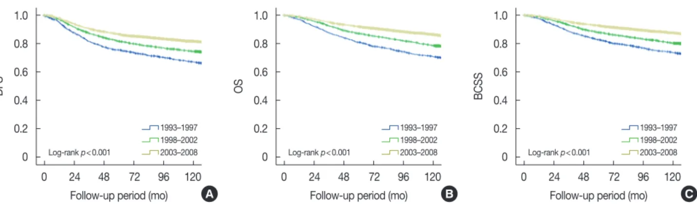
Figure 1. Chronological changes of survival in patients with primary breast cancer. Disease-free survival (DFS) (A), overall survival (OS) (B), and breast cancer-specific survival (BCSS) (C) of breast cancer according to periods at diagnosis in overall series.
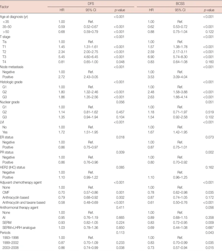
Table 2. Multivariate analysis for DFS and BCSS
Factor DFS BCSS
HR 95% CI p-value HR 95% CI p-value
Age at diagnosis (yr) <0.001 <0.001
<35 1.00 Ref. 1.00 Ref.
35–50 0.59 0.52–0.67 <0.001 0.62 0.53–0.72 <0.001
>50 0.68 0.59–0.79 <0.001 0.88 0.75–1.04 0.122
T stage <0.001 <0.001
Tis 1.00 Ref. 1.00 Ref.
T1 1.45 1.31–1.61 <0.001 1.57 1.38–1.78 <0.001
T2 2.34 2.00–2.75 <0.001 2.59 2.17–3.11 <0.001
T3 5.45 4.60–6.45 <0.001 6.90 5.74–8.30 <0.001
T4 0.81 0.65–1.00 0.048 0.83 0.64–1.08 0.160
Node metastasis <0.001 <0.001
Negative 1.00 Ref. 1.00 Ref.
Positive 2.72 2.43–3.05 3.53 3.09–4.04
Histologic grade <0.001 <0.001
G1 1.00 Ref. 1.00 Ref.
G2 1.80 1.32–2.46 <0.001 2.48 1.58–3.88 <0.001
G3 1.86 1.35–2.56 <0.001 2.63 1.66–4.14 <0.001
Nuclear grade 0.056 0.051
G1 1.00 Ref. 1.00 Ref.
G2 1.14 0.81–1.62 0.457 1.18 0.71–1.97 0.519
G3 1.35 0.94–1.94 0.104 1.54 0.92–2.58 0.102
LVI <0.001 <0.001
No 1.00 Ref. 1.00 Ref.
Yes 1.72 1.51–1.95 1.67 1.42–1.95
ER status 0.018 0.073
Negative 1.00 Ref. 1.00 Ref.
Positive 0.86 0.75–0.97 0.87 0.75–1.01
PR status 0.009 0.002
Negative 1.00 Ref. 1.00 Ref.
Positive 0.86 0.76–0.96 0.80 0.70–0.92
HER2 (IHC) status 0.085 0.162
Negative 1.00 Ref. Ref.
Positive 1.10 0.99–1.22 1.10 0.96–1.25
Adjuvant chemotherapy agent <0.001 <0.001
None 1.00 Ref. 1.00 Ref.
CMF 0.70 0.57–0.86 0.001 0.78 0.62–0.98 0.035
Anthracyclin based 0.79 0.68–0.92 0.002 0.87 0.74–1.05 0.172
Anthracyclin and taxane based 0.58 0.48–0.69 <0.001 0.61 0.50–0.76 <0.001
Antihormonal therapy agent 0.411 0.056
None 1.00 Ref. 1.00 Ref.
AI 0.95 0.76–1.19 0.665 0.89 0.68–1.15 0.358
SERM 0.93 0.82–1.05 0.224 0.83 0.72–0.95 0.009
SERM+LHRH analogue 1.03 0.78–1.36 0.850 0.69 0.44–1.08 0.687
Periods 0.113 0.043
1993–1998 1.00 Ref. 1.00 Ref.
1999–2002 0.87 0.70–1.09 0.233 0.83 0.70–0.99 0.035
2003–2008 0.86 0.74–0.99 0.038 0.73 0.57–0.94 0.015
DFS=disease-free survival; BCSS=breast cancer-specific survival; HR=hazard ratio; CI=confidence interval; ref.=reference; LVI=lymphovascular invasion;
ER=estrogen receptor; PR=progesterone receptor; HER2=human epidermal growth factor receptor 2; IHC=immunohistochemistry; CMF=cyclophosphamide+
methotrexate+fluorouracil; AI=aromatase inhibit; SERM=selective estrogen receptor modulator; LHRH=luteinizing hormone-releasing hormone.
with early stage breast cancer (stages 0 and I), as measured by T stage and nodal status, also increased from P1 to P3 (33.8%
in P1 vs. 50.0% in P3, p<0.001).
Survival
Survival was examined for 10,988 patients with breast can- cer first diagnosed during 1993–2008 and followed up through
1.0 0.8 0.6 0.4 0.2 0 1.0 0.8 0.6 0.4 0.2 0
1.0 0.8 0.6 0.4 0.2 0
1.0 0.8 0.6 0.4 0.2 0
1.0 0.8 0.6 0.4 0.2 0 1.0 0.8 0.6 0.4 0.2 0
1.0 0.8 0.6 0.4 0.2 0
1.0 0.8 0.6 0.4 0.2 0
1.0 0.8 0.6 0.4 0.2 0 1.0 0.8 0.6 0.4 0.2 0
1.0 0.8 0.6 0.4 0.2 0
1.0 0.8 0.6 0.4 0.2 0 0 24 48 72 96 120
0 24 48 72 96 120
0 24 48 72 96 120
0 24 48 72 96 120
0 24 48 72 96 120 0 24 48 72 96 120
0 24 48 72 96 120
0 24 48 72 96 120
0 24 48 72 96 120 0 24 48 72 96 120
0 24 48 72 96 120
0 24 48 72 96 120 Follow-up period (mo)
Follow-up period (mo)
Follow-up period (mo)
Follow-up period (mo)
Follow-up period (mo) Follow-up period (mo)
Follow-up period (mo)
Follow-up period (mo)
Follow-up period (mo) Follow-up period (mo)
Follow-up period (mo)
Follow-up period (mo)
DFS (local)DFS (local)DFS (local)DFS (local) DFS (regional)DFS (regional)DFS (regional)DFS (regional) DFS (systemic)DFS (systemic)DFS (systemic)DFS (systemic)
Log-rank p=0.250 Log-rank p=0.372
Log-rank p=0.579
Log-rank p=0.009
Log-rank p=0.313 Log-rank p=0.766
Log-rank p=0.587
Log-rank p=0.019
Log-rank p=0.002 Log-rank p<0.001
Log-rank p<0.001
Log-rank p<0.001
A A
D
G
B B
E
H
C C
F
I
1993–1997 1993–1997
1993–1997
1993–1997
1993–1997 1993–1997
1993–1997
1993–1997
1993–1997 1993–1997
1993–1997
1993–1997 1998–2002
1998–2002
1998–2002
1998–2002
1998–2002 1998–2002
1998–2002
1998–2002
1998–2002 1998–2002
1998–2002
1998–2002 2003–2008
2003–2008
2003–2008
2003–2008
2003–2008 2003–2008
2003–2008
2003–2008
2003–2008 2003–2008
2003–2008
2003–2008
Figure 4. Disease-free survival (DFS) according to recurrence type in patients with breast cancer by stage. DFS according to periods at diagnosis in stage I (A-C), stage II (D-F), and stage III (G-I).
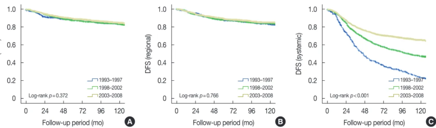
Figure 3. Disease-free survival (DFS) according to recurrence type in patients with breast cancer. DFS according to periods at diagnosis in overall se- ries. Local DFS (A), regional DFS (B), and systemic DFS (C).
August 31, 2014. Among the 10,988 patients, 372 were lost to follow-up (3.4%). The follow-up rates at 1 year, 3 years, and 5 years after surgery were 99.6%, 99.1%, and 98.2%, respectively.
The median follow-up period for the entire cohort was 98.7 months (range, 0–269.5 months), and the median duration of follow-up during P1, P2, and P3 was 166.7 months, 131.8 months, and 86.8 months, respectively.
During the follow-up period, 1,678 breast cancer-specific mortalities and 200 non-cancer-related deaths occurred. The 5-year OS rate was 89.8% for the entire cohort. We observed that survival outcomes had improved recently (Figure 1). The 5-year DFS increased from 75.6% in P1 to 86.6% in P3 (p<0.001). The 5-year OS increased from 81.0% in P1 to 92.0% in P3 (p<0.001). The 5-year BCSS also increased, from 82.8% in P1 to 92.6% in P3 (p<0.001).
We analyzed survival according to tumor stage to discover the influencing chronological changes on improved survival outcome (Figure 2). An improvement in 5-year BCSS of 80%
to 98.0% was observed in all stages. Otherwise, the 5-year DFS was improved in stage II (78.5% in P1 vs. 88.0% in P3) and stage III (43.4% in P1 vs. 68.2% in P3) patients.
We performed multivariate Cox proportional hazards re- gression analysis to identify the factors influencing DFS and BCSS (Table 2). This analysis demonstrated that the time fac- tor was significantly and independently associated with only BCSS (p=0.043). We also observed that age at diagnosis, T stage, node metastasis, histologic grade, lymphovascular inva- sion (LVI), PR status, and chemotherapeutic agent were asso- ciated with DFS and BCSS; ER status was significantly associ- ated with DFS, but not BCSS.
Recurrence
We performed an analysis of DFS according to time period for each type of recurrence (Figure 3). There were no signifi- cant differences in local or regional DFS according to time pe- riod. However, systemic DFS recently increased to a signifi- cant degree. We performed an additional analysis of DFS in each stage according to time period (Figure 4). There were no significant differences in stage 0 patients (data not shown).
While distant DFS was improved in all stages, local and re- gional DFS improved only in stage III patients. We analyzed the changes of adjuvant treatment according to time period in stage III patients. The administration of all adjuvant treat- ments, including radiotherapy, chemotherapy, and hormonal therapy, increased from P1 to P3. From P1 to P3, radiotherapy administration increased from 47.7% to 94.6%, chemotherapy administration increased from 78.5% to 97.1%, and hormonal therapy administration increased from 55.4% to 63.9%. More- over, the agents of chemotherapy and hormonal therapy
changed. In P1, the most frequently used chemotherapy was CMF (29.4%), but in P3, anthracycline/taxane-based agents (79.9%) were the most used. For hormonal therapy, the use of aromatase inhibitors increased from P1 to P3 (Supplementary Table 5, available online).
DISCUSSION
Our present chronological study indicated improvements in the survival of Korean patients with breast cancer during the study period from 1993 to 2008. The 5-year BCSS was 92.6% during 2003–2008, which was a significant improve- ment over the earlier periods (Figure 1). In the Surveillance, Epidemiology, and End Results database, the age-standardized 5-year relative survival rate for American patients with breast cancer diagnosed during 2003–2009 was 89.2% [13]. More- over, the 5-year survival rate of patients with breast cancer di- agnosed in 2006–2010 in the Korea central registration statis- tics was 91.1%, similar to that of the present study [14]. How- ever, the enrolled population in our study was based on a uni- form treatment environment at a single center, unlike the population registries in these previous studies.
There are many factors that can influence survival change in patients with breast cancer. Our data showed that more pa- tients were diagnosed with stage I disease since 2003 (40.0%) compared with the earlier periods. We expect that the detec- tion rate of early breast cancer in Korea has increased owing to societal generalization of organized screening programs and the development of early detection systems in cases of op- portunistic screening [15]. Therefore, the seeking of treatment in earlier stages of breast cancer may have resulted in better patient outcomes. Although all patients lived longer in later periods, survival also improved significantly more in patients with stage III breast cancer (Figure 2), which can likely be at- tributed to advancements in adjuvant systemic therapy. CMF was usually administered during P1, but its use gradually de- creased over the years between P1 and P3. New drugs, such as taxane- and anthracycline-containing regimens, have become available since the 2000s. Identical results have been reported in previous studies, suggesting that the administration of ad- juvant chemotherapy is associated with a better prognosis [10,16,17]. Henderson et al. [17] found that the risk reduc- tions of the addition of paclitaxel to AC (doxorubicin+
cyclophosphamide) were 17% for recurrence (p=0.001) and 18% for death (p=0.010). At 5 years, the DFS was 65% and 70% and the OS was 77% and 80% after AC alone or AC plus paclitaxel, respectively [18]. Our current study was not de- signed to identify which specific regimens led to survival im- provement, but we speculate that advances in adjuvant che-
motherapy in general have contributed to this clear trend.
When we performed multivariate survival analysis, the con- tributions of adjuvant chemotherapy to survival improvement were independent of other important factors such as age, tu- mor size, nodal status, and hormone receptor status (Table 2).
Another factor found to influence survival and recurrence was the use of anti-hormonal therapy. The use of newer endo- crine drugs could at least partially explain the increase in sur- vival over time. We initially mainly administered tamoxifen as an anti-hormonal treatment. Aromatase inhibitors and LHRH agonists subsequently gained widespread use in 2003. As ex- pected, there were major differences in the use of new aroma- tase inhibitors and/or LHRH agonists before and after 2003, because 1% of our patients treated during P1 but 28% treated during P3 received at least one of these drugs. The number of our patients administered aromatase inhibitors as adjuvant therapy was quite small, such that their impact on improve- ments over time appeared to be minimal. Trastuzumab has also improved the survival of HER2-positive patients [17], al- though our present findings do not significantly support this because the use of trastuzumab in the adjuvant setting for breast cancer was not covered by the Korean National Health Insurance during the period of this study.
In the second part of our present investigation, in which we analyzed DFS according to the type of recurrence, no change was seen in local and regional recurrence (LRR), but there was a difference in systemic recurrence in all patients among the three periods. Furthermore, there was a difference in sys- temic recurrence among the three periods in each stage of breast cancer for stages I–III (Figure 4). Hence, our results imply that advanced systemic therapy was sufficient to achieve better long-term survival rates, as mentioned above. Add- itionally, there was a significant difference in the LRR rates during the investigated period in stage III patients, but there were no significant differences in LRR for stage 0–II patients.
These results suggest that advancement in local management may be associated with reduced LRR rates of patients with breast cancer, particularly in patients with locally advanced breast cancer, and may have less of an effect on recurrence in early breast cancer. These outcomes are in line with those of a number of previous studies [19,20].
The survival benefit of post-mastectomy radiotherapy (PMRT) in patients with node-positive breast cancer has been well established through multiple randomized trials [19-21].
The results of the Early Breast Cancer Trialists’ Collaborative Group meta-analyses show that PMRT substantially reduces the risk of LRR [22]. These findings are congruent with other studies that confirmed the clinical benefit of adjuvant radio- therapy in patients who underwent breast-conserving surgery
[23,24]. There has been a benefit improvement from radio- therapy in patients with early breast cancer, which is assumed to have a relatively low absolute LRR risk (5-year local recur- rence rate P1: 1.5%, P2: 1.4%, and P3: 2.0% in stage I; P1:
2.5%, P2: 3.2%, and P3: 2.2% in stage II). To clarify the possi- bility of erroneous results in our present study in terms of lo- cal recurrence according to the type of surgery, we analyzed local recurrence among the subgroups according to surgical method. Although breast-conserving surgery has become more common, we found no significant difference in local re- currence according to the type of surgery (data not shown).
We wanted to identify the factors that exerted considerable influence, so we compared survival and recurrence between the P1, P2, and P3 time periods, including the time factor, by performing multivariate survival analysis. This analysis al- lowed us to conclude that age at diagnosis, tumor size, nodal status, histologic grade, LVI, and PR status had constant pro- portional effects on DFS and BCSS (Table 2). In addition, the effect of the period at diagnosis also had strong effects inde- pendent of other important factors; there was a 25% improve- ment in survival over the 16-year period. This increase is most likely a surrogate for improvements in detection, such as in- creases in screening, greater awareness of breast cancer, better preoperative diagnostic planning, better multidisciplinary de- cision making, and a thorough pathological investigation. The overall gains from the time effect were most likely due to a combination of other biological and social factors. Unfortu- nately, we did not have data on these factors and could only evaluate the effect of changes in treatment on survival.
Our present study had a number of limitations that should be considered when interpreting the results. As in all single- institution, retrospective, observational cohort studies, there was a potential for both referral and selection bias. In add- ition, the bias due to the drastic increase in the number of pa- tients in P3 and the associated change toward lower cancer stage would have had an impact on the survival results. Add- itionally, we speculated that differences in the follow-up peri- od between the investigated periods might be a limitation;
therefore, we adjusted the follow-up period to 87 months (median follow-up duration of patients diagnosed in P3), and similar results were obtained (p<0.001, data not shown).
In conclusion, this study of more than 10,000 patients re- vealed a marked improvement in survival for patients with breast cancer during the investigated period. Moreover, as the analyzed chronological change in recurrence rates of local, re- gional, and systemic recurrence differed from previous stud- ies, we identified a reduction in systemic recurrence for pa- tients with stage I–III breast cancer and in the LRR for those with stage III breast cancer during the most recent period. We
conclude that the recent improvement in Korean breast can- cer patient outcomes might be due to therapeutic advances in breast cancer treatment and a time effect, including intricate factors such as widespread screening and developments in di- agnostic planning and multidisciplinary decision making.
CONFLICT OF INTEREST
The authors declare that they have no competing interests.
REFERENCES
1. Jemal A, Bray F, Center MM, Ferlay J, Ward E, Forman D. Global cancer statistics. CA Cancer J Clin 2011;61:69-90.
2. Parkin DM, Bray F, Ferlay J, Pisani P. Global cancer statistics, 2002. CA Cancer J Clin 2005;55:74-108.
3. Ahn SH, Yoo KY; Korean Breast Cancer Society. Chronological changes of clinical characteristics in 31,115 new breast cancer patients among Koreans during 1996-2004. Breast Cancer Res Treat 2006;99:209-14.
4. Forouzanfar MH, Foreman KJ, Delossantos AM, Lozano R, Lopez AD, Murray CJ, et al. Breast and cervical cancer in 187 countries between 1980 and 2010: a systematic analysis. Lancet 2011;378:1461-84.
5. Shin HR, Joubert C, Boniol M, Hery C, Ahn SH, Won YJ, et al. Recent trends and patterns in breast cancer incidence among Eastern and Southeastern Asian women. Cancer Causes Control 2010;21:1777-85.
6. Ko SS; Korean Breast Cancer Society. Chronological changing patterns of clinical characteristics of Korean breast cancer patients during 10 years (1996-2006) using nationwide breast cancer registration on-line program: biannual update. J Surg Oncol 2008;98:318-23.
7. Park EH, Min SY, Kim Z, Yoon CS, Jung KW, Nam SJ, et al. Basic facts of breast cancer in Korea in 2014: the 10-year overall sur vival progress. J Breast Cancer 2017;20:1-11.
8. You JM, Kim YG, Moon HG, Nam SJ, Lee JW, Lim W, et al. Survival im- provement in Korean breast cancer patients due to increases in early- stage cancers and hormone receptor positive/HER2 negative subtypes:
a nationwide registry-based study. J Breast Cancer 2015;18:8-15.
9. Dawood S, Broglio K, Gonzalez-Angulo AM, Buzdar AU, Hortobagyi GN, Giordano SH. Trends in survival over the past two decades among white and black patients with newly diagnosed stage IV breast cancer. J Clin Oncol 2008;26:4891-8.
10. Early Breast Cancer Trialists’ Collaborative Group (EBCTCG). Effects of chemotherapy and hormonal therapy for early breast cancer on re- currence and 15-year survival: an overview of the randomised trials.
Lancet 2005;365:1687-717.
11. Trudeau M, Charbonneau F, Gelmon K, Laing K, Latreille J, Mackey J, et al. Selection of adjuvant chemotherapy for treatment of node-positive breast cancer. Lancet Oncol 2005;6:886-98.
12. Piccart-Gebhart MJ, Procter M, Leyland-Jones B, Goldhirsch A, Untch M, Smith I, et al. Trastuzumab after adjuvant chemotherapy in HER2- positive breast cancer. N Engl J Med 2005;353:1659-72.
13. Howlader N, Noone AM, Krapcho M, Garshell J, Neyman N, Altekruse SF, et al. SEER cancer statistics review, 1975-2010. National Cancer In- stitute. http://seer.cancer.gov/csr/1975_2010. Accessed Apr 11th, 2017.
14. Korea Central Cancer Registry, National Cancer Center. Annual report of cancer statistics in Korea in 2014. Sejong: Ministry of Health and Welfare; 2016. p.36.
15. Min SY, Kim Z, Hur MH, Yoon CS, Park EH, Jung KW, et al. The basic facts of Korean breast cancer in 2013: results of a nationwide survey and breast cancer registry database. J Breast Cancer 2016;19:1-7.
16. Goldhirsch A, Gelber RD, Coates AS. What are the long-term effects of chemotherapy and hormonal therapy for early breast cancer? Nat Clin Pract Oncol 2005;2:440-1.
17. Henderson IC, Berry DA, Demetri GD, Cirrincione CT, Goldstein LJ, Martino S, et al. Improved outcomes from adding sequential Paclitaxel but not from escalating Doxorubicin dose in an adjuvant chemothera- py regimen for patients with node-positive primary breast cancer. J Clin Oncol 2003;21:976-83.
18. Slamon DJ, Clark GM, Wong SG, Levin WJ, Ullrich A, McGuire WL.
Human breast cancer: correlation of relapse and survival with amplifi- cation of the HER-2/neu oncogene. Science 1987;235:177-82.
19. EBCTCG (Early Breast Cancer Trialists’ Collaborative Group), McGale P, Taylor C, Correa C, Cutter D, Duane F, et al. Effect of radiotherapy af- ter mastectomy and axillary surgery on 10-year recurrence and 20-year breast cancer mortality: meta-analysis of individual patient data for 8135 women in 22 randomised trials. Lancet 2014;383:2127-35.
20. Recht A, Gray R, Davidson NE, Fowble BL, Solin LJ, Cummings FJ, et al. Locoregional failure 10 years after mastectomy and adjuvant chemo- therapy with or without tamoxifen without irradiation: experience of the Eastern Cooperative Oncology Group. J Clin Oncol 1999;17:1689- 700.
21. Taghian A, Jeong JH, Mamounas E, Anderson S, Bryant J, Deutsch M, et al. Patterns of locoregional failure in patients with operable breast cancer treated by mastectomy and adjuvant chemotherapy with or without tamoxifen and without radiotherapy: results from five National Surgical Adjuvant Breast and Bowel Project randomized clinical trials. J Clin Oncol 2004;22:4247-54.
22. Overgaard M, Nielsen HM, Overgaard J. Is the benefit of postmastecto- my irradiation limited to patients with four or more positive nodes, as recommended in international consensus reports? A subgroup analysis of the DBCG 82 b&c randomized trials. Radiother Oncol 2007;82:247- 53.
23. Poortmans PM, Collette S, Kirkove C, Van Limbergen E, Budach V, Struikmans H, et al. Internal mammary and medial supraclavicular ir- radiation in breast cancer. N Engl J Med 2015;373:317-27.
24. Krug D. Regional nodal irradiation in early-stage breast cancer with 0-3 positive nodes. Strahlenther Onkol 2015;191:889-91.