대한족부족관절학회지: 제9권 제2호 2005
J Korean Foot Ankle Soc. Vol. 9. No. 2. pp.220-223, 2005
- 220 -
서 론
색소 융모 결절성 활액막염(pigmented villonodular synovitis: PVNS)은 신체의 어느 관절에나 발생할 수 있는 병인이 밝혀지지 않은 활막 증식성 질환으로 전반적으로 드 문 질환으로 보고되고 있다4,6). 특히 족관절에 발생하는 경 우는 매우 드물며2,3,6,10), 슬관절을 흔히 침범하는 것으로 보
고 되고 있다. 국소형과 미만형으로 분류되며 국소형은 활액 막의 일부에 결절(nodule) 또는 자루 형태(pedunculated)의 형태로 발견되며, 미만형의 경우에는 이환된 관절의 전 활 액막을 침범하는 것으로 알려져 있고 국소형보다 재발이 흔 한 것으로 알려져 있다. 저자들은 재발한 비교적 큰 크기의 미만형 색소 융모 결절성 활액막염을 경험하였기에 문헌 고 찰과 함께 보고하고자 한다.
증 례
47세 남자 환자로 5년 전부터 좌측 족관절에 발생한 종 물을 주소로 2002년 10월 타병원에서 색소 융모 결절성 활 액막염 진단하에 종물 제거술을 시행 받은 과거력이 있는
∙Address for correspondence Taik-Seon Kim, M.D.
Department of Orthopaedic Surgery, Seoul Veterans Hospital 6-2, Dunchon 2-dong, Gangdong-gu, Seoul, 134-791, Korea Tel : +82-2-2225-1352 Fax : +82-2-487-0754
E-mail : zenyjr@yahoo.co.kr
재발한 족관절의 미만성 색소 융모 결절성 활액막염
서울보훈병원 정형외과, 한림대학교 의과대학 강남성심병원 정형외과학교실*, 성애병원 김학준․김택선․서동훈․윤광섭․정국진*․전승주†
Recurred Diffuse Pigmented Villonodular Synovitis of Ankle Joint - Case Report -
Hak Jun Kim, M.D., Taik-Seon Kim, M.D., Dong-Hun Suh, M.D., Kwang Sup Yoon, M.D., Kuuk-Jin Chung, M.D.*, and Seung Ju Jeon, M.D.†
Department of Orthopedic Surgery, Seoul Veterans Hospital, Seoul;
Department of Orthopedic Surgery, College of Medicine, Hallym University, Kangnam Sacred Heart Hospital*, Seoul;
Department of Orthopedic Surgery, Sung Ae Hospital†, Seoul, Korea
=Abstract=
Pigmented villonodular synovitis (PVNS) in ankle is relatively uncommon. This disorder results in increased proliferation of synovium causing villous or nodular changes containing histiocytes, fibroblasts, multinucleated giant cell, and hemosiderin. PVNS is classified into two different type : localized and diffuse. Diffuse type of PVNS in ankle is more common than localized type. Also, recurrence of diffuse type is more frequent. We report a case of diffuse type of PVNS which was recurred soon after the excision.
Key Words: Ankle, PVNS, Recurred, Diffuse, Synovitis
재발한 족관절의 미만성 색소 융모 결절성 활액막염
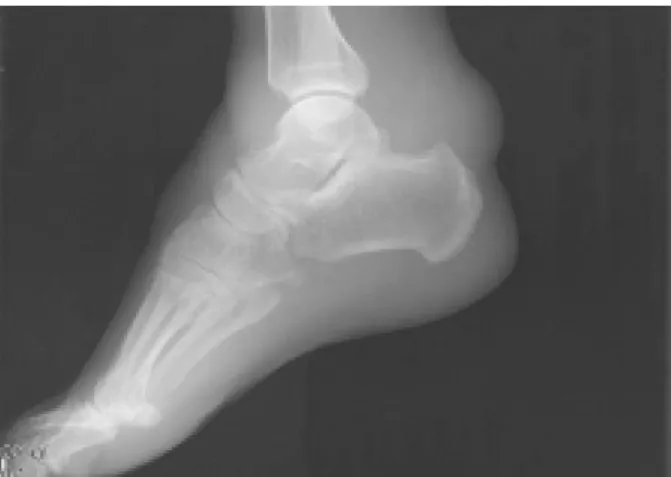
- 221 - Figure 1. Plain lateral view of left ankle shows diffuse swelling around the ankle without bony erosion.
환자로 수술후 1년내에 종물이 촉지되고 서서히 커지는 양 상을 보여 본원에 내원하였다. 과거력상 종물 제거 수술이 외의 특이 소견이 없었으며, 내원 당시 이학적 검사상 좌측 족관절의 내측에 이전 수술의 상흔이 관찰되었으며, 족관절 의 전방, 내측, 후방에 걸쳐 종물이 촉지되었고, 염증의 소
견은 없었다. 단순 방사선사진 소견상 족관절 전체에 걸친 연부 조직의 종창 및 종골 후면 상방에 종물의 압박에 의한 미란성(bony erosion) 변화와 골경화(sclerosis) 소견이 관 찰되었다(Fig. 1). 자기 공명 영상 검사상 T1 강조 시상면 영상상 족관절 전방과 후방에 비균질의 저신호 강도의 종물 이 관찰되었으며. T2 강조 관상면 영상상 족관절 후면에 넓 게 퍼져 있는 비균질의 저신호 강도의 종물이 관찰되었고 T1 지방 억제 시상면 영상상 중등도 신호 강도의 종물과 주 변의 고신호 강도를 보이고 있었다(Fig. 2). 전방 도달법 및 후내측 도달법을 이용하여 활액막 전절제술을 시행하였고, 절제된 종물은 10×10 cm 크기의 황갈색의 미만성 활액막 증식 소견과 함께 종물의 군데 군데에는 혈색소의 침착으로 보이는 적갈색의 부분이 관찰되었다 (Fig. 3-A). 조직 검사 상 섬유성 기질의 증식, 조직구와 다핵성 거대 세포의 침윤, 혈철소의 침착 , 활액막 조직내의 거대세포와 유사 조직구 세포 등이 보여 미만성 색소 융모 결절성 활액막염에 합당한 소견을 보였다(Fig. 3-B). 환자는 술 후 1년 추시 관찰에서 종물의 촉지, 동통 등의 다른 임상 증상이 발생하지 않았다.
Figure 2. MRI images of left ankle. (A) T1 weighted sagittal image shows large sized mass of low signal intensity in anterior aspect of ankle and cortical erosion at posterior aspect of calcaneus. (B) T2 weighted saggital image shows large size mass of high signal intensity with focal intermediate signal intensity. (C) T1 weighted fat suppressed sagittal image with enhance shows mass of low signal intensity surrounding high signal intensity.
김학준․김택선․서동훈․윤광섭․정국진․전승주
- 222 -
고 찰
색소 융모 결절성 활액막염은 연간 백만명당 1.8명 정도 가 발생하는 비교적 드문 질환으로 알려져 있고, 비교적 큰 관절에서 발생하는 미만성 색소 융모 결절성 활액막염은 국 소성 보다 그 발생 빈도가 높으나 슬관절에 발생하는 비율 은 66%에 달하나 족부 및 족관절에서 발생하는 비율은 5%
이하로 무척 드문 것으로 보고되고 있다2,3,6).
병인에 대해서는 이론의 여지가 많지만, 현재까지는 반 복적인 외상에 의한 발생9), 활액막 섬유 조직구의 기원의 양성 종양에서 발생한다는 설1), 원인 모를 자극에 의해서 활액막에 발생한 염증에 의한다는 설7), 지방대사의 이상에 의해 발생한다는 설5) 등이 있으나 그 정확한 병인에 대해서 는 알려진 바가 없으며 본 증례에서도 과거력상의 특이한 외상 등의 병력은 없었다.
일반적으로 환자들은 동통과 재발되는 관절의 부종 및 종물의 존재를 호소하는 것으로 알려져 있고 때로는 증식된 활액막에 의해 골 파괴나 관절 연골의 침식이 관찰되며 이 로 인해 관절의 동통이 악화된다는 보고8)가 있으나, 본 증 례에서는 종물의 존재를 호소하며 방사선상 종골 후면 상방 에서 종물의 압박에 의한 미란성 변화와 골경화 이외의 동
통이나 관절 연골의 파괴는 보이지 않았다.
미만형의 색소 융모 결절성 활액막염은 재발률이 높은 것으로 알려져 있는데2) 그 원인으로는 부적절한 활액막의 제거에 의해 병변의 완전한 제거가 이루어지지 않아서 재발 하는 것으로 알려져 있으며, 재발된 미만형 색소 융모 결절 성 활액막염의 조직학적 소견과 처음 발생한 조직과의 소견 이 다르지 않다고 보고3)되고 있다. 슬관절과 같이 비교적 큰 관절에서는 주로 관절경을 이용하여 활액막을 충분히 제 거할 수 있지만 족관절은 관절의 용적이 작고 격자 구조 (mortise)에 싸여 있으며 삽입구(portal)가 제한적이어서 활액막을 모두 관절경으로 관찰하고 제거하는 것이 쉽지않 으므로 절개하여 개방적으로 활액막을 제거하는 것이 더 유 용하다고 생각된다. 본 증례에서도 첫번째 활액막 제거술 시행후 1년 내에 재발하였는데 이는 부적절한 활액막 제거 가 그 원인에 있다고 생각된다. 이전 타병원의 수술에서 후 내측 도달법에 의한 개방적 제거술을 시행하였으므로, 저자 들은 전방 도달법 및 후내측 도달법에 의한 개방적 절제술 을 시행하여 활액막 전절제술을 시행하였으며, 조직학적 소 견상 세포의 이형성증이 동반되지 않은 다핵성 거대 세포 및 지방 함유 대식 세포에 헤모시데린 침착을 보이는 조직 구가 많이 침착된 활액막의 증식을 관찰할 수 있었다. 미만 Figure 3. Gross and microscopic findings of the mass. (A) Gross finding after excision shows yellowish-brown color mass with localized dark brownish portion. (B) Microscopic finding shows proliferation of fibrous matrix, multinucleated giant cells and pigmentation (H&E, × 100).
재발한 족관절의 미만성 색소 융모 결절성 활액막염
- 223 - 형 색소 융모 결절성 활액막염은 재발률이 높아 재발 방지 를 위해 방사선 조사, 화학적 처리 등의 방법2,10)이 제시되 고 있으나 방사선 조사 후 관절 강직이나 젊은 환자에서는 종양의 발생 등의 부작용10)이 있고 그 유용성이 확실하지 않으므로 사용에 주의가 요구된다. 본 증례에서는 전방 도 달법과 후내측 도달법을 이용하여 관절내의 활액막을 완전 히 제거 할 수 있었다. 그러므로 족관절에 발생한 미만형 색 소 융모 결절성 활액막염에서는 관절경적 활액막 제거술 이 나 활액막 부분 절제술보다는 여러 절개선을 이용한 개방적 활액막 제거술을 시행함으로써 활액막의 완전한 제거를 시 행함으로써 재발을 막을 수 있다고 생각된다.
REFERENCES
1) Choong PF, Willen H, Nilbert M, et al: Pigmented villonodular synovitis. Monoclonality and metastasis--a case for neoplastic origin? Acta Orthop Scand, 66: 64-68, 1995.
2) de Visser E, Veth RP, Pruszczynski M, Wobbes T and Van de Putte LB: Diffuse and localized pigmented villonodular synovitis: evaluation of treatment of 38 patients. Arch Orthop Trauma Surg, 119: 401-404, 1999.
3) Ghert MA, Scully SP and Harrelson JM: Pigmented villonodular synovitis of the foot and ankle: a review of six
cases. Foot Ankle Int, 20: 326-330, 1999.
4) Johansson JE, Ajjoub S, Coughlin LP, Wener JA and Cruess RL: Pigmented villonodular synovitis of joints.
Clin Orthop Relat Res. 159-166, 1982.
5) Klompmaker J, Veth RP, Robinson PH, Molenaar WM and Nielsen HK: Pigmented villonodular synovitis. Arch Orthop Trauma Surg, 109: 205-210, 1990.
6) Myers BW and Masi AT: Pigmented villonodular synovitis and tenosynovitis: a clinical epidemiologic study of 166 cases and literature review. Medicine (Baltimore), 59:
223-238, 1980.
7) Sakkers RJ, de Jong D and van der Heul RO: X- chromosome inactivation in patients who have pigmented villonodular synovitis. J Bone Joint Surg Am, 73: 1532- 1536, 1991.
8) Saxena A and Perez H: Pigmented villonodular synovitis about the ankle: a review of the literature and presentation in 10 athletic patients. Foot Ankle Int, 25: 819-826, 2004.
9) Singh R, Grewal DS and Chakravarti RN: Experimental production of pigmented villonodular synovitis in the knee and ankle joints of rhesus monkeys. J Pathol, 98: 137-142, 1969.
10) Sung JH, Kim WY, Han CW, Yoon JK and Kim JY:
Simulatenous Pigmented Villonodular Synovitis and Synovial Chondreomatosis in the Ankle Joint. J Kor Orthop Assoc, 33: 477-483, 1998.