http://dx.doi.org/10.4174/astr.2014.87.2.94 Annals of Surgical Treatment and Research
Pancreaticoduodenectomy for secondary periampullary cancer following extrahepatic bile duct cancer resection
Dong Hun Kim, Dong Wook Choi1, Seong Ho Choi1, Jin Seok Heo1
Department of Surgery, Dankook University Hospital, Cheonan, 1Department of Surgery, Samsung Medical Center, Sungkyunkwan University School of Medicine, Seoul, Korea
INTRODUCTION
Cholangiocarcinoma is a relatively rare tumor with a poor prognosis and few long-term survivors [1]. Surgical resection remains the only potentially curative treatment, but many patients develop tumor recurrence [2]. Another biliary cancer, that is, multiple biliary cancer, have been found rarely after surgical treatment of cholangiocarcinoma, rather than recur- rence. Some multiple gastrointestinal cancers have been reported. However, multiple cancers in the extrahepatic
bile duct (EHBD) have not been reported frequently. While synchronous multiple biliary cancers occur in 3.7%−7.4% of all surgically resected biliary tumors, metachronous multiple biliary cancers have not been reported frequently [3-5].
Moreover, treatment or outcome of metachronous biliary cancer has not been found precisely.
Local recurrence following resection of extrahepatic bile duct cancer is generally regarded as incurable, and secondary curative surgery as virtually impossible. However, metachronous biliary cancers with about 10 cases reported in the world literature
Received March 7, 2014, Revised April 14, 2014, Accepted April 15, 2014
Corresponding Author: Dong Wook Choi
Department of Surgery, Samsung Medical Center, Sungkyunkwan University School of Medicine, 81 Irwon-ro, Gangnam-gu, Seoul 135-710, Korea
Tel: +82-2-3410-3462, Fax: +82-2-3410-6980 E-mail: dwchoi@skku.edu
This paper was presented at the Oral Session in the 22nd World Congress of the International Association of Surgeons, Gastroenterologists and Oncologists, IASGO 2012 (December 7, 2012).
Copyright ⓒ 2014, the Korean Surgical Society
cc Annals of Surgical Treatment and Research is an Open Access Journal. All articles are distributed under the terms of the Creative Commons Attribution Non- Commercial License (http://creativecommons.org/licenses/by-nc/3.0/) which permits unrestricted non-commercial use, distribution, and reproduction in any medium, provided the original work is properly cited.
Purpose: This study addressed the feasibility and effect of surgical treatment of metachronous periampullary carcinoma after resection of the primary extrahepatic bile duct cancer. The performance of this secondary curative surgery is not well-documented.
Methods: We reviewed, retrospectively, the medical records of 10 patients who underwent pancreaticoduodenectomy (PD) for secondary periampullary cancer following extrahepatic bileduct cancer resection from 1995 to 2011.
Results: The mean age of the 10 patients at the second operation was 61 years (range, 45−70 years). The primary cancers were 7 hilar cholangiocarcinomas, 2 middle common bile duct cancers, and one cystic duct cancer. The secondary cancers were 8 distal common bile duct cancers and 2 carcinomas of the ampulla of Vater. The second operations were 6 Whipple procedures and 4 pylorus-preserving pancreaticoduodenectomies. The mean interval between primary treatment and metachronous periampullary cancer was 20.6 months (range, 3.4−36.6 months). The distal resection margin after primary resection was positive for high grade dysplasia in one patient. Metachronous tumor was confirmed by periampullary pathology in all cases. Four of the 10 patients had delayed gastric emptying (n = 2) or pancreatic fistula (n = 2) after reoperation. There were no perioperative deaths. Median survival after PD was 44.6 months (range, 8.5−120.5 months).
Conclusion: Based on the postoperative survival rate, PD may provide an acceptable protocol for resection in patients with metachronous periampullary cancer after resection of the extrahepatic bile duct cancer.
[Ann Surg Treat Res 2014;87(2):94-99]
Key Words: Extrahepatic bile duct, Cholangiocarcinoma, Metachronous neoplasms, Pancreaticoduodenectomy
were treated with curative resection [6-8]. Thus few studies have addressed the surgical treatment of secondary periampullary carcinoma after resection of the extrahepatic bile duct cancer and the role of this reresection is not well-documented or established.
This study was conducted to examine the feasibility and effect of secondary curative surgery for metachronous periam- pullary cancer in the extrahepatic bile duct.
METHODS
Between October 1995 and December 2011, 455 patients with EHBD cancer underwent surgical resection in a tertiary institution. The EHBD cancers in the present study did not include gallbladder (GB) cancer or distal common bile duct (CBD) cancer. Lymph node dissection was performed for at least the hepatoduodenal ligament in all EHBD cancers. We excluded 40 patients who underwent palliative surgery for EHBD cancer in this study. Among the 415 patients who underwent surgical resection with curative intent for EHBD cancer, 245 patients (59%) had recurrence, including local recurrences (n = 104), intrahepatic metastases (n = 61), and systemic metastases (n
= 80). Instances of local recurrence occurred in the following areas: hepaticojejunostomy site (n = 34), hepatic pedicle lymphadenectomy area (n = 43), liver resection margin (n = 15) and periampullary region (n = 12). In two of the 12 patients with periampullary recurrence, local recurrences consequently revealed duodenal cancer of primary origin, regardless of biliary tract involvement. Those two patients underwent palliative surgery, such as gastrojejunostomy, due to aggressive vascular invasion. Ten patients underwent pancreaticoduodenectomy (PD) for secondary periampullary cancer following EHBD cancer resection.
Follow-up studies after EHBD cancer resection included routine laboratory tests, serum CA 19-9, and abdominal CT in the first month after surgery and every 3–6 months for 2 years after surgery, every 6 months until 5 years, and then annually thereafter. All periampullary lesions were first detected with abdominal CT during follow-up for EHBD cancer resection.
We checked preoperative magnetic resonance imaging scans and PET to evaluate resectability and seek evidence of distant metastasis. The criteria for resectability for secondary periampullary tumor were as follows; (1) absence of distant metastasis on imaging scans, (2) absence of intrahepatic metastasis, and (3) absence of clinically and/or radiologically evident lymphadenopathy in the para-aortic region.
We reviewed medical records, including surgeon’s notes, radiologic images and pathology reports retrospectively. A pathologic examination was performed for all of the resected specimens. Preoperative interventions such as endoscopic retrograde cholangiopancreatography (ERCP), endoscopic
ultrasonography (EUS) and esophagogastroduodenoscopy (EGD) were performed to confirm preoperative pathologic status. Intraoperative frozen biopsy was performed to confirm margin status. The diagnosis of periampullary cancer was based on the histology obtained from the postoperative biopsy of the resected specimen. Time to metachronous disease was calculated from the date of EHBD cancer resection to the date when metachronous tumor was first diagnosed with interventional biopsy or imaging studies.
Tumor stage was based on criteria from the 7th edition of the American Joint Committee on Cancer (AJCC) cancer staging manual for ampulla of Vater (AoV) cancer, distal CBD cancer and hilar cholangiocarcinoma. Curative resection (R0 resection) was defined as no-residual-tumor status, whereas microscopic (R1 resection) and macroscopic residual tumor (R2 resection) were defined as noncurative resection. Postoperative pancreatic fistula was classified by definition of the International Study Group of Pancreatic Fistula [9]. The cumulative survival rates were calculated using the Kaplan-Meier method.
RESULTS
Clinicopathologic classification of the 10 extrahepatic bile duct cancers
The study group included 7 men and 3 women of mean age 61 years (range, 45−70 years). The EHBD cancers were 7 hilar cholangiocarcinomas, 2 middle CBD cancers, and one cystic duct cancer. In accordance with tumor location, 5 patients with hilar cholangiocarcinoma underwent hepatectomy with caudate lobectomy; 4 patients with hilar cholangiocarcinoma and middle CBD cancer and one with cystic duct cancer underwent bile duct segmental resection with hepaticojejunostomy. The TNM classifications and tumor stages of the EHBD cancers, based on the AJCC cancer staging manual, are shown in Table 1. Lymph node involvement was observed in only one patient.
Perineural invasion was noted in 4 patients (40%). An R0 resection was achieved in 9 patients and an R1 resection in one.
No patient underwent adjuvant therapy following EHBD cancer resection.
Clinicopathologic characteristics of
the metachronous periampullary cancers
Clinical features of the 10 patients who underwent PD are shown in Table 1. Only in patient number 6, who had hilar cholangiocarcinoma, was margin status of the distal bile duct positive after EHBD resection, i.e., with focal high grade dysplasia. After 25.8 months, however, the metachronous cancer in this patient was detected in the AoV, not in the remnant bile duct. Resection margins at the first operation were free of tumor in 9 patients. Preoperative CA 19-9 before PD was high in 5 patients with 4 hilar cholangiocarcinomas and in one
with middle CBD cancer. The metachronous cancers included 8 distal CBD cancers and 2 AoV cancers. Interventional biopsies using ERCP, EUS, and EGD were preoperatively performed in 6 patients and cancer was confirmed. Following detection of the metachronous cancer progression, 10 patients underwent PD, including 6 Whipple procedures and 4 pylorus-preserving pancreaticoduodenectomies. In the 10 patients who underwent PD, the mean number of lymph nodes harvested was 6 (range, 0−13) while the mean number of lymph nodes involved was 0.8 (range, 0−6). The volume of blood loss was 586 ± 243 mL (range, 300−1150 mL). No patient underwent homologous blood transfusion during the perioperative period. The postoperative complications after PD included delayed gastric emptying in 2 patients and pancreatic fistula as grade B and C in 2 others.
There were no intra- or postoperative deaths in this series.
The mean interval between extrahepatic bile duct cancer resection and detection of the metachronous periampullary cancer was 20.6 months (range. 3.4−36.6 months). The follow- up period after PD ranged from 8.5 to 120.5 months. Five of the 10 patients died during the follow-up period. Patients No.
2 and 3 died as cancer progression with multiple metastases to the lymph nodes, detected at 35 and 17 months after PD, respectively. Patients No. 5, 6, and 10 died through systemic recurrence with hepatic metastasis and peritoneal seeding, detected at 3, 5, and 16 months after PD, respectively. Patient No. 3 and 5, who had positive nodal disease, underwent adjuvant chemotherapy (5-fluorouracil based regimens). Patient No. 10, who also had positive node status, refused treatment with adjuvant chemotherapy. Palliative radiotherapy was administered to Patient No. 2 due to local recurrence around the hepaticojejunostomy site 35.5 months after the second operation.
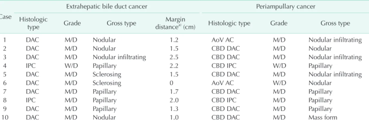
Comparison of the histopathological findings
The histologic types, cell differentiation and gross types between the EHBD cancer and the metachronous periam- pul lary cancer were compared in Table 2. The degrees of cell dif ferentiation were identical in the primary and secondary cancers of 9 patients but not in the cancers of Patient No. 6.
Among the 10 patients, the gross tumor types were papillary in 4 patients. Mean distance from the resection margin of distal bile duct to the EHBD cancer was 1.7 cm (range, 1.0−2.5 cm) except in one case in which the resection margin was positive for tumor.
Survival
Median survival after EHBD cancer resection including the survival period after PD was 56.1 months (range, 18.6−152.7 months). Median survival after PD was 44.6 months (range, 8.5−120.5 months) (Fig. 1).
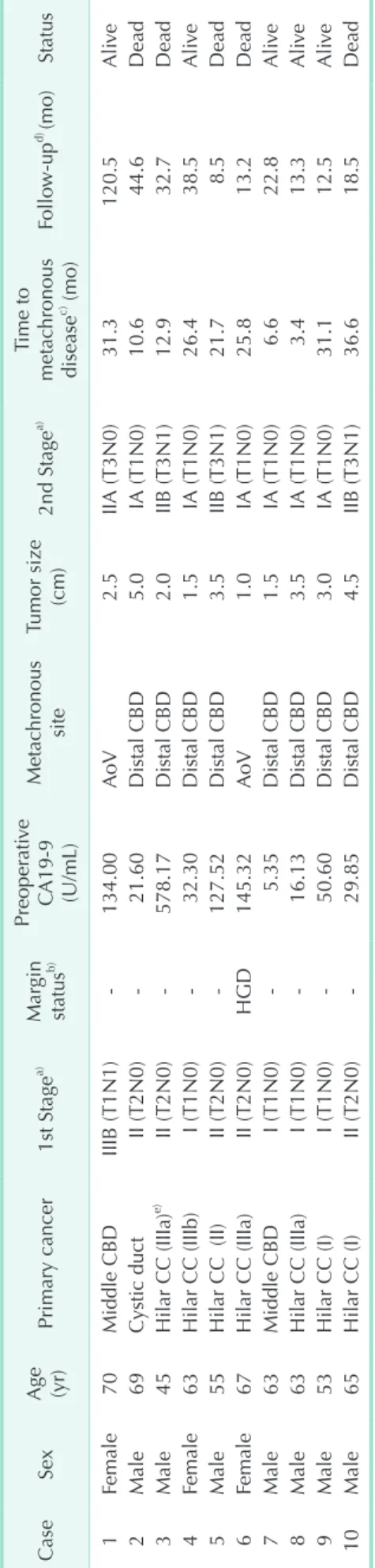
Table 1. Clinical profiles of 10 patients who underwent pancreaticoduodenectomy CaseSex Age (yr)
Primary cancer1st Stagea)
Margin status
b)Preoperative CA19-9 (U/mL)
Metac
hronous site
Tumor size (cm)2nd Stagea)Time to metachronous diseasec) (mo)Follow-upd) (mo)Status 1Female70Middle CBDIIIB (T1N1)-134.00AoV2.5IIA (T3N0)31.3 120.5Alive 2Male69Cystic ductII (T2N0)-21.60Distal CBD5.0IA (T1N0)10.6 44.6Dead 3Male45Hilar CC (IIIa)e) II (T2N0)-578.17Distal CBD2.0IIB (T3N1)12.9 32.7Dead 4Female63Hilar CC (IIIb)I (T1N0)-32.30Distal CBD1.5IA (T1N0)26.4 38.5Alive 5Male55Hilar CC (II)II (T2N0)-127.52Distal CBD3.5IIB (T3N1)21.7 8.5Dead 6Female67Hilar CC (IIIa)II (T2N0)HGD145.32AoV1.0IA (T1N0)25.8 13.2Dead 7Male63Middle CBDI (T1N0)-5.35Distal CBD1.5IA (T1N0) 6.6 22.8Alive 8Male63Hilar CC (IIIa)I (T1N0)-16.13Distal CBD3.5IA (T1N0) 3.4 13.3Alive 9Male53Hilar CC (I)I (T1N0)-50.60Distal CBD3.0IA (T1N0)31.1 12.5Alive 10Male65Hilar CC (I)II (T2N0)-29.85Distal CBD4.5IIB (T3N1)36.6 18.5Dead CBD, common bile duct; AoV, ampulla of Vater; CC, cholangiocarcinoma; HGD, high grade dysplasia. a) Based on the American Joint Committee on Cancer 7th ed. b) Distal bile duct resection margin after primary resection.c) Interval between primary treatment and detection of the metachronous periampullary cancer. d) Follow-up period after pancreatico duodenectomy. e) Bismuth classification of the hilar cholangicarcinoma.
DISCUSSION
Surgical resection is the only accepted curative treatment for cancer of the EHBD. Nagakawa et al. [10] reported the resection rate and the curative rate for bile duct cancer as 67.0%
and 30.4%, respectively. Multiple cancers in the EHBD have not been found frequently. In many such instances, there are double cancers of the CBD and the GB [3,6]. But, double cancers of the CBD are rare. Although the actual incidence of multiple malignant tumors in the bile duct is unknown, it is reported to occur in approximately 5%−10% of bile duct cancers [4,5].
When metachronous cancers are identified, the distinction between secondary primary cancer and recurrence is often clinically difficult. Warren and Gates [11] proposed the three still generally accepted criteria that had to be met in order to consider malignant tumors as multiple primary tumors rather than recurrence: (1) each tumor must present a definite
picture of malignancy, (2) each tumor must be distinct, (3) the possibility of one being a metastasis of the other must be ruled out. Gertsch et al. [5] suggested that the criteria for double primary cancer in the biliary tract was their location at separate sites with normal intervening tissue, together with histologic evidence sufficient to rule out submucosal to periductal lymphatic spread. Kobayashi et al. [12] demonstrated that multicentric adenocarcinoma of the biliary tract is more likely to be early cancer and papillary carcinoma with extensive dysplastic epithelium and is less likely to have lymph node metastasis.
In the present study, the sites of metachronous cancers in 2 patients were the AoV, not biliary tract. Conceivably, those two AoV cancers may be regarded as metachronous multiple tumors or derived by intraluminal tumor implantation rather than recurrence. Local recurrence usually originates from re si dual cancer cells at the surgical margins [13]. Based on Table 2. Comparison of pathologic finding for extrahepatic bile duct cancers and subsequent periampullary cancers
Case
Extrahepatic bile duct cancer Periampullary cancer
Histologic
type Grade Gross type Margin
distancea) (cm) Histologic type Grade Gross type
1 DAC M/D Nodular 1.2 AoV AC M/D Nodular infiltrating
2 DAC M/D Nodular 1.5 CBD DAC M/D Nodular
3 DAC M/D Nodular infiltrating 2.5 CBD DAC M/D Nodular infiltrating
4 IPC W/D Papillary 2.2 CBD IPC W/D Papillary
5 DAC M/D Sclerosing 1.5 CBD DAC M/D Nodular infiltrating
6 DAC M/D Sclerosing 0 AoV AC W/D Nodular
7 DAC M/D Papillary 1.7 CBD DAC M/D Papillary
8 IPC M/D Papillary 2.0 CBD IPC M/D Papillary
9 DAC M/D Papillary 1.3 CBD DAC M/D Papillary
10 DAC M/D Nodular 1.0 CBD DAC M/D Mass form
DAC, ductal adenocarcinoma; M/D, moderately differentiated; AoV AC, ampulla of Vater adenocarcinoma; CBD, common bile duct;
IPC, intraductal papillary adenocarcinoma; W/D, well-differentiated.
a)Distance from distal resection margin.
Fig. 1. Overall survival rates (A) after extrahepatic bile duct resection and (B) after pancreaticoduodenectomy for secondary periampullary cancer.
evi dence from pathologic reviews, all of the primary tumors in this study were more than 1.0 cm distant from the distal resection margin except in one case. Although histologic types and grades in the primary and secondary tumors of 8 patients were matched, we could not rule out the occurrence of multiple malignant tumors solely on the following evidence: (1) negative resection margins of distal bile duct in fist pathology, (2) distance from primary tumor to resection margin, (3) secondary tumor sites in remnant distal bile duct being far from original resection margin. Moreover, as stated by Joo et al. [8] and Kobayashi et al. [12], the bile duct cancer resembled a primary tumor from evidence of dysplastic changes near the main mass. Extensive dysplasia in the biliary tract suggests that multicentric adenocarcinomas might arise from the car- cino genetic background epithelial dysplasia. Metachronous tumors of 5 patients in this study developed within 2 years following EHBD resection. In view of the recurrence, it should be noted that the remnant bile duct recurrence developed in a patient with synchronous multicentric cancer despite negative resection margin. This recurrence might be considered to be metachronous tertiary cancer even though it developed within 2 years after the prior surgery [12].
The cause of multiple biliary cancers has not been defined, but it is generally accepted that the anomalous pancrea tico- biliary duct union (APBDU) plays an important role in the develop ment of multiple biliary cancers. Previous studies describe the occurrence of multiple malignant tumors in asso- ciation with APBDU [6,8,14-17]. It is reported that about 50% of cases of synchronous double biliary cancers were asso ciated with APBDU [6]. The presence of APBDU was not evaluated in this series. Others have speculated that familial and genetic factors are associated with occurrence of multiple malignant tumors [7]. Three of the patients in this study had family histories of cancer; and two of these cancers involved gastrointestinal tissues, suggesting genetic predisposition. Upper biliary tract cancers and metachronous double biliary cancers might have a strong genetic predisposition such as loss of heterozygosity, point mutation of k-ras oncogene, or overexpression of the tumor suppressor gene p53 [3,8,18]. Multiple malignant tumors are rare, hence it is proposed that bile duct cancers are usually advanced by the time of diagnosis, and that when surgery is possible with curative intent, this should be performed with a wide surgical margin [7,19]. Papillary carcinomas of the EHBD tend to grow multiply and spread fairly long distances superficially, and appear to have better prognosis than do other types of EHBD cancer [12,20-22]. Kobayashi et al. [12]
reported that early papillary adenocarcinoma in the biliary tract were associated with multicentric cancer. Although 4 of the tumors in this study were grossly of papillary type, we would not conclude from this that papillary carcinomas of bile duct are more likely than ductal adenocarcinomas to develop
metachronous cancer after surgical resection. Moreover, 4 of 5 survivors in this study had papillary carcinomas according to the gross tumor type and this may have contributed to the favorable survival rates observed.
Similar cases of metachronous bile duct cancer are reported [6-8,12]. In all of those reported cases, the metachronous bile duct cancers were treated successfully by surgery. Jung et al.
[23] reported median overall survival after biliary tract cancer resection as 48.8 months. These results are similar with our results, although we observed longer median survival after the bile duct cancer resection. Previous studies report five-year survival rates of 11.0%−28.6% for hilar or superior bile duct cancer [24-26] and 21.0%−32.7% for middle and distal bile duct cancer after resection [2,27-29]. Median survival times after EHBD resection and the secondary operations were 56.1 and 44.6 months, respectively, in the present study. Based on these results, our suggestion is that development of surgical treatment permitting long-term survival for metachronous periampullary cancer is both feasible and necessary.
There is a wide variation in practice patterns between indi- viduals and institutions, because no studies have compared high- versus low-intensity surveillance in patients after surgical resection for EHBD cancer. Follow-up management after EHBD cancer resection may contribute to prolonged survival. Triphasic CT scan of the liver (abdomen and pelvis) was performed as the usual follow-up imaging modality every 3−6 months for the first 2 years after resection and every 6 months thereafter. As for the follow-up of those patients enough to have a pro longed survival after surgical resection for metachronous periam- pullary cancer, we suggest an intensive role for CT scanning, especially in papillary carcinoma of the EHBD. Based on the overall survival rate after EHBD cancer resection in this series, follow-up duration should be maintained for at least 5 years.
Analysis for factors related to long-term survival was limited by the small number of patients in our study. However, the findings support the hypothesis that PD for secondary periampullary cancer, although a relatively aggressive treat- ment, may potentially extend patient survival.
In conclusion, because local recurrence after EHBD cancer resection is common, to achieve long-term survival in patients with extrahepatic bile duct cancer, and papillary carcinoma in particular, a second curative surgery should be considered. In metachronous periampullary cancer, following resection of the extrahepatic bile duct cancer, PD may provide a feasible and acceptable treatment.
CONFLICTS OF INTEREST
No potential conflict of interest relevant to this article was reported.
1. Lindell G, Hansson L, Dawiskiba S, Ander- sson R, Axelson J, Ihse I. Operations for extrahepatic bile duct cancers: are the results really improving? Eur J Surg 2000;
166:535-9.
2. Miyakawa S, Ishihara S, Horiguchi A, Takada T, Miyazaki M, Nagakawa T. Biliary tract cancer treatment: 5,584 results from the Biliary Tract Cancer Statistics Registry from 1998 to 2004 in Japan. J Hepatobiliary Pancreat Surg 2009;16:1-7.
3. Hori H, Ajiki T, Fujita T, Okazaki T, Suzuki Y, Kuroda Y, et al. Double cancer of gall bladder and bile duct not associated with anomalous junction of the pancreatico- biliary duct system. Jpn J Clin Oncol 2006;36:638-42.
4. Kurosaki I, Watanabe H, Tsukada K, Hata keyama K. Synchronous primary tumors of the extrahepatic bile duct and gallbladder. J Surg Oncol 1997;65:258-62.
5. Gertsch P, Thomas P, Baer H, Lerut J, Zimmer mann A, Blumgart LH. Multiple tumors of the biliary tract. Am J Surg 1990;159:386-8.
6. Fujii T, Kaneko T, Sugimoto H, Okochi O, Inoue S, Takeda S, et al. Metachronous double cancer of the gallbladder and common bile duct. J Hepatobiliary Pan- creat Surg 2004;11:280-5.
7. Merenda R, Portale G, Sturniolo GC, Mar- ciani F, Faccioli AM, Ancona E. A rare sur gical case of metachronous double carci noma of the biliary tract. Scand J Gastro enterol 2007;42:1265-8.
8. Joo HJ, Kim GH, Jeon WJ, Chae HB, Park SM, Youn SJ, et al. Metachronous bile duct cancer nine years after resection of gallbladder cancer. World J Gastroenterol 2009;15:3440-4.
9. Bassi C, Dervenis C, Butturini G, Fingerhut A, Yeo C, Izbicki J, et al. Postoperative pancreatic fistula: an international study group (ISGPF) definition. Surgery 2005;
138:8-13.
10. Nagakawa T, Kayahara M, Ikeda S, Futa- kawa S, Kakita A, Kawarada H, et al. Bili- ary tract cancer treatment: results from the Biliary Tract Cancer Statistics Regi stry in Japan. J Hepatobiliary Pancreat Surg 2002;9:569-75.
11. Warren S, Gates O. Multiple primary malignant tumors: a survey of literature and statistical study. Am J Cancer 1932;16:
1358-414.
12. Kobayashi S, Konishi M, Kato Y, Gotohda N, Takahashi S, Kinoshita T, et al. Surgical outcomes of multicentric adenocar cino- mas of the biliary tract. Jpn J Clin Oncol 2011;41:1079-85.
13. Ogura Y, Takahashi K, Tabata M, Mizu- moto R. Clinicopathological study on carcinoma of the extrahepatic bile duct with special focus on cancer invasion on the surgical margins. World J Surg 1994;
18:778-84.
14. Komi N, Takehara H, Kunitomo K. Chole- dochal cyst: anomalous arrange ment of the pancreaticobiliary ductal sys tem and biliary malignancy. J Gastroen terol Hepatol 1989;4:63-74.
15. Hasumi A, Matsui H, Sugioka A, Uyama I, Komori Y, Fujita J, et al. Precancerous conditions of biliary tract cancer in pa- tients with pancreaticobiliary maljunc- tion: reappraisal of nationwide survey in Japan. J Hepatobiliary Pancreat Surg 2000;7:551-5.
16. Okamura K, Hayakawa H, Kuze M, Taka- hashi H, Kosaka A, Mizumoto R, et al.
Triple carcinomas of the biliary tract asso ciated with congenital choledochal dila tation and pancreaticobiliary maljunc- tion. J Gastroenterol 2000;35:465-71.
17. Kasuya K, Nagakawa Y, Matsudo T, Ozawa T, Tsuchida A, Aoki T, et al. p53 gene mutation and p53 protein overexpression in a patient with simultaneous double cancer of the gallbladder and bile duct associated with pancreaticobiliary mal- junction. J Hepatobiliary Pancreat Surg 2009;16:376-81.
18. Funabiki T, Matsubara T, Miyakawa S, Ishihara S. Pancreaticobiliary maljunction and carcinogenesis to biliary and pan- creatic malignancy. Langenbecks Arch Surg 2009;394:159-69.
19. Yoon YS, Kim SW, Jang JY, Park YH.
Curative reoperation for recurrent cancer of the extrahepatic bile duct: report of two cases. Hepatogastroenterology 2005;
52:381-4.
20. Henson DE, Albores-Saavedra J, Corle D.
Carcinoma of the extrahepatic bile ducts.
Histologic types, stage of disease, grade, and survival rates. Cancer 1992;70:1498- 501.
21. Hoang MP, Murakata LA, Katabi N, Hen- son DE, Albores-Saavedra J. Invasive papil lary carcinomas of the extrahepatic bile ducts: a clinicopathologic and immu- no histochemical study of 13 cases. Mod Pathol 2002;15:1251-8.
22. Okamoto A, Tsuruta K, Matsumoto G, Taka hashi T, Kamisawa T, Egawa N, et al.
Papillary carcinoma of the extrahepatic bile duct: characteristic features and implications in surgical treatment. J Am Coll Surg 2003;196:394-401.
23. Jung SJ, Woo SM, Park HK, Lee WJ, Han MA, Han SS, et al. Patterns of initial dise- ase recurrence after resection of biliary tract cancer. Oncology 2012;83:83-90.
24. Klempnauer J, Ridder GJ, von Wasielewski R, Werner M, Weimann A, Pichlmayr R.
Resectional surgery of hilar cholangiocar- cinoma: a multivariate analysis of prog- nostic factors. J Clin Oncol 1997;15:947-54.
25. Launois B, Reding R, Lebeau G, Buard JL.
Surgery for hilar cholangiocarcinoma:
French experience in a collective survey of 552 extrahepatic bile duct cancers. J Hepatobiliary Pancreat Surg 2000;7:128- 34.
26. Lillemoe KD, Cameron JL. Surgery for hilar cholangiocarcinoma: the Johns Hop- kins approach. J Hepatobiliary Pan creat Surg 2000;7:115-21.
27. Fong Y, Blumgart LH, Lin E, Fortner JG, Bren nan MF. Outcome of treatment for distal bile duct cancer. Br J Surg 1996;83:
1712-5.
28. Cheng Q, Luo X, Zhang B, Jiang X, Yi B, Wu M. Distal bile duct carcinoma:
prognostic factors after curative surgery:
a series of 112 cases. Ann Surg Oncol 2007;14:1212-9.
29. DeOliveira ML, Cunningham SC, Came- ron JL, Kamangar F, Winter JM, Lillemoe KD, et al. Cholangiocarcinoma: thirty-one- year experience with 564 patients at a single institution. Ann Surg 2007;245:755- 62.