Brief Report
Vol. 31, No. 6, 2019 685
Received March 13, 2019, Revised May 7, 2019, Accepted for publication June 1, 2019
Corresponding author: Young Ho Won, Department of Dermatology, Chonnam National University Medical School, 42 Jebong-ro, Dong-gu, Gwangju 61469, Korea. Tel: 82-62-220-6681, Fax: 82-62-222-4058, E-mail: yhwon@jnu.ac.kr
ORCID: https://orcid.org/0000-0003-4640-4337
This is an Open Access article distributed under the terms of the Creative Commons Attribution Non-Commercial License (http://creativecommons.org/li- censes/by-nc/4.0) which permits unrestricted non-commercial use, distribution, and reproduction in any medium, provided the original work is properly cited.
Copyright © The Korean Dermatological Association and The Korean Society for Investigative Dermatology bacterium chelonae infection in a hospitalized patient. J Am
Acad Dermatol 2014;71:e248-e250.
4. Yoo JS, Huh JW, Kim MS, Jue MS, Choi KH, Park HJ.
Cutaneous atypical mycobacterial infection in a body scrub- ber (“Ddaemirri”). Korean J Dermatol 2017;55:156-157.
https://doi.org/10.5021/ad.2019.31.6.685
A Case of Recalcitrant Erythema Nodosum Associated with Pancreatic Cancer
In Soon Jung, Sook Jung Yun, Jee-Bum Lee, Seung-Chul Lee, Young Ho Won
Department of Dermatology, Chonnam National University Medical School, Gwangju, Korea
Dear Editor:
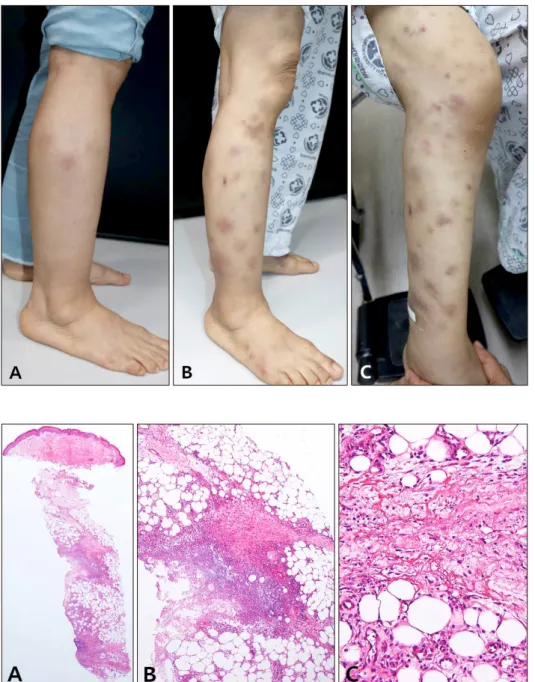
A 56-year-old female presented with erythematous nod- ules on arms and legs, which first appeared on legs two weeks prior (Fig. 1). The patient had not previously experi- enced similar symptoms. She had no specific medication, medical, or family history and showed no systemic symp- toms. Routine blood examination showed no specific find- ings. Skin biopsy showed fibrous interlobular septum with widening and septal infiltrate by inflammatory cells, in- cluding histiocytes, lymphocytes, and eosinophils. Infiltr- ate extended to adjacent fat lobules near the septa. Von Kossa stain was negative (Fig. 2). Although the patient was administered pentoxyfylline, colchicine, zaltoprofen and prednisolone (10 mg/day), the number of lesions continued to increase (Fig. 1). Further investigations were conducted to identify the reason for treatment resistance. Laboratory tests showed an increase in lipase (14,139 U/L), α-fetopro- tein (829.9 IU/ml), and carbohydrate antigen 19-9 (86.08 U/ml). Abdominal computed tomography showed malig- nant pancreatic cancer with metastasis to the liver and kid- neys. The prednisolone dose was increased to 40 mg/day, but there was no improvement. She died of tumor lysis syndrome 5 days after diagnosis of malignancy
Erythema nodosum (EN) can be idiopathic or secondary to infection, medication, inflammatory disease, or malignancy1. Although majority of cases associated with malignancy have been reported in relation to hematologic malignancies2, there are rare reports of EN secondary to solid tumors, such as lung cancer, colon cancer, and parathyroid can- cer. Cases associated with pancreatic cancer are especially rare3,4. We were unable to find any reported cases of EN in pancreatic cancer patients in Korean literature. In the present case, the patient had no factors except pancreatic cancer that could have caused EN. Moreover, the patient developed EN at 56-year-old and showed no improve- ment in spite of over 2 months of appropriate treatment.
There is no clear difference in clinicopathological features between idiopathic and paraneoplastic EN. It is difficult to distinguish between these two states by morphologic find- ings and distribution pattern. The most helpful clue is dis- ease course and response to treatment. Paraneoplastic EN shows poor response to treatment and relapses more fre- quently than idiopathic EN. Chowaniec et al.1 reported that malignancy must be considered as a cause of EN in cases with clinical symptoms such as weight loss, age over 50 years and poor response to treatment. In pancreatic
Brief Report
686 Ann Dermatol
Fig. 1. Erythematous subcutaneous nodules on the right leg at first visit (A), 4 weeks (B), and 6 weeks later (C). The skin lesions aggravated des- pite intensive therapy including oral colchicines, analgesics, and oral ster- oid. We received the patient’s consent form about publishing all photographic materials.
Fig. 2. Skin biopsy form leg nodule revealed fibrous interlobular septum with widening and septal infiltrate by inflammatory cells, including his- tiocytes, lymphocytes, and eosino- phils. The infiltrate extends into ad- jacent fat lobules (H&E: A, scanning view; B, ×100; C, ×200).
cancer patients with panniculitis, it is important to differ- entiate between EN and pancreatic panniculitis. It is clin- ically similar to EN, with erythematous nodules mostly ap- pearing on the legs5. However, it can be differentiated from EN histologically due to the presence of a lobular panniculitis pattern with ghost cells, which occur when adipose cells lose their nuclei due to calcium deposits, leaving only an outline. Von Kossa stain can be used to identify calcium deposits, which was negative in the pres- ent case.
We experienced a rare case of EN in a patient with pan- creatic cancer, and we believe this is the first report in Korea. This case shows the importance of considering pos- sibility of malignancy, including pancreatic cancer, in cas-
es of EN that do not show improvement following treat- ment.
CONFLICTS OF INTEREST
The authors have nothing to disclose.
ORCID
In Soon Jung, https://orcid.org/0000-0002-0548-7159 Sook Jung Yun, https://orcid.org/0000-0003-4229-5831 Jee-Bum Lee, https://orcid.org/0000-0002-1477-4037 Seung-Chul Lee, https://orcid.org/0000-0002-4428-3837 Young Ho Won, https://orcid.org/0000-0003-4640-4337
Brief Report
Vol. 31, No. 6, 2019 687
Received March 13, 2019, Revised May 9, 2019, Accepted for publication June 3, 2019
Corresponding author: Dong-Youn Lee, Department of Dermatology, Samsung Medical Center, Sungkyunkwan University School of Medicine, 81 Irwon-ro, Gangnam-gu, Seoul 06351, Korea. Tel: 82-2-3410-6578, Fax: 82-2-3410-3869, E-mail: dylee@skku.edu
ORCID: https://orcid.org/0000-0003-0765-9812
This is an Open Access article distributed under the terms of the Creative Commons Attribution Non-Commercial License (http://creativecommons.org/li- censes/by-nc/4.0) which permits unrestricted non-commercial use, distribution, and reproduction in any medium, provided the original work is properly cited.
Copyright © The Korean Dermatological Association and The Korean Society for Investigative Dermatology
REFERENCES
1. Chowaniec M, Starba A, Wiland P. Erythema nodosum - review of the literature. Reumatologia 2016;54:79-82.
2. Matsuoka LY. Neoplastic erythema nodosum. J Am Acad Dermatol 1995;32(2 Pt 2):361-363.
3. Virshup AM, Sliwinski AJ. Polyarthritis and subcutaneous nodules associated with carcinoma of the pancreas. Arthritis
Rheum 1973;16:388-392.
4. Durden FM, Variyam E, Chren MM. Fat necrosis with features of erythema nodosum in a patient with metastatic pan- creatic carcinoma. Int J Dermatol 1996;35:39-41.
5. Arbeláez-Cortés A, Vanegas-García AL, Restrepo-Escobar M, Correa-Londoño LA, González-Naranjo LA. Polyarthritis and pancreatic panniculitis associated with pancreatic carcinoma:
review of the literature. J Clin Rheumatol 2014;20:433-436.
https://doi.org/10.5021/ad.2019.31.6.687
Repigmentation of Eyebrow Leukotrichia in Segmental Vitiligo Treated with Suction Blister Epidermal Grafting Following Hair Plucking
Se Jin Oh, Cho Rok Kim
1, Ji-Hye Park, Dong-Youn Lee, Dokyoung Yoon
Department of Dermatology, Samsung Medical Center, Sungkyunkwan University School of Medicine, 1Kye Dermatology Clinic, Seoul, Korea
Dear Editor:
Vitiligo is generally classified into two clinical categories, nonsegmental vitiligo and segmental vitiligo (SV)1. SV on hairy areas such as the scalp and eyebrows is frequently associated with leukotrichia2. SV with overlying leuko- trichia is refractory to conventional medical treatments be- cause leukotrichia itself suggests a deficient melanocyte reservoir within the hair follicles1. Thus surgical manage- ment including epidermal grafting should be considered and preferred as an early intervention for the SV with leu- kotrichia, however only few literature showed improve- ment following surgical treatment. Herein, we present two cases of SV on the eyebrow successfully treated with suc- tion blister epidermal grafting (SBEG) after hair plucking.
An 11-year-old boy showed SV and leukotrichia on his left eyelid and forehead which had developed 8 months ago
(Fig. 1A). A 16-year-old girl had SV with leukotrichia on her right eyebrow and eyelid (Fig. 2A). Both cases did not respond to 6 months of conventional combination treat- ment including 308-nm excimer laser, topical steroid, and a calcineurin inhibitor. SBEG was performed under the consent of both the patients. Hairs of the eyebrow were pulled with forceps as a whole, including the hair follicle.
Thigh skin was used as a donor for epidermal grafting.
Vacuum suction was performed to donor sites at a pres- sure of 200 to 250 mmHg, and blisters formed after 2 hours. Epidermis on the recipient site was removed with defocusing, superpulsed mode CO2 laser. Subsequently, the roofs of the bullae were carefully excised and grafted.
The recipient areas were almost completely pigmented and leukotrichia was significantly improved after 1∼2 years of clinical follow-up (Fig. 1B, 2B).