This is an Open Access article distributed under the terms of the Creative Commons Attribution Non-Commercial License (http://creativecommons.org/licenses/by-nc/4.0/) which permits unrestricted non-commercial use, distribution, and reproduction in any medium, provided the original work is properly cited.
Copyright © 2017. Anatomy & Cell Biology
retinacula have been exhaustively described in the standard textbooks of anatomy [2, 3].
While these retinacula effectively prevent bowstringing near the ankle joint, the extensor tendons run a long course on the dorsum of the foot before insertion into toes. To pre- vent the bowstringing effect and for a more effective action of these long extensor tendons on the toes, there is a possibility of presence of an additional extensor retinaculum at the mid- foot region distal to the lower band of inferior extensor reti- naculum. So, an attempt was made to look for the presence of such restraining band of deep fascia.
Materials and Methods
Fifty lower limbs from adult embalmed cadavers in the Department of Anatomy of K. S. Hegde Medical Academy, Deralakatte, Mangalore, India were used for the study. Limbs were already disarticulated from the hip joint. So, the sex of the study population could not be determined. The samples
Introduction
Retinacula are thickenings of deep fascia in the region of joints, to keep the tendons in place as they cross the joints.
Many retinacula are present in the vicinity of ankle joint.
These retinacula are transverse bands across the ankle that holds down the tendons that cross from the leg to the foot.
When we contract our leg muscles to move our foot or toes, the pull of the muscles on their tendons would raise these tendons away from the ankle, a phenomenon known as bowstringing which would weaken the ability of the muscles to move the foot or toes [1]. Superior and inferior extensor
Original Article
https://doi.org/10.5115/acb.2017.50.3.171 pISSN 2093-3665 eISSN 2093-3673
Corresponding author:
Swathi
Department of Anatomy, K. S. Hegde Medical Academy, Deralakatte, Mangalore 575018, Karnataka, India
Tel: +91-9845910244, Fax: +91-824-2204162, E-mail: swathishriya@
gmail.com
Mid-foot retinaculum: an unrecognized entity
Swathi
1, Geetha Gangadaran Nellithala
2, Sunita Arvind Athavale
31Department of Anatomy, K. S. Hegde Medical Academy, Deralakatte, Mangalore, 2Department of Anatomy, Academy of Medical Sciences, Pariyaram, Kannur, 3Department of Anatomy, All India Institute of Medical Sciences, Bhopal, India
Abstract: Retinacula are thickenings of deep fascia in the region of joints that hold down the tendons preventing them from bowing out of position. In the region of ankle, number of such retinacula have been described. Retinacula like superior and inferior extensor retinacula have been described which hold down the tendons of leg muscles passing to the foot beneath them.
As the extensor tendons of the leg have more distal attachment to the toes, the present study was conducted to ascertain the presence of any additional retinaculum in the mid-foot region, which would tie down the tendons for their effective action at the distal joints. The aim was also to determine the attachments of the retinaculum, if present as well as the structures passing beneath them. Fifty cadaveric feet were dissected carefully for this purpose. Presence of an additional extensor retinaculum distal to the inferior band of inferior extensor retinaculum in the mid-foot region was found in 22 feet. Besides the extensor tendons, medial terminal branch of deep peroneal nerve and dorsalis pedis artery was found to pass beneath the retinaculum.
A partial or complete mid-foot retinaculum existed in the mid-foot region covering the tarsometatarsal joints in about half of study population. Functionally, this retinaculum may prevent bowstringing of the extensor tendons, clinically it may predispose to entrapment of deep peroneal nerve mimicking anterior tarsal tunnel syndrome.
Key words: Extensor retinaculum, Foot, Entrapment
Received September 21, 2016; Revised April 13, 2017; Accepted June 2, 2017
Anat Cell Biol 2017;50:171-174 Swathi, et al
172
www.acbjournal.org https://doi.org/10.5115/acb.2017.50.3.171
with any congenital or acquired anomaly of foot were ex- cluded from the study. The skin of the dorsum of the foot was reflected. Superficial nerves and vessels in the dorsum of the foot were retracted. The deep fascia of the dorsum of the foot was carefully dissected to determine the borders of the reti- naculum. The attachments of the additional retinaculum were noted, so also the structures passing beneath them.
Results
Of the 50 lower limbs, 26 were of right side and the rest were of left side. In 22 limbs (11 right, 11 left), an additional retinaculum (mid-foot retinaculum) other than the superior and inferior extensor retinacula at about the middle of the foot was found. The additional retinaculum was found to be of two types—complete and partial.
Complete retinaculum
In 8 lower limbs (3 right, 5 left), this retinaculum was found distal to inferior extensor retinaculum extending across the width of the foot over the mid-foot region. Medially this retinaculum blended with plantar fascia covering the abduc- tor hallucis muscle and laterally with the fascia over the base of 5th metatarsal (Fig. 1). Proximo-distally the retinaculum was about two fingers wide and covered the distal half of cuneiforms, cuboid and proximal thirds of metatarsals (i.e., over the tarsometatarsal joints). In two limbs (1 right, 1 left)
the medial end of the retinaculum extended distally till the first metatarso-phalangeal joint (Fig. 2). The retinaculum had septations extending plantarwards to the metatarsals and the fascia over the dorsal interossei forming six osseofibrous compartments as shown in Fig. 3. The contents of the com- partments (I to VI compartments) from medial to lateral side are shown in the Table 1. It was also noted that the septations as well as the retinaculum was thicker medially than laterally.
Partial retinaculum
In 14 limbs (8 right, 6 left), the retinacular band did not extend completely across the width of the foot from its medial border to lateral border. It was limited to medial third of the
SER
MFR IER
Fig. 1. Complete mid foot retinaculum. Superior extensor retinaculum (SER), inferior extensor retina culum (IER), and a complete midfoot retinaculum (MFR).
MFR IER
I MTP
Medial Distal Lateral Proximal
Fig. 2. The medial end of midfoot retinaculum (MFR) blending with plan tar fascia (black arrows). Proximally, it is seen merging with the distal limb of inferior extensor retinaculum (IER) and distally it is seen ex tend ing up to first metatarsophalangeal joint (I MTP) marked in white arrow.
I
II
III EHB
Medial Distal Lateral Proximal
Fig. 3. Different compartments deep to midfoot retinaculum. First, second, and third compartments (I , II, III), and extensor hallucis brevis (EHB).
Mid-foot retinaculum: an unrecognised entities
https://doi.org/10.5115/acb.2017.50.3.171
Anat Cell Biol 2017;50:171-174
173
www.acbjournal.org
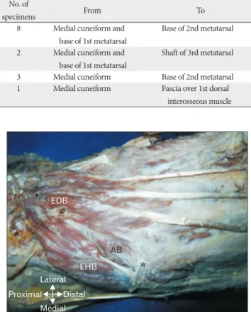
foot (Fig. 4A, B). A single septum divided this band into two compartments corresponding to the first two compartments of the complete retinaculum and having similar contents. At- tachments of partial retinacula are shown in Table 2. In one limb, an additional belly of extensor digitorum brevis (EDB) passed beneath the retinacular band along with extensor hal- lucis brevis (EHB), terminal branch of deep peroneal nerve (DPN), and the dorsalis pedis artery (DPA) (Fig. 5).
Discussion
In the present study, 22 specimens (44%) showed the pres- ence of midfoot retinaculum, compared to 64.2% of 151 spec- imens by Kaneff and his colleagues (as quoted by Sarrafian) [4], which they called as retinaculum musculorum imum in man. Aktan Ikiz et al. [5] have also described a similar band crossing the DPN, DPA, and EHB in 61% of the 38 specimens.
Lawrence and Botte [6] in their study of 17 specimens found a similar fibrous band although the prevalence was only 12%.
The presence of septations between the structures passing beneath the retinaculum has not been described in any study to the best of author’s knowledge. Human foot is still under- going evolutionary changes upon assumption of erect posture.
The functional demands of bipedal gait with plantigrade foot are manifested in evolving musculature of human foot. Pre- venting bowstringing is imperative to ensure optimum action of long tendons on the distal joints of foot. This explains pro- vision of additional retinaculum. The foot is more extensive in length medially. Provision of partial retinacula limited to A
B
*
EHB IER *
EHL
DPA DPN
Fig. 4. (A, B) Partial retinaculum (asterisk) in midfoot region. (B) The dor salis pedis artery (DPA), deep peroneal nerve (DPN), extensor hallu cis longus (EHL), extensor hallucis brevis (EHB) deep to the retina culum. IER, inferior extensor retinaculum.
Table 1. Six compartments deep to midfoot retinaculum and structures passing beneath it
Compartment Content
I Extensor hallucis longus
II Extensor hallucis brevis, dorsalis pedis artery, medial terminal branch of deep peroneal nerve
III 1st and 2nd tendons of extensor digitorum longus, 2nd tendon of extensor digitorum brevis
IV 3rd tendon of extensor digitorum longus, 3rd tendon of extensor digitorum brevis
V 4th tendon of extensor digitorum longus, 4th tendon of extensor digitorum brevis
VI Tendon of peroneus tertius
Table 2. Extent of partial retinaculum No. of
specimens From To
8 Medial cuneiform and base of 1st metatarsal
Base of 2nd metatarsal 2 Medial cuneiform and
base of 1st metatarsal
Shaft of 3rd metatarsal 3 Medial cuneiform Base of 2nd metatarsal 1 Medial cuneiform Fascia over 1st dorsal
interosseous muscle
*
EHB
Medial Distal Lateral Proximal
EDB
AB
Fig. 5. An additional belly (AB) of extensor digitorum brevis (EDB) passing deep to the retinaculum (asterisk). EHB, extensor hallucis brevis.
Anat Cell Biol 2017;50:171-174 Swathi, et al
174
www.acbjournal.org https://doi.org/10.5115/acb.2017.50.3.171
medial aspect may be explained thus. Even the inferior exten- sor retinaculum is more expansive medially by splitting itself in a “y” like pattern. The retinaculum and septations probably enhance the stability of extensor tendons at the tarsometatar- sal joints and prevent bow stringing.
The importance of knowing the presence of such a retinac- ulum lies in the fact that the retinaculum crosses DPA and the medial terminal branch of DPN and the EHB tendon, which are present within the compartment. This can give rise to en- trapment neuropathy of the DPN if the nerve is stretched or compressed within it. This is more likely if an additional belly of EDB also passes through the second compartment as was observed in one limb in the present study.
Several studies have described anterior tarsal tunnel syn- drome which is entrapment neuropathy of DPN beneath the inferior extensor retinaculum of the foot giving rise to either motor symptoms, sensory symptoms or both depending on the site of entrapment of the nerve [5-10].
Dellon [11] has described an area of “anatomic tightness”
where EHB tendon crossed the DPN in the mid-foot region.
In this area, the nerve is prone to entrapment in cases of in- jury in this area, high arched foot, swelling of the foot or a footwear with the band crossing this area [11]. Kanbe et al. [12]
have also reported the entrapment neuropathy of DPN by the EHB. Presence of a retinacular band in this region, with septations, would thus predispose to entrapment of the nerve lying within and mimic symptoms of anterior tarsal tunnel syndrome.
To conclude, a partial or complete mid-foot retinaculum existed in the mid-foot region covering the tarsometatarsal joints in about half of study population. Functionally this reti- naculum may prevent bowstringing of the extensor tendons, clinically it may predispose to entrapment of DPN mimicking
anterior tarsal tunnel syndrome.
Limitations of the study is that sex of the specimen was not known. Hence, variations of retinaculum with respect to gen- der could not be assessed.
References
1. Muscolino JE. Kinesiology: the skeletal system and muscle func- tion. 2nd ed. St. Louis, MO: Elsevier; 2011. p.306-12.
2. Standring S, Ellis H, Healy JC, Johnson D, Williams A, Collins P, Wigley C. Gray’s anatomy: the anatomical basis of clinical prac- tice. 39th ed. Edinburgh: Churchill Livingstone; 2005. p. 1507.
3. Sinnatamby CS. Last’s anatomy: regional and applied. 12th ed.
Edinburgh: Churchill Livingstone; 2011. p.145.
4. Sarrafian SK. Anatomy of foot and ankle: descriptive, topo- graphic, functional. 2nd ed. Philadelphia, PA: J. B. Lippincott Co.; 1993. p.123.
5. Aktan Ikiz ZA, Ucerler H, Uygur M. Dimensions of the anterior tarsal tunnel and features of the deep peroneal nerve in relation to clinical application. Surg Radiol Anat 2007;29:527-30.
6. Lawrence SJ, Botte MJ. The deep peroneal nerve in the foot and ankle: an anatomic study. Foot Ankle Int 1995;16:724-8.
7. Borges LF, Hallett M, Selkoe DJ, Welch K. The anterior tarsal tunnel syndrome: report of two cases. J Neurosurg 1981;54:89- 92.
8. Kennedy JG, Baxter DE. Nerve disorders in dancers. Clin Sports Med 2008;27:329-34.
9. Liu Z, Zhou J, Zhao L. Anterior tarsal tunnel syndrome. J Bone Joint Surg Br 1991;73:470-3.
10. Seçıl Y, Tokuçoğlu F, Beckmann Y, Arici Ş, Eryaşar G. Anterior tarsal tunnel syndrome: electrophysiological and clinical evalua- tion of five cases. J Neurol Sci 2012;29:819-25.
11. Dellon AL. Deep peroneal nerve entrapment on the dorsum of the foot. Foot Ankle 1990;11:73-80.
12. Kanbe K, Kubota H, Shirakura K, Hasegawa A, Udagawa E. En- trapment neuropathy of the deep peroneal nerve associated with the extensor hallucis brevis. J Foot Ankle Surg 1995;34:560-2.