Copyrights © 2017 The Korean Society of Radiology
138
Case Report
pISSN 1738-2637 / eISSN 2288-2928 J Korean Soc Radiol 2017;76(2):138-141 https://doi.org/10.3348/jksr.2017.76.2.138
INTRODUCTION
Follicular carcinoma cannot be differentiated from a follicular adenoma based on clinical, cytologic and ultrasonography (US) features. Follicular neoplasms are indeterminate lesions and pres- ent a diagnostic challenge to clinicians. Currently, these patients are advised to undergo a hemithyroidectomy and isthmectomy for accurate diagnosis (1, 2). Endoscopic thyroidectomy is con- sidered appropriate for follicular neoplasms because of its out- standing cosmetic results. However, it occasionally leads to unex- pected complications such as brachial plexus injury, Horner’s syndrome, chyle leaks and operative track seeding (1, 3). Follicu- lar thyroid neoplasm seeding around the operative bed and along
the port insertion site is very rare with only 4 cases reported to date. We present a case of follicular thyroid adenoma seeding af- ter endoscopic thyroidectomy for a follicular neoplasm, both in the subcutaneous tunnel of the upper chest wall and in the opera- tive bed.
CASE REPORT
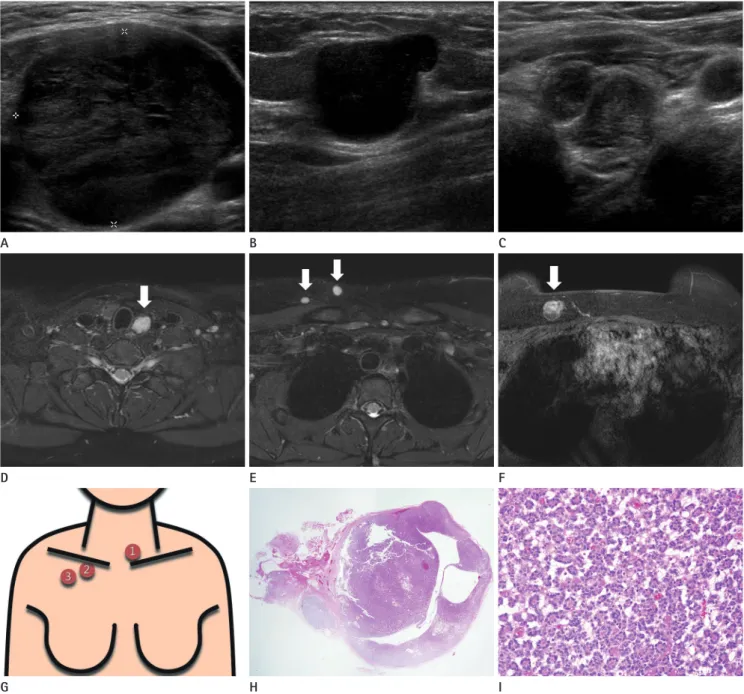
A 14-year-old female visited our hospital because of a palpable neck mass. US using 5–17 MHz linear probe (iU 22; Philips, Bothell, WA, USA) showed a 3.8 cm sized hypoechoic mass with smooth margins in the left lobe of the thyroid gland (Fig. 1A).
The diagnosis resulting from fine-needle aspiration biopsy was
Simultaneous Seeding of Follicular Thyroid Adenoma Both Around the Operative Bed and Along the Subcutaneous Tunnel of the Upper Chest Wall after Endoscopic Thyroidectomy
갑상선 여포종양의 내시경적 갑상선 절제술 후 갑상선 부위 및 유방의 피하터널로의 파종
Jo Sung Jung, MD
1, Shin Young Kim, MD
1*, Hae Yoen Jung, MD
2, Seon Wook Han, MD
3, Jong Eun Lee, MD
3, Deuk Young Lee, MD
4Departments of 1Radiology, 2Pathology, 3Surgery, Soonchunhyang University Cheonan Hospital, Cheonan, Korea
4Department of Surgery, YonseiAngelot Hospital, Cheonan, Korea
Endoscopic thyroidectomy is considered appropriate for follicular neoplasms, but on occasion, it leads to unexpected complications such as seeding along the port inser- tion site. Only 4 cases of operative track seeding after endoscopic thyroidectomy have been reported. Furthermore, simultaneous seeding at both operative track of upper chest wall and operative bed is also very rare. We present a case of thyroid follicular adenoma seeding at both the subcutaneous tunnel of the upper chest wall and the operative bed after endoscopic thyroidectomy, with an emphasis on mag- netic resonance imaging and ultrasonography with pathologic correlations.
Index terms Thyroid Neoplasm Follicular Adenoma Thyroidectomy Neoplasm Seeding
Received March 9, 2016 Revised June 7, 2016 Accepted August 8, 2016
*Corresponding author: Shin Young Kim, MD Department of Radiology, Soonchunhyang University Cheonan Hospital, 31 Suncheonhyang 6-gil, Dongnam-gu, Cheonan 31151, Korea.
Tel. 82-41-570-3540 Fax. 82-41-572-4655 E-mail: [email protected]
This is an Open Access article distributed under the terms of the Creative Commons Attribution Non-Commercial License (http://creativecommons.org/licenses/by-nc/3.0) which permits unrestricted non-commercial use, distri- bution, and reproduction in any medium, provided the original work is properly cited.
139
Jo Sung Jung, et al
jksronline.org J Korean Soc Radiol 2017;76(2):138-141 follicular neoplasm. The patient wanted endoscopic surgery for cosmetic reasons. She elected to undergo an endoscopic gas in- sufflation left hemithyroidectomy with a bilateral axillo-breast
approach (BABA). The final diagnosis of the resected mass was a follicular adenoma. About 3 years after the first surgery, the pa- tient revisited our hospital due to palpable nodules on the right
Fig. 1. Simultaneous seeding of follicular thyroid adenoma around both the operative bed and along the subcutaneous tunnel of the upper chest wall after endoscopic thyroidectomy in a 14-year-old female. Preoperative US shows a 3.8 cm sized, smooth, oval, heterogeneous, hypoechoic mass in the left thyroid bed (A). Postoperative follow-up, transverse, US images show an approximately 1.9 cm sized, circumscribed, oval hy- poechoic nodule along the right upper chest wall close to the right clavicle and along the subcutaneous tract of the prior endoscopic thyroidec- tomy (B), and about 1.1 cm sized, smooth, oval, heterogeneous, isoechoic nodule in the left operative bed (C). Post-gadolinium enhanced axial MRI images (D-F) show variable-sized, well-enhancing masses in the left thyroid operative bed (arrow on D), in the right infraclavicular area (ar- rows on E), and in the subcutaneous layer of the right upper chest wall (arrow on F). Schematic illustration shows seeded nodules in the opera- tive bed and along the subcutaneous tunnel of the upper chest wall (G). Microscopic examination of the mass resected from the upper chest wall shows a well-encapsulated nodular lesion (× 20) (H). Higher magnification shows that the tumor cells form microfollicles and are without nucle- ar atypia (× 40) (I).
MRI = magnetic resonance imaging, US = ultrasonography A
D
G
B
E
H
C
F
I
140
Simultaneous Seeding of Follicular Thyroid Adenoma Both the Operative Bed and Subcutaneous Tunnel after Endoscopic Thyroidectomy
jksronline.org
J Korean Soc Radiol 2017;76(2):138-141 upper chest wall. The nodules were 1–2 cm in size and freely mo-
bile on physical exam. Mammography demonstrated an about 1.7 cm sized, circumscribed, oval, isodensity nodule in the right up- per chest wall. On US examination, multiple 0.5–1.9 cm sized circumscribed, oval, hypoechoic nodules were noted in a row along the putative, right, subcutaneous track of the prior endo- scopic thyroidectomy, and in the left thyroid bed to level VII (Fig.
1B, C). Magnetic resonance imaging (MRI) (Ingenia 3.0 T; Phil- ips, Best, the Netherlands) performed to determine the exact sur- gical extent, also showed multiple nodules along the endoscopic track and thyroidectomy bed. These nodules were slightly high to iso-signal intensity on T2 weighted images and iso-signal intensi- ty on T1 weighted images (not shown) with homogenous nodu- lar or peripheral rim enhancement on contrast-enhanced, fat- suppressed axial T1-weighted images (Fig. 1D-F). Schematic illustration showed seeded nodules in the operative bed, infracla- vicular area and upper chest wall (Fig. 1G). The patient under- went the surgical resection. The pathological examination of the resected nodules revealed follicular adenoma with the same pat- tern as the previous hemithyroidectomy specimen (Fig. 1H, I).
DISCUSSION
Recently, endoscopic techniques have been introduced for thy- roidectomy instead of classical thyroidectomy (1, 2). Shimazu et al. (4) described the axillo-bilateral-breast approach using both axillary and breast incisions, and Choe et al. (5) included another incision to the contralateral axilla, the BABA. The benefit of en- doscopic thyroid surgery is a better cosmetic result compared to conventional open surgery. However, endoscopic thyroid surgery has limits for optimal visualization and complete surgery, and has some complications including rare cases of operative track tumor seeding (5).
Kim et al. (3) reported a patient with papillary thyroid carcino- ma recurrence around the operative bed and the subcutaneous tunnel after endoscopic thyroidectomy. Kim et al. (3) suggested that seeding at the trocar site is due to spillage of tumor during tumor manipulation. Traumatic handling of the tumor and inad- equate surgical skill are suspected essential factors for port site seeding. Our case is thought to be similar to Kim et al. (3) because tumor seeding occurred along the subcutaneous tunnel of port insertion site. Hur et al. (6) also reported computed tomography
findings of follicular thyroid cancer recurrence with multiple, en- hancing, bean-sized soft tissue masses around the operative bed and along the port insertion site after endoscopic thyroidectomy.
Keiko et al. (7) reported the following mammographic, US and MRI findings of port-site implantation of benign thyroid adeno- ma after endoscopic thyroidectomy: a polygonal mass with un- clear margins on mammography, a heterogenous echoic mass with no interruption of boundary on ultrasound, and a polygonal nodular lesion with clear boundary and peak enhancement in the early phase under contrast on MRI. In our case, two nodules with clear margins were seen on mammography, and US showed well-circumscribed, oval or round hypoechoic lesions. The MRI image showed well-marginated, oval or round nodules with ho- mogenous or rim enhancements along the port insertion site and around the operative bed.
In conclusion, based on US and MRI findings, we report a case of follicular thyroid adenoma, simultaneously seeded along the subcutaneous tunnel of the upper chest wall and around the op- erative bed. This is a rare, but possible complication after endo- scopic thyroidectomy.
REFERENCES
1. Beninato T, Kleiman DA, Scognamiglio T, Fahey TJ, Zarnegar R. Tract recurrence of a follicular thyroid neoplasm follow- ing transaxillary endoscopic thyroidectomy. Thyroid 2012;
22:214-217
2. McHenry CR, Phitayakorn R. Follicular adenoma and carci- noma of the thyroid gland. Oncologist 2011;16:585-593 3. Kim JH, Choi YJ, Kim JA, Gil WH, Nam SJ, Oh YL, et al. Thy-
roid cancer that developed around the operative bed and subcutaneous tunnel after endoscopic thyroidectomy via a breast approach. Surg Laparosc Endosc Percutan Tech 2008;
18:197-201
4. Shimazu K, Shiba E, Tamaki Y, Takiguchi S, Taniguchi E, Ohashi S, et al. Endoscopic thyroid surgery through the axillo-bilateral-breast approach. Surg Laparosc Endosc Percutan Tech 2003;13:196-201
5. Choe JH, Kim SW, Chung KW, Park KS, Han W, Noh DY, et al. Endoscopic thyroidectomy using a new bilateral axillo- breast approach. World J Surg 2007;31:601-606
6. Hur SM, Kim SH, Lee SK, Kim WW, Choi JH, Kim JH, et al. Is
141
Jo Sung Jung, et al
jksronline.org J Korean Soc Radiol 2017;76(2):138-141
갑상선 여포종양의 내시경적 갑상선 절제술 후 갑상선 부위 및 유방의 피하터널로의 파종
정조성
1· 김신영
1* · 정혜연
2· 한선욱
3· 이종은
3· 이득영
4갑상선 여포종양은 내시경적 갑상선 절제술의 좋은 적응증이다. 그러나 내시경 포트 삽입구를 따라 종양이 파종되는 예상치 못한 합병증을 야기할 수 있다. 내시경 갑상선 절제술 후 종양이 수술 트랙을 따라 파종된 예가 4증례만이 보고 되었다.
하지만, 흉벽의 피하터널과 갑상선 제거 부위에서 동시에 파종된 예는 매우 드물다. 저자들은 내시경적 갑상선 절제술 후 갑상선 절제 부위 및 상부 흉벽의 피하터널로 파종된 갑상선 여포성 선종 1예를 초음파, 자기공명영상 그리고 병리소견을 중심으로 증례 보고한다.
순천향대학교 천안병원 1영상의학과, 2병리과, 3외과, 4연세앙즈로병원 외과 a thyroid follicular neoplasm a good indication for endo-
scopic surgery? Surg Laparosc Endosc Percutan Tech 2011;
21:e148-e151
7. Keiko K, Hiroshi N, Kazushige F, Masato O, Michiko H, Toshio
O, et al. A case of port-site implantation in the breast after endoscopic thyroid surgery. J Japan Surgl Assoc 2010;71:
25-30