http://dx.doi.org/10.5534/wjmh.2014.32.1.1
Review Article
Received: Oct 14, 2013; Accepted: Oct 24, 2013 Correspondence to: Ashok Agarwal
Center for Reproductive Medicine & Andrology Center, Glickman Urological and Kidney Institute, Cleveland Clinic, Desk X11, 10681 Carnegie Avenue, Cleveland, Ohio 44195, USA.
Tel: +1-216-444-9485, Fax: +1-216-445-6049, E-mail: agarwaa@ccf.org Copyright © 2014 Korean Society for Sexual Medicine and Andrology
This is an Open Access article distributed under the terms of the Creative Commons Attribution Non-Commercial License (http://creativecommons.
org/licenses/by-nc/3.0) which permits unrestricted non-commercial use, distribution, and reproduction in any medium, provided the original work is properly cited.
Effect of Oxidative Stress on Male Reproduction
Ashok Agarwal1, Gurpriya Virk1, Chloe Ong1, Stefan S du Plessis2
1Center for Reproductive Medicine, Cleveland Clinic, Cleveland, OH, USA, 2Medical Physiology, Faculty of Medicine and Health Sciences, Stellenbosch University, Tygerberg, South Africa
Infertility affects approximately 15% of couples trying to conceive, and a male factor contributes to roughly half of these cases.
Oxidative stress (OS) has been identified as one of the many mediators of male infertility by causing sperm dysfunction. OS is a state related to increased cellular damage triggered by oxygen and oxygen-derived free radicals known as reactive oxygen species (ROS). During this process, augmented production of ROS overwhelms the body’s antioxidant defenses. While small amounts of ROS are required for normal sperm functioning, disproportionate levels can negatively impact the quality of spermatozoa and impair their overall fertilizing capacity. OS has been identified as an area of great attention because ROS and their metabolites can attack DNA, lipids, and proteins; alter enzymatic systems; produce irreparable alterations; cause cell death; and ultimately, lead to a decline in the semen parameters associated with male infertility. This review highlights the mechanisms of ROS production, the physiological and pathophysiological roles of ROS in relation to the male reproductive system, and recent advances in diagnostic methods; it also explores the benefits of using antioxidants in a clinical setting.
Key Words: Antioxidants; Infertility, male; Oxidative stress; Reactive oxygen species; Spermatozoa
INTRODUCTION
For most couples, procreating is a natural part of life that involves neither special planning nor intervention.
Unfortunately, when trying to conceive, 15% to 25% of the couples struggle and, consequently, seek medical ad- vice on how to improve their chances of fertilization and successful pregnancy [1]. According to the World Health Organization [2] guidelines, in approximately half of these cases, the male factor is the cause of infertility when an
“alteration in sperm concentration, motility, and/or mor- phology is present in at least one sample of two sperm
analyses, collected 1 to 4 weeks apart” [3]. This problem is further compounded when no identifiable reason can be found. Currently, oxidative stress (OS) is believed to be an important and plausible cause of idiopathic male infertility.
“OS is a condition that reflects an imbalance between the systemic manifestation of reactive oxygen species (ROS) and a biological system’s ability to readily detoxify (antioxidant defenses) the reactive interm1ediates or to re- pair the resulting damage” [4,5]. In a healthy body, pro-ox- idants and antioxidants remain in balance. Spermatozoa are equipped with antioxidant defense mechanisms and
are likely to quench ROS, thereby protecting gonadal cells and mature spermatozoa from oxidative damage [6].
However, under pathological conditions, the uncon- trolled production of ROS exceeds the antioxidant ca- pacity of the seminal plasma, resulting in OS [1,6].
Statistics from the United States indicate that OS is one of the major causes of male infertility; that is, 30% to 40%
of infertile men have elevated levels of ROS in their semi- nal plasma [7]. Spermatozoa were the first cell type re- ported to show potential susceptibility to OS. In some sit- uations, the damage caused by oxidants may be repaired.
Unfortunately, spermatozoa are unable to restore the damage induced by OS because they lack the necessary cytoplasmic-enzyme repair systems. This is one of the fea- tures that make spermatozoa unique in their susceptibility to oxidative insult [8]. This is predominantly due to the fact that their cell membranes are rich in polyunsaturated fatty acids (PUFAs), rendering them highly susceptible to oxy- gen-induced damage and hence, lipid peroxidation (LPO).
Subsequently, a rapid loss of intracellular adenosine tri-phosphate (ATP) from LPO causes axonemal damage, decreased sperm viability, and increased mid-piece sperm morphological defects, all of which contribute to de- creased sperm motility [9,10].
OS has become an area of great concern for clinicians and scientists because of the fact that this pathway of pro- grammed deterioration has also resulted in poor fertiliza- tion, poor embryonic development, pregnancy loss, birth defects (including autism), and childhood cancer [11-14].
Over the years, the literature on this topic has increased in volume with the addition of several original contributions concerning the role of OS in male reproduction.
In this paper, we will provide a comprehensive over- view of the latest evidence regarding the mechanism of ROS production, the physiological roles of ROS, and the patho-physiology of ROS, as well as the impact of OS on male infertility in humans. Furthermore, we will elaborate on different techniques used to measure ROS as well as different treatment strategies implemented by physicians to reduce OS levels in the seminal plasma of infertile men, which hopefully contribute to increased potential for natu- ral fertilization and conception.
REACTIVE OXYGEN SPECIES
ROS, also known as free radicals, have at least one un- paired electron. They are oxidizing agents generated as byproducts from the metabolism of oxygen. Due to the un- paired electron in the outer shell, they form highly reactive molecules [6,15]. ROS represents a collection of a broad range of radicals (e.g., hydroxyl ion [OH-], superoxide ion [O2-], nitric oxide [NO], peroxyl [RO2], lipid peroxyl [LOO], and Thiyl [RS-]) and non-radical molecules (singlet oxygen [-1O2], hydrogen peroxide [H2O2], hypochloric acid [HOCL], lipid peroxide [LOOH], and ozone [O3]) [9].
1. Generation of reactive oxygen species
Research has shown that ROS causes electron leakage from actively respiring spermatozoa, mediated by intra- cellular redox activities. The generation of ROS in sperma- tozoa may occur via two methods: (1) the nicotinamide adenine dinucleotide phosphate oxidase system at the lev- el of the sperm plasma membrane and/or (2) the nic- otinamide adenine dinucleotide-dependent oxido-reduc- tase reaction at the mitochondrial level. The latter mecha- nism appears to be the main source of ROS. Spermatozoa are rich in mitochondria because a constant supply of en- ergy is required for their motility [6]. Therefore, the pres- ence of dysfunctional spermatozoa in the semen sig- nificantly elevates the production of ROS, which in turn af- fects its mitochondrial function and subsequently, sperm function such as motility.
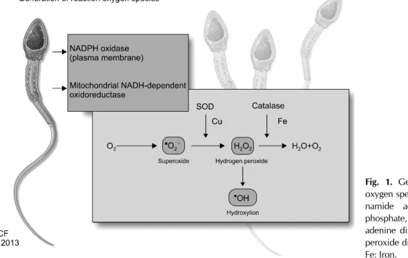
A majority of ROS generated in human spermatozoa is O2-. This electron-reduced product of O2- reacts with itself via dismutation to generate H2O2. In the presence of tran- sition metals such as iron and copper, H2O2 and O2- under- go the Haber-Weiss reaction to generate the extremely re- active and destructive OH- (Fig. 1). OH- radicals are excep- tionally potent initiators of the LPO cascade and can lead to a loss of sperm function from the disruption of mem- brane fluidity [16-18].
A recent study describing the production of O2- in sper- matozoa revealed that the presence of a calcium-depend- ent NADPH oxidase called NOX5 (encoded by the NOX5 gene) has been established within human spermatozoa, particularly in the acrosomal and midpiece regions [19].
NOX5 was initially detected in the human testis and is acti-
Fig. 1. Generation of reactive oxygen species. NADPH: nicoti- namide adenine dinucleotide phosphate, NADH: nicotinamide adenine dinucleotide, SOD: su- peroxide dismutase, Cu: copper, Fe: Iron.
vated when Ca2 binds to its cytosolic N-terminal EF-hand domain. This binding causes conformational changes to the cell, thereby inducing OS. This finding provides further evidence that NOX5 is a major source of ROS generation in human spermatozoa. Whether NOX5 is overexpressed in the spermatozoa of patients exhibiting infertility associated with OS has not yet been determined [16].
SOURCES OF REACTIVE OXYGEN SPECIES IN SEMINAL PLASMA
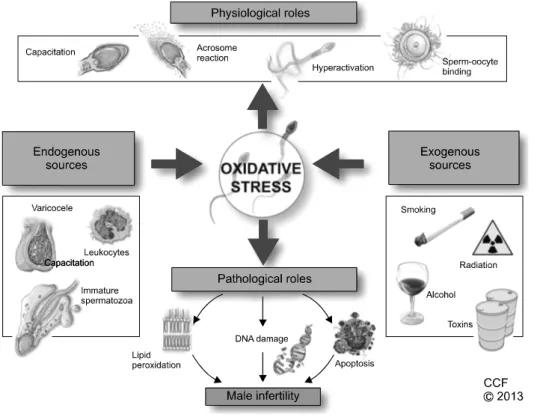
ROS found in seminal plasma originates from various endogenous and exogenous sources. The human ejacu- late consists of different types of cells, including mature and immature cells, round cells from different stages of spermatogenesis, leukocytes, and epithelial cells. Of these, leukocytes-mainly neutrophils and macrophages- and immature spermatozoa are considered the main en- dogenous sources of ROS, while several lifestyle factors such as excessive smoking and alcohol consumption, and environmental factors such as radiation and toxins can contribute to exogenous ROS [10,20,21]. Fig. 2 summa- rizes the relationship of increasing ROS production in se- men with male infertility.
1. Endogenous sources of reactive oxygen species 1) Leukocytes
Peroxidase-positive leukocytes include polymorpho- nuclear leukocytes (50%∼60%) and macrophages (20%
∼30%) [22]. A large proportion of these peroxidase-pos- itive leukocytes originate from the prostate and seminal vesicles. When these major sources of ROS are activated by various intracellular or extracellular stimuli, such as in- fection or inflammation, they can discharge up to 100 times more ROS than normal and increase the NADPH production via the hexose monophosphate shunt [23,24].
An increase in proinflammatory cytokines, such as inter- leukin (IL)-8, and a decrease in the antioxidant superoxide dismutase (SOD) can result in a respiratory burst, pro- duction of high levels of ROS, and ultimately, OS. OS will cause sperm damage if seminal leukocyte concentrations are abnormally high as is the case in leukocytospermia [25], which the World Health Organization defines as the presence of more than one million peroxidase-positive cells per milliliter of semen [2].
Over the years, extensive research has been carried out to establish a link between the presence of leukocytes in the ejaculate and a male factor as the cause of infertility.
Fig. 2. Oxidative stress in male reproduction.
Various studies point to a correlation between decreased sperm function and seminal plasma with abnormally ele- vated levels of ROS, IL-6, IL-8, and tumor necrosis factor, all of which result in increased sperm cell membrane LPO [23,26].
2) Immature spermatozoa
During spermatogenesis, developing spermatozoa ex- trude their cytoplasm in order to prepare for fertilization.
However, damaged spermatozoa retain excess cytoplasm around the midpiece due to an arrest in spermiogenesis;
this condition is known as excess residual cytoplasm (ERC). ERC activates the NADPH system by means of the hexose-monophosphate shunt, which spermatozoa use as a source of electrons for ROS generation and potentially, OS [27]. Hence, ERC ultimately affects sperm motility, morphology, and fertilization potential, which may lead to male infertility [5].
3) Varicocele
Varicocele is defined as an abnormal dilation of veins in the pampiniform plexus around the spermatic cord. Since varicocele is detected in about 40% of male partners of all
infertile couples, it is considered the leading cause of male factor infertility [28]. It has been shown that the level of seminal ROS is associated with the grade of varicocele;
that is, the higher the grade of varicocele is, the greater is the level of ROS detected [29].
2. Exogenous sources of reactive oxygen species 1) Radiation
Radiation, a natural source of energy, has significant clinical effects on humans. With respect to male re- productive health, several studies have implicated radia- tion emitted from mobile phones in the increase of the pro- duction of ROS in human semen with impaired semen quality [30,31]. In vitro studies have demonstrated that electromagnetic radiation induces ROS production and DNA damage in human spermatozoa, which further de- creases the motility and vitality of sperm cells as well as their concentration depending on the duration of exposure to radiation [32]. These radiofrequency electromagnetic waves can negatively affect the electron flow along the in- ternal membranes of the cell as a result of the numerous charged molecules within the cytosol, thus disrupting nor- mal cellular and organelle function [23].
2) Toxins
Toxins released from structural materials or industrial products accumulate in the human body and increase ROS production in the testes, negatively impacting the sperm structure and function [33]. Phthalates, found in a variety of plastic objects used for domestic and industrial purposes, have been studied in great detail [34,35]. They have been found to impair spermatogenesis and induce sperm DNA damage [36]. Furthermore, it was demon- strated that workers who were regularly exposed to toxins in the form of metals such as cadmium, chromium, lead, manganese, and mercury were more likely to have de- creased sperm quality, count, volume, and density [37].
3) Smoking
Tobacco is known to be one of the major preventable causes of death worldwide. Cigarettes contain more than 4,000 chemical compounds including alkaloids, nitros- amines, and inorganic molecules. Some of the chemicals were shown to cause an imbalance between ROS and anti- oxidants in the semen of smokers [23]. This ROS and anti- oxidant disproportion affects the overall semen quality.
Smoking has been shown to result in a 48% increase in seminal leukocyte concentrations and a 107% increase in seminal ROS levels [38]. Moreover, smokers have de- creased levels of seminal plasma antioxidants such as vita- min E and vitamin C, placing their sperm at the additional risk of oxidative damage. This has been confirmed by a sig- nificant increase in the levels of 8-OHdG, another bio- marker of oxidative damage, in the seminal plasma of smok- ers [21]. A study on the semen profiles of smokers versus non-smokers showed that spermatozoa from smokers were significantly more sensitive to acid-induced DNA denatura- tion than those of non-smokers and resulted in higher levels of DNA strand breaks [39]. Another study performed on smokers revealed that the increased cadmium and lead con- centrations in their blood and semen led to increased ROS production with an accompanying decrease in sperm mo- tility [40]. Further, it was proven that prolonged exposure to tobacco smoke is linked to an increase in sperm DNA dam- age and apoptosis, leading to increased male infertility.
4) Alcohol consumption
Alcohol is known as a promoter of ROS production and
interferes with the body’s antioxidant defense mecha- nism, particularly in the liver. Many factors are involved in causing alcohol-induced OS. When acetaldehyde, one of the by-products of ethanol metabolism, interacts with pro- teins and lipids, ROS is formed. This results in molecular damage to proteins, lipids, and DNA. Therefore, excessive alcohol consumption is associated with a decreased per- centage of normal spermatozoa in asthenozoospermic pa- tients [41]. A study of 46 alcoholic men of reproductive age has reported a significant increase in serum LPO by-products and a decrease in antioxidants, providing fur- ther evidence of ethanol-induced OS within the testes [4].
PHYSIOLOGICAL ROLES OF ROS IN SEMINAL PLASMA
Although high concentrations of ROS cause sperm path- ologies (ATP depletion) in the form of inadequate ax- onemal phosphorylation or LPO, resulting in a loss of sperm motility and viability, many studies have demon- strated that low and controlled (physiological levels) con- centrations of ROS play an important role in normal phys- iological processes such as capacitation, hyperactivation, acrosome reaction, and sperm-oocyte fusion in order to ensure appropriate fertilization [8].
1. Capacitation
Capacitation is the penultimate process in the matura- tion of spermatozoa and is required to render them com- petent to successfully fertilize the ovum [20]. Controlled ROS production occurs in spermatozoa during the capaci- tation process, initiating various molecular modifications.
The first step involves an increase in cyclic adenosine 3',5'-monophosphate (cAMP). The cAMP pathway is nec- essary for many living organisms and life processes as it can activate enzymes and regulate gene expression [42].
This pathway involves the activation of protein kinase A (PKA) and the phosphorylation of PKA substrates (arginine, serine, and threonine). This subsequently leads to the phosphorylation of MEK (extracellular signal regu- lated kinase)-like proteins and threonine-glutamate-ty- rosine, and finally tyrosine phosphorylation of fibrous sheath proteins. This increase in cAMP causes hyper- activation of the spermatozoa. Only hyperactivated sper-
matozoa have increased motility to undergo acrosome re- action and acquire the characteristics required for success- ful fertilization. From these observations, it was deduced that ROS was involved [16,43,44].
2. Hyperactivation
Hyperactivation is a specific state of sperm motility when spermatozoa become highly motile. The process of hyperactivation is essential for successful fertilization and is considered a subcategory of capacitation. Hyperactive spermatozoa exhibit features of high amplitude, asym- metric flagellar movement, increased side-to-side head displacement, and non-linear motility [45]. The role of ROS in the initiation of hyperactivation has been well documented in vitro as was shown when spermatozoa were incubated with low concentrations of OH- induced hyperactivation [46].
3. Acrosome reaction
Once the hyperactivated spermatozoon passes the cu- mulus oophorus, it binds to the zona pellucida (ZP) of the oocyte and initiates an exocytotic release of proteolytic enzymes, creating a pore in ZP’s extracellular matrix. The spermatozoa then penetrate this physical zona barrier and fuse with the oocyte [44]. The molecular events of the ac- rosome reaction overlap substantially with those of ca- pacitation, including phosphorylation of similar tyrosine proteins, influx of Ca2+, and increased cAMP and PKA levels. The role of ROS in the in vivo acrosome reaction in- volves the spermatozoa’s actions on the ZP via phosphor- ylation of three plasma membrane proteins. In vitro activa- tion of the AR was also observed when physiological con- centrations of O2-, H2O2, and NO were added to the semi- nal plasma [9]
4. Sperm-oocyte fusion
For successful fertilization, the spermatozoa must pene- trate the ZP and fuse with the oocyte. High amounts of PUFAs, particularly docosahexaenoic acid (DHA), play a major role in regulating membrane fluidity in sperm. In studies of human spermatozoa, ROS has been shown to increase the membrane fluidity and rates of sperm-oocyte fusion, which occurs during the biochemical cascade of capacitation and acrosome reaction. Throughout capaci-
tation, ROS inhibits protein tyrosine phosphatase activity and prevents dephosphorylation and deactivation of phos- pholipase A2 (PLA2). PLA2 cleaves the secondary fatty acid from the triglycerol backbone of the membrane phospho- lipid and increases the membrane’s fluidity [47,48].
PATHOLOGICAL ROLES OF ROS
When the highly potent ROS overcomes the antioxidant defense systems and disrupts the intricate balance be- tween ROS and antioxidants, pathological defects occur.
Depending on the nature, amount, and duration of the ROS insult, these defects cause significant damage to bio- molecules such as lipids, proteins, nucleic acids, and sug- ars [41].
1. Lipid peroxidation
Lipids are responsible for the fluidity of membrane lay- ers and the changes that occur during capacitation in the female reproductive tract [49]. The plasma membrane of mammalian spermatozoa is markedly different from mam- malian somatic cells in terms of its lipid composition. The plasma membrane contains high levels of lipids in the form of PUFAs. These lipids contain unconjugated double bonds separated by methylene groups. The placement of a double bond adjacent to a methylene group weakens the methyl carbon-hydrogen bond, consequently making hy- drogen extremely susceptible to abstraction and oxidative damage. When the levels of ROS within the cell are high, ROS will attack PUFA, causing a cascade of chemical re- actions called LPO [46]. Approximately 50% of the fatty acids in human spermatozoa are composed of DHA with 22-carbon chains and six cis double bonds. DHA is thought to play a major role in regulating spermatogenesis and membrane fluidity [13]. As the LPO cascade proceeds in the sperm, almost 60% of the fatty acid is lost from the membrane, hence affecting its function by decreasing its fluidity, increasing non-specific permeability to ions, and inactivating membrane-bound receptors and enzymes.
Since LPO is an autocatalytic self-propagating reaction as- sociated with abnormal fertilization, it is critical to under- stand the mechanism behind this process, which can be conveniently separated into three main steps, namely ini- tiation, propagation, and termination [11].
Initiation involves the abstraction of hydrogen atoms as- sociated with carbon-carbon double bonds, which results in free radical formation. These free radicals react with fat- ty acid chains and form lipid radicals, which then react with oxygen to form the peroxyl radicals. These peroxyl radicals, which can abstract hydrogen from lipid mole- cules, particularly in the presence of metals such as copper and iron, cause an autocatalytic chain reaction. The radi- cals eventually react with hydrogen to form lipid per- oxides [4]. This reaction characterizes the propagation stage. These radicals act on additional lipids, forming cyto- toxic aldehydes due to hydroperoxide degradation.
Peroxyl and alkyl radicals are regenerated in a cyclical fashion in the propagation step until they react with anoth- er radical to form a stable end product called malondialde- hyde (MDA) during the third step of termination. Thus, MDA is used in biochemical arrays to monitor the degree of peroxidative damage to spermatozoa [18,49]. Another byproduct of LPO is 4-hydroxynonenal, which is formed from low-density lipoproteins. Hydroxynonenals are hy- drophilic and can cause severe cell dysfunction at both ge- nomic and proteomic levels [5].
2. DNA damage
Semen parameters such as concentration, motility, and morphology are commonly used to determine the fertil- ization potential of sperm from an ejaculate. Although this provides a general overview of the quality of sperm, it does not provide information on one of the most important components of the reproductive outcome, DNA. Single- or double-stranded DNA breaks can be a source of differ- ences in reproductive potential between fertile and in- fertile men [50].
It has been reported that chromatin in the sperm nu- cleus is vulnerable to oxidative damage, leading to base modifications and DNA fragmentation [50]. The chroma- tin of human spermatozoa has a highly condensed and or- ganized structure. This is further packaged into nucleo- somes and coiled into a solenoid. During the process of spermiogenesis, sperm chromatin undergoes a series of modifications in which histones are replaced with tran- sition proteins and subsequently, protamines. DNA strands are condensed by the protamines and form the ba- sic packaging unit of sperm chromatin called toroid.
Toroids are further compacted by intra- and inter-molec- ular disulfide cross-links. This DNA compaction and or- ganization help protect sperm chromatin from oxidative damage, making them particularly resistant to DNA dam- age [51]. However, in some cases where poor compaction and incomplete protamination of sperm chromatin exist, DNA is more vulnerable to OS and produces base-free sites, deletions, frame-shift mutations, DNA cross-links, and chromosomal rearrangements. Damaged DNA has been observed in testicular, epididymal, and ejaculated human spermatozoa [52]. Single- and double-stranded DNA breaks can be detected by using either the TUNEL or Comet Assay. Single-strand breaks are a direct result of ox- idative damage on sperm DNA, while double-strand breaks may arise from exposure to 4-hydroxyl-2-nonenal- a major product of LPO [53]. It was discovered that 8-hy- droxy-2-deoxyguanosine and two ethenonucleosides (1, N6-ethenoadenosine and 1, N6-ethenoguanosine) are the two major DNA adducts found in human sperm DNA, both of which have been considered key biomarkers of DNA damage caused by OS [54,55]. Despite these find- ings, DNA damage is not a cause for concern during intra- uterine insemination and in vitro fertilization (IVF), be- cause the coexisting LPO damage by ROS eliminates the possibility of fertilization. However, if normal natural se- lection is bypassed during intracytoplasmic sperm in- jection (ICSI), sperm with significant amounts of DNA damage have the opportunity to fertilize the oocyte [46].
When DNA is minimally damaged, spermatozoa can un- dergo self-repair and potentially regain the ability to fertil- ize the oocyte and proceed with development [56]. In fact, the oocyte is also capable of repairing damaged sperm DNA. In cases where the oocyte repair machinery is not sufficient to repair DNA damage, the embryo may fail to develop or implant in the uterus and can be naturally aborted. In other cases, the oocyte may successfully repair sperm DNA-strand breaks before the initiation of the first cleavage division, thereby producing normal offspring. It has been reported that 80% of the structural chromosomal aberrations are of paternal origin in humans [54]. DNA damage is a contributory factor to apoptosis, poor fertiliza- tion rate, high frequency of miscarriage, and morbidity in offspring [16].
3. Apoptosis
Another theory regarding sperm DNA damage and im- paired fertilization is that of unsuccessful apoptosis.
Apoptosis, also known as programmed cell death, is a physiological phenomenon characterized by cellular morphological and biochemical modifications that cause cells to die in a controlled manner [46]. During early de- velopment, apoptosis is important in the ontogeny of the germ line as a means of regulating the germ cell to Sertoli cell ratio. In adulthood, apoptosis plays a vital role in se- lectively destroying the premeiotic spermatogonia during the first round of spermatogenesis by preventing the over- production of germ cells from seminiferous tubules in re- sponse to ROS [11]. During this process, the human ejacu- late expresses various apoptotic markers that initiate apop- tosis, some of which include Fas, phosphatidylserine (PS), Bcl-Xl, and p53. Fas is a type I membrane protein that be- longs to the tumor necrosis factor-nerve growth factor re- ceptor family and is secreted by the Sertoli cells located on the germ cell surface [24]. To further support this theory, the same study reported that the percentage of Fas-positive spermatozoa was as high as 50% in men with abnormal sperm parameters [24]. In addition, this apoptotic pathway activates the inner and outer mitochondrial membranes to cause the release of the signaling molecule cytochrome C, which triggers caspases, such as caspases 3 and 9, and an- nexin-V binding (Annexins are calcium-dependent phos- pholipid-binding proteins, which bind to PS). This path- way eventually leads to sperm apoptosis [57]. In an earlier study, it was reported that annexin-V staining was used to study the externalization of PS−a marker for early apoptosis. It was observed that mature spermatozoa from infertile patients with increased ROS levels had sig- nificantly higher levels of apoptosis than mature spermato- zoa from the control group [58].
IMPAIRED SEMEN PARAMETERS
OS and the excessive production of ROS have been as- sociated with impaired sperm motility, concentration, and morphology. These parameters are the most important predictors of an individual’s potential to produce viable sperm [59]. Impaired motility may be the result of a cas-
cade of events that occur during LPO, such as decreased axonemal protein phosphorylation and sperm immobili- zation. Another hypothesis for the cause of reduced sperm motility proposed is that when H2O2 diffuses across the cell membrane, enzyme activity is inhibited, decreasing NADPH levels and further limiting the antioxidant-de- fense mechanism of spermatozoa and membrane perox- idation [9,38,41]. A study conducted by du Plessis et al [60] revealed that human spermatozoa incubated and ex- posed to artificial H2O2 have detrimental effects on sperm motility and give rise to a significant increase in the overall ROS and NO levels. Furthermore, ROS generated by leu- kocytes or granulocytes have harmful effects on human spermatozoa, causing a marked loss of sperm motility and morphology and hence, reducing hyperactivation and oo- cyte penetration [61]. An in vitro study conducted by Aitken et al [62] demonstrated that artificial addition of lip- id aldehydes, such as 4-hydroxy-2-nonenal (4HNE: α,β- unsaturated hydroxyalkenal, produced by LPO) and acro- lein, resulted in the loss of motility and progressive mo- tility in human spermatozoa. This is due to the ability of electrophilic lipid aldehydes to directly adduct onto pro- teins that regulate sperm movement. Another study eval- uated the potential damage that radio frequency electro- magnetic radiation (RF-EMR) emitted by mobile phones can cause on sperm parameters. It was found that the sper- matozoa exposed to RF-EMR for the longest time periods decreased motility (particularly progressive motility), con- centration, and viability [63].
The impact of higher levels of NO has been shown to be damaging to various sperm parameters. Various resear- chers have reported that the number of sperm bound to the ZP, and sperm motility and viability considerably de- creased with the addition of sodium nitroprusside, as com- pared to the control group [64]. Studies have also demon- strated that there is a higher concentration of NO in the seminal plasma of infertile men than in that of fertile men, thereby resulting in the inhibition of capacitation and sperm oocyte binding [46,65].
Patients with decreased sperm concentrations (oligozo- ospermia) have minimal chances of fertilization, which can be correlated to increased ROS levels in their seminal plasma. As discussed earlier, pathological levels of ROS are known to cause apoptosis of spermatozoa, resulting in
cell death and a reduced sperm count. A number of studies have indicated a higher percentage of apoptotic spermato- zoa in oligoasthenozoospermic subjects than in normo- zoospermic men [16,66,67]. In fact, elevated levels of ROS, known to induce apoptosis, were reported in mature spermatozoa from infertile men as compared to fertile donors. In addition, the presence of ROS in patients with varicocele revealed a negative relationship between ROS and sperm concentration [57].
REACTIVE OXYGEN SPECIES IN AN ASSISTED REPRODUCTIVE SETTING
Advances in infertility treatment have led to the devel- opment of new and improved procedures. However, suc- cess rates have remained low because IVF settings are still unable to mimic the physiological conditions of an in vivo microenvironment. Three factors contributing to the accu- mulation of ROS in vitro are (1) the lack of endogenous de- fense mechanisms, (2) the exposure of spermatozoa to var- ious manipulations, and (3) an environment that is suscep- tible to OS.
A potential source of ROS in the assisted reproductive setting (ART) media occurs during the preparation of se- men, which can cause the activation of ROS production by immature spermatozoa. This occurs during centrifugation in the absence of antioxidant-rich seminal plasma or from contamination by leukocytes, oxygen partial pressure, light, culture media, cryopreservation, and thawing [68].
1. Culture media
Different culture media designed to mimic the physio- logical environment are used in ART treatments. The com- position of the commercially available culture media, such as Ham’s F-10, contains metallic ions (e.g., Fe2+ and Cu2+) that can independently facilitate ROS generation within spermatozoa and damage DNA by participating in the Fenton and Haber-Weiss reactions. Moreover, media additives like serum albumin contain high levels of amine oxidase, which leads to an increase in the H2O2 pro- duction [22,68].
2. Laboratory environment
Increased ROS production by spermatozoa can be ini-
tiated by and originate from various possible sources pres- ent in the ART laboratory during procedures. For instance, approximately 95% of the gas intake by incubators is di- rectly from the environmental air, causing the partial pres- sure for oxygen (PO2) in the culture media to be much higher than what it normally is in vivo at the tissue level.
It has been shown that at 37oC, the O2 concentration in a medium equilibrated with atmospheric oxygen inside an incubator is 20 times higher than the physiological intra- cellular O2 concentration. This higher-than-normal PO2
level activates various intracellular oxidase systems, which contribute to increased ROS generation [69].
Visible light may also lead to a higher-than-normal ROS production. Visible light induces photodynamic stress, causing potential oxidative damage to unsaturated lipids and cholesterol in the sperm membrane as well as DNA damage. Some researchers speculate that the generation of pathological ROS levels within spermatozoa depends on the duration of light exposure. Light exposure of more than 5 min has been shown to cause a significant increase in the H2O2 levels [70].
3. Assisted reproductive setting techniques
During ART, the gametes are manipulated and prepared in order to perform various types of fertilization proce- dures. This is one of the reasons why the cellular sources of ROS in conventional IVF are different from those of ICSI. Spermatozoa are routinely centrifuged during prepa- ration. The centrifugation process has been shown to in- crease ROS production and cause OS in male gametes. In a study conducted by Agarwal et al [24], it was reported that the production of ROS by human spermatozoa in- creased significantly when spermatozoa were exposed to repeated cycles of centrifugation. Interestingly, the re- searchers observed that the duration of centrifugation was more important than the force of centrifugation, hence causing more DNA fragmentation with adverse con- sequences on ART.
Furthermore, it is known that the cryopreservation of spermatozoa can induce cellular damage and cause cells to lose their antioxidant defense systems [68]. After freeze-thawing cycles, the SOD activity was reduced by 50% in human spermatozoa, thus further enhancing the susceptibility of membranes to LPO by ROS [9,68].
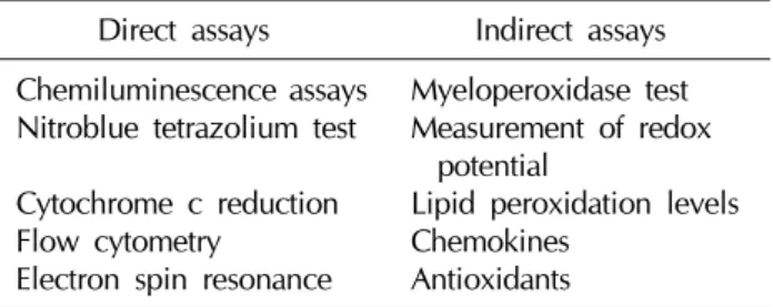
Table 1. Various direct and indirect assays to measure reactive oxygen species human semen
Direct assays Indirect assays Chemiluminescence assays
Nitroblue tetrazolium test Cytochrome c reduction Flow cytometry
Electron spin resonance
Myeloperoxidase test Measurement of redox
potential
Lipid peroxidation levels Chemokines
Antioxidants
MEASUREMENT OF OXIDATIVE STRESS
Over the last decade, research has provided growing support to indicate that excess ROS production leads to abnormal semen parameters and increased sperm damage. Standard semen analysis continues to be the backbone of clinical evaluation of male infertility. Studies have shown that ROS-mediated damage to sperm is a con- siderable contributing pathology in 30% to 80% of un- selected infertile patients [11,46,68]. Therefore, it would be reasonable to expect the screening of all infertile men for the presence of increased ROS levels. However, the detection of the levels and sources of excess ROS pro- duction in semen is currently not included in the routine evaluation of subfertile men [67,71]. Some of the reasons include mere inconvenience, cost-effective and efficient assays, and, perhaps most importantly, the lack of a uni- versally accepted analysis method. All three contribute to the widespread limitations when measuring ROS as part of a male infertility assessment, despite its importance. At present, over 30 different assays are used to measure ROS and the presence of OS in the semen of men (Table 1).
1. Indication of sperm oxidative stress from routine semen analysis
Routine semen analyses have allowed clinicians to make a fairly accurate diagnosis of OS. A reduction in any of the semen parameters (count, motility, and morphology) is more frequently seen in men with OS. Asthenozoosper- mia is most likely the best surrogate marker for OS in a rou- tine semen analysis. The hyperviscosity of seminal plasma is also associated with increased levels of seminal plasma MDA and reduced seminal plasma antioxidant status [72], making impaired viscosity a reasonable surrogate marker
of OS [11]. In addition, infection of the semen with the bac- terial microorganism Ureaplasma urealyticum is asso- ciated with increased seminal plasma viscosity and an in- crease in ROS production [73]. It is possible that these in- fections may damage the prostate and seminal vesicles, al- tering the substrates involved in maintaining normal se- men viscosity. The indication and the presence of a large number of round cells imply possible OS caused by leukocytospermia. However, these round cells may be im- mature spermatozoa rather than leukocytes. For this rea- son, an accurate identification of these cells requires ancil- lary tests such as the peroxidase test, CD45 (transmem- brane glycoprotein expressed at high levels on the cell sur- face) antibody staining or measurement of seminal elastase [74]. Abnormal sperm morphology related to ERC and cy- toplasmic droplets are principal features of anomalous spermatozoa generating high levels of ROS. Lastly, poor sperm membrane integrity, which may be assessed by the hypo-osmotic swelling test (HOST), has been linked to the presence of OS [75].
2. Laboratory assessment of oxidative stress As mentioned earlier, OS results from an imbalance be- tween ROS production and the intracellular/extracellular antioxidants present in seminal plasma. Direct assays of OS measures the net oxidative result of this imbalance by detecting and measuring the amount of oxidation in the sperm cell membrane. MDA, which is one of the final products of membrane LPO, can be measured via the thio- barbituric acid assay, which is one of the oldest and most widely used direct assays for assessing sperm membrane oxidation. Various authors have reported that increased levels of MDA are associated with decreased sperm mo- tility and sperm-oocyte fusion [41,67].
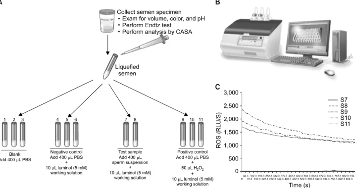
Chemiluminescence assays are most commonly used for measuring seminal ROS. A luminometer is used in con- junction with a chemiluminescent probe such as luminol (5-amino-2,3,-dihydro-1,4-phthalazinedione; Sigma-Aldrich, St. Louis, MO, USA). Aliquots of liquefied semen are cen- trifuged at 300× g for 7 minutes. Seminal plasma is ali- quoted and frozen at −20oC for a later measurement of the total antioxidant levels. The pellet is washed with phos- phate-buffered saline (PBS, pH 7.4) and re-suspended in the same washing medium at a concentration of 2×10⁶
Fig. 3. Measurement of reactive oxygen species (ROS) in sperm suspensions by chemiluminescence assay. (A) Preparation of the samples for ROS measurement. A total of 11 tubes are labeled from S1-S11: Blank, negative control, patient sample, and positive control. Luminol is added to all tubes except blank. Hydrogen peroxide is added only to the positive control. (B) Autolumat 953 plus luminometer used in the measurement of ROS by chemiluminescence assay. Multiple tubes can be loaded simultaneously for measuring ROS. The luminometer is connected to a computer and a monitor, and all the steps can be observed on the screen. (C) A typical graph showing the ROS levels in the 11 tubes (S1-S11). As can be seen, only positive controls have significantly higher levels of ROS. Those producing low levels (Tubes S1-S8) of ROS are seen very close to the X axis. ROS levels measured by the luminometer are expressed as relative light units/s (or RLU/s).
sperm/mL. 400-μL aliquots of the resulting cell suspen- sions containing sperm and leukocytes are used to assess the basal ROS levels. A negative control is prepared by add- ing 10 mL of 5-mM luminol to 400 mL of PBS. Luminol (5-amino-2,3,-dihydro-1,4-phthalazinedione; Sigma), which is prepared as 5-mM stock in dimethyl sulfoxide, is added to the mixture and serves as a probe. All the test tubes are load- ed into the luminometer for 15 minutes to assess the ROS lev- els [8]. Luminol is extremely sensitive and reacts with a varie- ty of ROS at neutral pH [76]. It has the ability to measure both intracellular and extracellular ROS. The free radicals in the semen sample combine with luminol to generate a light sig- nal that is converted to an electric signal (photon) by the lu- minometer (Fig. 3). The number of free radicals produced is measured as relative light units/s/106 sperm. Normal ROS levels in washed sperm suspensions range from 0.10 to 1.03×10⁶ cpm per 20×106 sperm [77].
Luminol can also be used to indirectly measure the total
antioxidant capacity (TAC) within the seminal plasma.
TAC is then quantified against a vitamin E analog Trolox (a water-soluble tocopherol analog). The results are ex- pressed as an ROS-TAC score, and this gives an indication of the combined antioxidant activities of all constituents, including vitamins, proteins, and lipids. This assay ap- pears to be the best established method for analyzing the balance between ROS and the antioxidant protection of sperm [78]. However, the cost of performing this techni- que remains a hurdle to its use in clinical laboratories [66].
An assay that has gained popularity due to its cost-effec- tiveness and user-friendliness is nitroblue tetrazolium (NBT). This assay provides information on the source(s) of ROS. This technique involves only a light microscope and accurately predicts whether ROS has been produced by spermatozoa or leukocytes. When NBT interacts with the O2-. present within spermatozoa or leukocytes, it is con- verted into a blue pigment called diformazam. With the
aid of a light microscope, the amount of diformazam can be observed, measured, and correlated to the intracellular ROS concentration [66,67].
Both direct and indirect assays have been used to quan- tify the levels of ROS. However, all assays of OS in the sperm are relatively expensive and time consuming as compared to a routine semen analysis. Therefore, many clinicians continue to avoid testing their patients for OS and by default offer therapeutic plans with the hope of re- lieving OS and improving the overall semen quality. It can be seen that an OS test may precisely distinguish between fertile and infertile men as well as clinically diagnose male factor infertility. Moreover, such tests can help identify subgroups of infertile patients suffering from OS that may be treated with antioxidant supplementation [71].
PREVENTION AND MANAGEMENT OF OXIDATIVE STRESS
As mentioned earlier, there are innate mechanisms in place to prevent OS from occurring in healthy males.
However, in instances where these natural defenses fail to maintain the fine balance between ROS and antioxidants, measures can be taken to alleviate OS, such as lifestyle changes and antioxidant supplementation (both enzy- matic and non-enzymatic).
1. Prevention of oxidative stress
In healthy males, sperm DNA is protected from OS by two main mechanisms. Firstly, the DNA is tightly coiled and packaged into chromatin such that the genetic materi- al is minimally exposed to attack by ROS [69]. Secondly, natural antioxidants in the seminal plasma and spermato- zoa assist in minimizing the ROS production to normal levels. Some natural antioxidants include enzymes like catalase and SOD as well as non-enzymatic compounds like vitamins C and E and carotenoids. These antioxidants react with and neutralize ROS, assisting in preventing OS onset and preserving the spermatozoa function [79].
Spermatozoa also contain the antioxidants lactoferrin and coenzyme Q10 [7]. A third lesser-mentioned protection mechanism is that of the prostasomes from the prostate.
The presence of prostasomes in the seminal plasma results in a decreased ability of neutrophils to produce super-
oxide radicals [7].
To remain healthy, a sufficient amount of antioxidants must be consumed in one’s diet to prevent OS from occur- ring [7]. However, in some patients who suffer from in- fertility, there may be either an overproduction of ROS or an underproduction of antioxidants, which disrupts the in- tricate balance and results in OS.
2. Management of oxidative stress
In the management of OS, the first step to take is to ascer- tain the underlying cause of the imbalance and treat it [80].
For instance, chlamydia infections can be treated with anti- biotics and anti-inflammatory medication, while varicocele can be corrected by surgery [11]. Thereafter, antioxidant treatment may be given to supplement the natural anti- oxidants and increase the ability of the seminal plasma to combat OS [80]. The following section explores the differ- ent methods, including lifestyle changes and antioxidant supplements, which can be employed to help reduce OS.
1) Lifestyle changes
Modernization, affluence, and the accompanying stresses of the society have resulted in an increase in negative behav- iors, including, but not limited to, smoking, substance abuse, obesity, and an unbalanced diet. All these have been shown to contribute to OS, and therefore, minimizing such detrimental behavior is likely to aid in alleviating OS [11].
It is also recognized that exposure to heat, pollution, tox- ins, and heavy metals play a role in the development of OS.
In addition, any activity that may cause the scrotum’s tem- perature to increase, such as hot baths, saunas, extended periods of driving, and long and sedentary office hours should be avoided. Lastly, adequate protective equipment and aeration should be ensured at work places to limit ex- posure to any chemical or vapor that may cause OS [11].
Undertaking these lifestyle changes can contribute to the reduction of the ROS production and help correct the imbalance causing OS.
2) Antioxidants
Another precautionary measure that can be taken is an- tioxidant supplementation. Antioxidants work by halting the oxidative chain reaction-eliminating, taking up, or re- ducing the formation of ROS [9]. They can be divided into
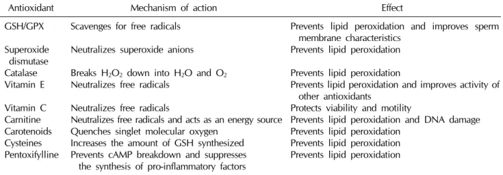
Table 2. Mechanisms of action and effects of various antioxidants
Antioxidant Mechanism of action Effect
GSH/GPX Superoxide
dismutase Catalase Vitamin E Vitamin C Carnitine Carotenoids Cysteines Pentoxifylline
Scavenges for free radicals Neutralizes superoxide anions Breaks H2O2 down into H2O and O2
Neutralizes free radicals Neutralizes free radicals
Neutralizes free radicals and acts as an energy source Quenches singlet molecular oxygen
Increases the amount of GSH synthesized Prevents cAMP breakdown and suppresses
the synthesis of pro-inflammatory factors
Prevents lipid peroxidation and improves sperm membrane characteristics
Prevents lipid peroxidation Prevents lipid peroxidation
Prevents lipid peroxidation and improves activity of other antioxidants
Protects viability and motility
Prevents lipid peroxidation and DNA damage Prevents lipid peroxidation
Prevents lipid peroxidation Prevents lipid peroxidation
GSH: glutathione reductase, GPX: glutathione peroxidase, cAMP: cyclic adenosine 3',5'-monophosphate.
two types on the basis of their actions: (1) preventive anti- oxidants are metal chelators or binding proteins, such as lactoferrin and transferrin, which prevent the formation of ROS; and (2) scavenging antioxidants, like vitamins C and E, remove the ROS that is already present [69]. There have been various studies conducted to elucidate the effective- ness of each individual antioxidant. However, results have been inconclusive as most experiments have a small sample size, differ in dosage and duration of therapy, and lack controls [81].
Moreover, antioxidants work cooperatively, and thus, it is extremely challenging to measure the effect of any sin- gle one alone [79]. This is supported in theory because a suitable combination of antioxidants with their different profiles will neutralize any ROS in its vicinity, hence re- sulting in an additive effect on the decrease in the total OS level of the body [10].
For the purpose of this review, antioxidants are catego- rized as enzymatic and non-enzymatic. Some enzymatic oxidants, or natural oxidants, include glutathione reduc- tase (GSH), SOD, and catalase, while some non-enzy- matic oxidants include vitamins C, E, and B; carotenoids;
carnitines; cysteines, pentoxifylline, metals, taurine, hy- potaurine, and albumin [9]. The non-enzymatic oxidants are acquired from fruits or vegetables containing the sup- plements [69]. Table 2 summarizes the mechanisms of ac- tion and the effects of the more important antioxidants used clinically.
(1) Enzymatic antioxidants
① Glutathione reductase and glutathione peroxidase:
GSH/glutathione peroxidase are the main reducing agents in the body and act as scavenging antioxidants in the epi- didymis and testes [82]. Their modification of the sperma- tozoa membrane confers protection on the lipid con- stituents, thus preserving sperm viability and motility [7].
Previous in vitro studies have shown that GSH preserves the tail-beat frequency, reduces LPO, and improves the sperm membrane characteristics [83].
② Superoxide dismutase and catalase: SOD protects sperm from superoxide anions by catalyzing the con- version of superoxide into oxygen and H2O2, thereby pre- venting LPO and improving motility [80]. On the other hand, catalase aids in the decomposition of H2O2 into wa- ter and oxygen [82]. Thus, both SOD and catalase assist in removing ROS that has the potential to damage sperm.
(2) Non-enzymatic antioxidants
① Vitamin E: Vitamin E (α-tocopherol) is a chain- breaking antioxidant found in the sperm’s cell membrane and acts by neutralizing H2O2 and quenching free radicals [9], hence halting chain reactions that produce lipid per- oxides and protecting the membrane from the damage in- duced by ROS [69]. Furthermore, it improves the activity of other scavenging oxidants [82]. In these ways, vitamin E helps to preserve both sperm motility and morphology [80].
② Vitamin C: Vitamin C (ascorbate) is another
chain-breaking antioxidant that plays a significant role (up to 65%) in combatting OS in the seminal plasma [79]. It re- acts with OH-, O2-, and H2O2 in the extracellular fluid, thus protecting sperm viability and motility [69].
However, vitamin C is only a weak ROS scavenger in the cell membrane and, hence, has almost no effect within the cell [7].
③ Carnitine: Carnitine is a water-soluble antioxidant commonly attained from dietary sources. It may partic- ipate in sperm motility as a fuel source by assisting free fat- ty acid utilization and preventing lipid oxidation [82].
Therefore, carnitine protects the sperm DNA and mem- branes from oxidative damage, and maintains the sperm viability and motility [79].
④ Carotenoids: Although carotenoids have short half-lives, they are very effective and efficient singlet molec- ular oxygen quenchers [84]. Two major carotenoids worth mentioning are β-carotene, which prevents lipids in the cell membrane from being peroxidized [9], and lycopene, which is the most potent and readily available carotenoid that prevents peroxidation in the seminal plasma [69].
⑤ Cysteines: Cysteines are precursors of intracellular GSH and therefore, increase the amount of GSH synthesized. GSH subsequently scavenges oxidants and prevents oxidative damage to the cell membrane and DNA [9]. N-acetyl-L-cysteine works via two mechanisms:
(1) by boosting the amount of reducing agent produced and (2) ridding the spermatozoa of free radicals [82], there- by proserving sperm motility [10].
⑥ Pentoxifylline: Pentoxifylline acts as a competitive phosphodiesterase inhibitor and prevents the breakdown of intracellular cyclic adenosine monophosphate (cAMP).
It also suppresses the synthesis of tumor necrosis factor-α (TNF-α) and leukotrienes and as a result, decreases the in- flammation levels [82]. When the amount of superoxide produced by spermatozoa is decreased, less LPO occurs and sperm motility is preserved [81]. A study has also shown that a 1,200-mg dose of oral pentoxifylline daily re- sults in increased motility and beat cross frequency [79].
(3) Other antioxidants
There are a few other minor antioxidants that contribute to relieving OS, such as albumin, taurine/hypotaurine, in- ositol, and some metals. Albumin, a plasma protein, also
has antioxidative properties. It interacts with peroxyl radi- cals and prevents the chain reactions that generate more free radicals, hence reducing the ROS production and pro- serving sperm motility and viability [79]. Taurine is another non-enzymatic antioxidant that scavenges ROS, while in- ositol is known to enhance GSH activity and preserve nor- mal sperm morphology [9]. Furthermore, certain metals have antioxidant properties. For example, selenium is an important component in the regular development and ma- turation of the testes and contributes to the protection of sperm DNA and cell membranes, particularly when used as an adjunct to vitamin E [85]. Additionally, zinc acts as a chelator and binds ROS [84], while manganese enhances sperm motility and viability [9].
3) Surgery
Varicocele refers to the abnormal dilation and elonga- tion of the pampiniform plexus of veins around the sper- matic cord [86]. Corrective surgery, involving the occlu- sion of these dilated veins, may be performed on subfertile males or men suffering from testicular aches and pains [87]. This has been shown to decrease the ROS levels in se- men, thereby protecting the sperm membrane and DNA from oxidative damage [11]. Surgical repair also improves other biomarkers of infertility, including sperm parame- ters and pregnancy rates [87].
CONCLUSION
OS has been considered a major contributory factor to male infertility. Studies have demonstrated that low and controlled concentrations of ROS play an important role in normal sperm physiological processes such as capacita- tion, hyperactivation, acrosome reactions, and signaling processes to ensure appropriate fertilization. Moreover, there is growing evidence that an increase in OS sig- nificantly impairs sperm function. These impairments have resulted in male infertility via mechanisms involving the induction of peroxidative damage to the sperm plasma membrane, DNA damage, and apoptosis. ROS must be maintained at appropriate levels to ensure appropriate physiological function, while preventing pathological damage to the spermatozoa. When the natural balance be- tween ROS and antioxidants is disturbed, the first re-
storative measure to be taken should be changes in life- style, such as cessation of smoking, limiting substance use, and maintaining a healthy and balanced diet. Antioxidant supplementation may then be taken together to improve the patient’s health outcomes.
An accurate assessment of the seminal ROS levels should become an integral part of the andrology work-up of men in order to assist clinicians in elucidating the fertil- ity status and thereby providing an optimal treatment re- gime for these patients.
ACKNOWLEDGEMENTS
This research was supported by funds from The Center for Reproductive Medicine, Glickman Urological &
Kidney Institute, Cleveland Clinic (Cleveland, OH, USA).
REFERENCES
1. Trussell JC. Optimal diagnosis and medical treatment of male infertility. Semin Reprod Med 2013;31:235-6.
2. World Health Organisation. WHO Laboratory Manual for the Examination and Processing of Human Semen. 5th ed.
Geneva: World Health Organization; 2010.
3. Agarwal A, Sekhon LH. Oxidative stress and antioxidants for idiopathic oligoasthenoteratospermia: is it justified?
Indian J Urol 2011;27:74-85.
4. Saalu LC. The incriminating role of reactive oxygen species in idiopathic male infertility: an evidence based evaluation.
Pak J Biol Sci 2010;13:413-22.
5. Hampl R, Drábková P, Kanďár R, Stěpán J. Impact of oxida- tive stress on male infertility. Ceska Gynekol 2012;77:
241-5.
6. Henkel RR. Leukocytes and oxidative stress: dilemma for sperm function and male fertility. Asian J Androl 2011;13:
43-52.
7. Lanzafame FM, La Vignera S, Vicari E, Calogero AE.
Oxidative stress and medical antioxidant treatment in male infertility. Reprod Biomed Online 2009;19:638-59.
8. Saleh RA, Agarwal A. Oxidative stress and male infertility:
from research bench to clinical practice. J Androl 2002;
23:737-52.
9. Bansal AK, Bilaspuri GS. Impacts of oxidative stress and an- tioxidants on semen functions. Vet Med Int 2011;2011:
686137.
10. Gharagozloo P, Aitken RJ. The role of sperm oxidative stress in male infertility and the significance of oral anti- oxidant therapy. Hum Reprod 2011;26:1628-40.
11. Tremellen K. Oxidative stress and male infertility--a clinical perspective. Hum Reprod Update 2008;14:243-58.
12. De Iuliis GN, Wingate JK, Koppers AJ, McLaughlin EA, Aitken RJ. Definitive evidence for the nonmitochondrial production of superoxide anion by human spermatozoa. J Clin Endocrinol Metab 2006;91:1968-75.
13. Aitken RJ, Baker MA, De Iuliis GN, Nixon B. New insights into sperm physiology and pathology. Handb Exp Pharmacol 2010;(198):99-115.
14. Butler A, He X, Gordon RE, Wu HS, Gatt S, Schuchman EH.
Reproductive pathology and sperm physiology in acid sphingomyelinase-deficient mice. Am J Pathol 2002;161:
1061-75.
15. Miranda-Vilela AL, Alves PC, Akimoto AK, Pereira LC, Nazaré Klautau-Guimarães M, Grisolia CK. The effect of hy- drogen peroxide-induced oxidative stress on leukocytes de- pends on age and physical training in healthy human sub- jects carrying the same genotypes of antioxidant enzymes' gene polymorphisms. Am J Hum Biol 2010;22:807-12.
16. Chen SJ, Allam JP, Duan YG, Haidl G. Influence of reactive oxygen species on human sperm functions and fertilizing capacity including therapeutical approaches. Arch Gynecol Obstet 2013;288:191-9.
17. Hazout A, Menezo Y, Madelenat P, Yazbeck C, Selva J, Cohen-Bacrie P. Causes and clinical implications of sperm DNA damages. Gynecol Obstet Fertil 2008;36:1109-17.
18. Sikka SC. Relative impact of oxidative stress on male re- productive function. Curr Med Chem 2001;8:851-62.
19. Sabeur K, Ball BA. Characterization of NADPH oxidase 5 in equine testis and spermatozoa. Reproduction 2007;134:
263-70.
20. Choudhary R, Chawala VK, Soni ND, Kumar J, Vyas RK.
Oxidative stress and role of antioxidants in male infertility.
Pak J Physiol 2010;6:54-9.
21. Esteves SC. Effect of cigarette smoking on levels of seminal oxidative stress in infertile men: a prospective study. Int Braz J Urol 2002;28:484-5.
22. Saleh RA, Agarwal A, Nada EA, El-Tonsy MH, Sharma RK, Meyer A, et al. Negative effects of increased sperm DNA damage in relation to seminal oxidative stress in men with idiopathic and male factor infertility. Fertil Steril 2003;79 Suppl 3:1597-605.
23. Lavranos G, Balla M, Tzortzopoulou A, Syriou V, Angelo- poulou R. Investigating ROS sources in male infertility: a common end for numerous pathways. Reprod Toxicol 2012;34:298-307.
24. Agarwal A, Saleh RA, Bedaiwy MA. Role of reactive oxygen species in the pathophysiology of human reproduction.
Fertil Steril 2003;79:829-43.
25. Lu JC, Huang YF, Lü NQ. WHO Laboratory Manual for the Examination and Processing of Human Semen: its applic- ability to andrology laboratories in China. Zhonghua Nan Ke Xue 2010;16:867-71.
26. Nandipati KC, Pasqualotto FF, Thomas AJ Jr, Agarwal A.
Relationship of interleukin-6 with semen characteristics and oxidative stress in vasectomy reversal patients. Andrologia 2005;37:131-4.
27. Rengan AK, Agarwal A, van der Linde M, du Plessis SS. An investigation of excess residual cytoplasm in human sper- matozoa and its distinction from the cytoplasmic droplet.
Reprod Biol Endocrinol 2012;10:92.
28. Will MA, Swain J, Fode M, Sonksen J, Christman GM, Ohl D. The great debate: varicocele treatment and impact on fertility. Fertil Steril 2011;95:841-52.
29. Shiraishi K, Matsuyama H, Takihara H. Pathophysiology of varicocele in male infertility in the era of assisted re- productive technology. Int J Urol 2012;19:538-50.
30. Agarwal A, Deepinder F, Sharma RK, Ranga G, Li J. Effect of cell phone usage on semen analysis in men attending in- fertility clinic: an observational study. Fertil Steril 2008;89:
124-8.
31. Aitken RJ, Bennetts LE, Sawyer D, Wiklendt AM, King BV.
Impact of radio frequency electromagnetic radiation on DNA integrity in the male germline. Int J Androl 2005;28:
171-9.
32. De Iuliis GN, Newey RJ, King BV, Aitken RJ. Mobile phone radiation induces reactive oxygen species production and DNA damage in human spermatozoa in vitro. PLoS One 2009;4:e6446.
33. Esfandiari N, Saleh RA, Blaut AP, Sharma RK, Nelson DR, Thomas AJ Jr, et al. Effects of temperature on sperm motion characteristics and reactive oxygen species. Int J Fertil Womens Med 2002;47:227-33.
34. Pant N, Shukla M, Kumar Patel D, Shukla Y, Mathur N, Kumar Gupta Y, et al. Correlation of phthalate exposures with semen quality. Toxicol Appl Pharmacol 2008;231:
112-6.
35. Latini G, Del Vecchio A, Massaro M, Verrotti A, De Felice C. Phthalate exposure and male infertility. Toxicology 2006;226:90-8.
36. Kasahara E, Sato EF, Miyoshi M, Konaka R, Hiramoto K, Sasaki J, et al. Role of oxidative stress in germ cell apoptosis induced by di(2-ethylhexyl)phthalate. Biochem J 2002;365:
849-56.
37. Jurasović J, Cvitković P, Pizent A, Colak B, Telisman S.
Semen quality and reproductive endocrine function with re- gard to blood cadmium in Croatian male subjects.
Biometals 2004;17:735-43.
38. Saleh RA, Agarwal A, Sharma RK, Nelson DR, Thomas AJ Jr.
Effect of cigarette smoking on levels of seminal oxidative stress in infertile men: a prospective study. Fertil Steril 2002;78:491-9.
39. Jarow JP. Semen quality of male smokers and nonsmokers in infertile couples. J Urol 2003;170:675-6.
40. Kiziler AR, Aydemir B, Onaran I, Alici B, Ozkara H, Gulyasar T, et al. High levels of cadmium and lead in semi- nal fluid and blood of smoking men are associated with high oxidative stress and damage in infertile subjects. Biol Trace Elem Res 2007;120:82-91.
41. Agarwal A, Prabakaran SA. Mechanism, measurement, and prevention of oxidative stress in male reproductive physiology. Indian J Exp Biol 2005;43:963-74.
42. Tsai WW, Niessen S, Goebel N, Yates JR 3rd, Guccione E, Montminy M. PRMT5 modulates the metabolic response to fasting signals. Proc Natl Acad Sci U S A 2013;110:8870-5.
43. Kothari S, Thompson A, Agarwal A, du Plessis SS. Free radi- cals: their beneficial and detrimental effects on sperm function. Indian J Exp Biol 2010;48:425-35.
44. de Lamirande E, O'Flaherty C. Sperm activation: role of re- active oxygen species and kinases. Biochim Biophys Acta 2008;1784:106-15.
45. Suarez SS. Control of hyperactivation in sperm. Hum Reprod Update 2008;14:647-57.
46. Makker K, Agarwal A, Sharma R. Oxidative stress & male infertility. Indian J Med Res 2009;129:357-67.
47. Calamera J, Buffone M, Ollero M, Alvarez J, Doncel GF.
Superoxide dismutase content and fatty acid composition in subsets of human spermatozoa from normozoospermic, as- thenozoospermic, and polyzoospermic semen samples. Mol Reprod Dev 2003;66:422-30.
48. Khosrowbeygi A, Zarghami N. Fatty acid composition of human spermatozoa and seminal plasma levels of oxidative stress biomarkers in subfertile males. Prostaglandins Leukot Essent Fatty Acids 2007;77:117-21.
49. Sanocka D, Kurpisz M. Reactive oxygen species and sperm cells. Reprod Biol Endocrinol 2004;2:12.
50. Zribi N, Chakroun NF, Elleuch H, Abdallah FB, Ben Hamida AS, Gargouri J, et al. Sperm DNA fragmentation and oxidation are independent of malondialdheyde. Reprod Biol Endocrinol 2011;9:47.
51. Schulte RT, Ohl DA, Sigman M, Smith GD. Sperm DNA damage in male infertility: etiologies, assays, and outcomes.
J Assist Reprod Genet 2010;27:3-12.
52. Kemal Duru N, Morshedi M, Oehninger S. Effects of hydro- gen peroxide on DNA and plasma membrane integrity of human spermatozoa. Fertil Steril 2000;74:1200-7.
53. Badouard C, Ménézo Y, Panteix G, Ravanat JL, Douki T, Cadet J, et al. Determination of new types of DNA lesions in human sperm. Zygote 2008;16:9-13.
54. González-Marín C, Gosálvez J, Roy R. Types, causes, de- tection and repair of DNA fragmentation in animal and hu- man sperm cells. Int J Mol Sci 2012;13:14026-52.
55. Valavanidis A, Vlachogianni T, Fiotakis C. 8-hydroxy-2' -deoxyguanosine (8-OHdG): a critical biomarker of oxida- tive stress and carcinogenesis. J Environ Sci Health C Environ Carcinog Ecotoxicol Rev 2009;27:120-39.
56. Aitken RJ, Koppers AJ. Apoptosis and DNA damage in hu- man spermatozoa. Asian J Androl 2011;13:36-42.
57. Aitken RJ, Baker MA. Causes and consequences of apopto- sis in spermatozoa; contributions to infertility and impacts on development. Int J Dev Biol 2013;57:265-72.
58. Agarwal A, Said TM. Role of sperm chromatin abnormalities and DNA damage in male infertility. Hum Reprod Update 2003;9:331-45.
59. Kao SH, Chao HT, Chen HW, Hwang TI, Liao TL, Wei YH.
Increase of oxidative stress in human sperm with lower motility. Fertil Steril 2008;89:1183-90.