HIGHLIGHTS
• A postoperative intraventricular hemorrhage could injure the limbic system.
• Disconnection of the limbic system could manifest psychosis.
• Diffusion tensor tractography was valuable for evaluating the limbic system.
Brain Neurorehabil. 2020 Nov;13(3):e21 https://doi.org/10.12786/bn.2020.13.e21 pISSN 1976-8753·eISSN 2383-9910
Case Report
Received: Jun 5, 2020 Revised: Aug 21, 2020 Accepted: Aug 22, 2020 Correspondence to Youngkook Kim
Department of Rehabilitation Medicine, Yeouido St. Mary's Hospital, College of Medicine, The Catholic University of Korea, 10, 63-ro, Yeongdeungpo-gu, Seoul 07345, Korea.
E-mail: england2formac@gmail.com
So-youn Chang, Youngkook Kim
Reversible Psychosis Caused by
Disconnection of the Limbic System:
Clinical Reasoning Using Diffusion Tensor Tractography
Brain & NeuroRehabilitation
Copyright © 2020. Korean Society for Neurorehabilitation i
ABSTRACT
Injury to the limbic system can result in amnesia, language difficulties, behavioral
abnormalities, and psychological disorders. We present a patient who suffered psychosis related to disconnection of the limbic system after intraventricular and orbitofrontal hemorrhages following removal of a sellar meningioma. Diffusion tensor tractography was valuable for evaluating the structural integrity of the injured limbic tracts and determining the regeneration of tracts corresponding to neuropsychiatric recovery after cognitive rehabilitation.
Keywords: Intracranial hemorrhage; Psychosis; Limbic system; Diffusion tensor imaging
INTRODUCTION
The limbic system is a set of cortical and subcortical structures interconnected by limbic tracts related to cognition, emotion, and behavior [1]. Limbic dysfunction can manifest not only as amnesia, language difficulties, and emotional or behavioral changes, but also as various psychological conditions, such as schizophrenia, bipolar disorder, depression, and psychopathy [2]. Identifying the limbic structures in the living human brain is challenging because they are difficult to differentiate from adjacent structures in conventional magnetic resonance imaging (MRI). Diffusion tensor imaging (DTI) is a noninvasive MRI technique that allows the construction of white matter tracts using diffusion tensor tractography (DTT) and enables evaluation of the structural integrity of individual tracts. We report a patient who presented with psychosis after postoperative intraventricular and orbitofrontal hemorrhage following the removal of a sellar meningioma.
CASE REPORT
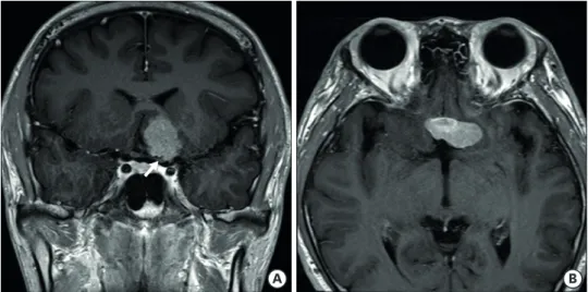
A 23-year-old male visited the ophthalmology clinic of our institution complaining of visual disturbance. Automated perimetry revealed left homonymous hemianopia, suggesting a left post-chiasmatic optic tract lesion. He was referred to the neurosurgery department and underwent MRI that revealed a 1.9 × 3.3 cm mass arising from the planum sphenoidale,
Case Report
Received: Jun 5, 2020 Revised: Aug 21, 2020 Accepted: Aug 22, 2020 Correspondence to Youngkook Kim
Department of Rehabilitation Medicine, Yeouido St. Mary's Hospital, College of Medicine, The Catholic University of Korea, 10, 63-ro, Yeongdeungpo-gu, Seoul 07345, Korea.
E-mail: england2formac@gmail.com Copyright © 2020. Korean Society for Neurorehabilitation
This is an Open Access article distributed under the terms of the Creative Commons Attribution Non-Commercial License (https://
creativecommons.org/licenses/by-nc/4.0) which permits unrestricted non-commercial use, distribution, and reproduction in any medium, provided the original work is properly cited.
ORCID iDs So-youn Chang
https://orcid.org/0000-0003-0040-8084 Youngkook Kim
https://orcid.org/0000-0003-3964-026X Conflict of Interest
The authors have no potential conflicts of interest to disclose.
So-youn Chang , Youngkook Kim
Department of Rehabilitation Medicine, Yeouido St. Mary's Hospital, College of Medicine, The Catholic University of Korea, Seoul, Korea
Reversible Psychosis Caused by
Disconnection of the Limbic System:
Clinical Reasoning Using Diffusion
Tensor Tractography
suggesting sellar meningioma (Fig. 1). He underwent a craniotomy, and the tumor was removed using the subfrontal transbasal approach. He was drowsy for 1 day after the surgery, and a computed tomography scan of his brain revealed a postoperative intraventricular hemorrhage (IVH) involving the left anterior horn of the lateral ventricle accompanying a left orbitofrontal hemorrhage. He underwent emergent bilateral external ventricular drainage.
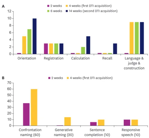
He was transferred to the rehabilitation department for cognitive rehabilitation 2 weeks after the surgery. He was drowsy at the initial evaluation and scored only 3 points on the Korean-Mini Mental State Examination (K-MMSE). The Western Aphasia Battery (WAB) revealed anomic aphasia with a language disorder due to cognitive dysfunction of aphasia quotient 65.2 (Fig. 2). He underwent cognitive rehabilitation with an occupational therapist and language therapy with a language therapist 5 days a week. Oral medications were given, including methylphenidate and donepezil for cognitive and language enhancement. He earned 21 points on the K-MMSE one week later and aphasia quotient 96 with generative naming difficulty on the WAB. However, bizarre behavior and hallucinations emerged unexpectedly after he achieved an alert mental status and fluent speech. He had no prior history of psychopathology before the hemorrhages and had no predisposing risk factors such as drug abuse or heavy alcohol consumption. He made statements such as “my headache was caused by a dead bird in my gut,” “the angel is fighting with the devil,” and “I lost a duck on the first floor of the hospital.” Moreover, he poured water into his right eye while complaining “my eye is burning” and wandered around the admissions ward “to catch flying bees” at dawn. After the emergence of psychosis, methylphenidate was withheld, and quetiapine was administered at a dose of 50 mg/day before bedtime with the impression of delirium. The patient also received non-pharmacological treatment for psychotic symptoms.
To maintain a familiar environment, the family took care of the patient, kept the lights soft at night, and the medical staffs were careful not to stimulate the patient. Besides, nursing personnel provided regular repeated education to allow the patient to recognize the current situation. However, the confusion, visual hallucinations, and bizarre behavior persisted, similar to those with schizophrenia patients.
MRI and DTI were performed for the differential diagnosis of psychosis caused by organic brain injury. DTI was conducted using a 3.0-T magnetic resonance imager (MAGNETOM®
2/7 https://doi.org/10.12786/bn.2020.13.e21
Psychosis caused by Limbic Disconnection Brain & NeuroRehabilitation
https://e-bnr.org
A B
Fig. 1. A 1.9 × 3.3 cm mass arising from the planum sphenoidale was noted and the left post-chiasmatic optic tract (arrow) was deviated by the mass in coronal (A) and axial T1-weighted images (B).
Verio; Siemens, Erlangen, Germany) equipped with a 6-channel head coil. Data were acquired in the form of single-shot spin-echo echo-planar images, with axial slices covering the whole brain across 70 interleaved slices of 2.0 mm thickness (no gap; repetition time/
echo time = 14,300/84 ms; field of view = 256 × 256 mm2; matrix = 128 × 128; voxel size = 2
× 2 × 2 mm3 [isotropic], number of excitations = 1). Diffusion-sensitizing gradients were applied in 30 noncollinear directions with a b-value of 1,000 ms/mm2. The b = 0 images were scanned before acquisition of the diffusion-weighted images, with 31 volumes acquired in total. The images were processed using the FMRIB Software Library (FSL; ver. 5.0.9; http://
www.fmrib.ox.ac.uk/fsl).
We conducted DTT to explore the whole structural integrity of the limbic system, including the fornix, the cingulum, the mammillothalamic tract, and the uncinate fasciculus [3,4].
The fornix is a projection bundle that connects the medial temporal lobe to the mamillary body and is involved in memory functions. The cingulum is a medial associative bundle that runs around the corpus callosum and is involved in attention, memory, and emotions. The mammillothalamic tract connects the mammillary body and the anterior thalamus and is involved in binding and association processes for memory storage. The uncinate fasciculus is a ventral associative bundle that connects the anterior temporal lobe with the orbitofrontal cortex and is involved in emotional processing, memory, and language functions.
12 10 8 6 4 2
0 Orientation Registration Calculation Recall Language &
judge &
construction
A 2 weeks 4 weeks (first DTI acquisition)
6 weeks 14 weeks (second DTI acquisition)
70 60 50 40 30 20 10
0 Confrontation naming (60)
Generative naming (20)
Sentence completion (10)
Responsive speech (10)
B 2 weeks 4 weeks (first DTI acquisition)
Fig. 2. The Korean-Mini Mental State Examination score of the patient improved after cognitive rehabilitation and administration of a cognitive-stimulating medication. A pronounced enhancement was noted on the orientation, calculation, and recall subscales 3 months after the postoperative hemorrhage (A). Generative naming in the naming subset of the Western Aphasia Battery remained abnormal but scores on other naming tasks were maximum 1 month after the postoperative hemorrhage (B).
DTI, diffusion tensor imaging.
Probabilistic tractography was performed using the FSL ProbtrackX program to reconstruct the limbic tracts. The fiber-tracking parameters were as follows: number of samples, 5,000;
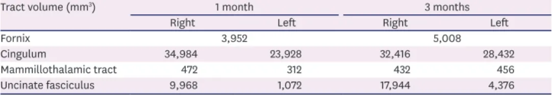
curvature threshold, 0.2 (cosine 78.5°); and step length, 0.5 mm. All region-of-interests (ROIs) were drawn on the axial slices of the color fractional anisotropy (FA) map [3,4]. The cingulum, mammillothalamic tract, and uncinate fasciculus were reconstructed by placing the ROIs on both left and right sides. Fornix was reconstructed by one ROI, including the left and right sides because it was impossible to define the exact boundary of the left and right fornix due to the low resolution of the DTI. To reconstruct the fornix, a single ROI was located on the body of the fornix within the lateral ventricles. To reconstruct the cingulum, a single ROI was located on the most dorsal part of the cingulum, cigar-shaped green fibers medial to the red fibers of the corpus callosum. A 2-ROI approach was used to reconstruct the mammillothalamic tract and the uncinate fasciculus. To reconstruct the mammillothalamic tract, the seed ROI was located on the mammillary body, and the way ROI was located on the anterior thalamus. To reconstruct the uncinate fasciculus, the seed ROI was located on the green fibers of the orbitofrontal cortex, and the way ROI was located on the green fibers of the anterior temporal lobe. The structural integrity of the reconstructed tracts was analyzed according to the continuity of tract by visual assessment and the tract volume by quantitative measurement. Continuity of the reconstructed tracts was verified based on neuroanatomical knowledge. Tract volume of each tract was calculated by multiplying the voxel volume by the number of traced voxels during fiber-tracking, based on a robust minimum intensity threshold of 300 for the fornix, 100 for the cingulum, 2,000 for the mammillothalamic tract, and 5 for the uncinate fasciculus, respectively.
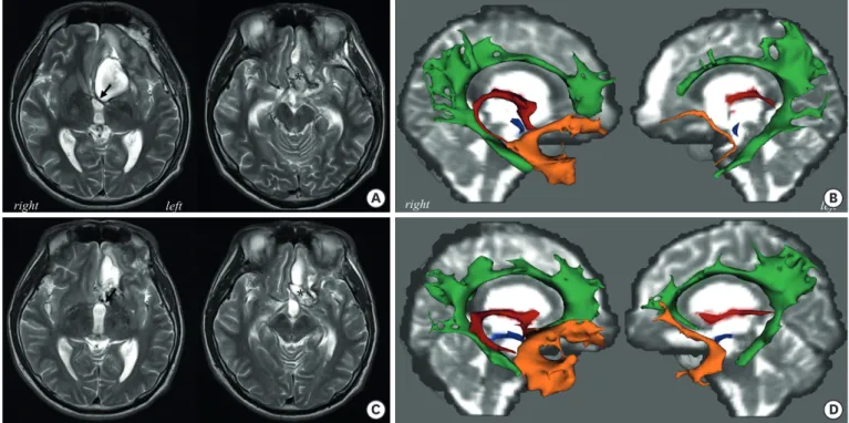
The results of DTT demonstrated loss of continuity in left column and crus of the fornix and loss of continuity and decreased volume in left mammillothalamic tract compared to those of the contralesional side. Left cingulum and uncinate fasciculus showed reduced tract volume compared to those of the contralesional side (Fig. 3A and B) (Table 1). This neuroanatomical deterioration was compatible with his psychotic features, anterograde amnesia, and anomic aphasia. We maintained symptomatic management for the psychiatric problems while waiting for the regeneration of the limbic tracts. He was discharged from the hospital 1 month after the hemorrhage. Four weeks later, his mother reported cessation of psychosis and improved memories from the first visit to the rehabilitation outpatient clinic. Brain MRI and DTT were conducted 3 months following the hemorrhages to ascertain recovery of the limbic system. The IVH, orbitofrontal hemorrhage, and perilesional edema were resolving, and DTT demonstrated the regeneration of the fornix, left cingulum, mammillothalamic tract, and uncinate fasciculus (Fig. 3C and D). Left cingulum, mammillothalamic tract, uncinate fasciculus, and the fornix, including left and right parts, expanded in volume compared to initial tractographic analysis. We also found an increased tract volume of the contralesional uncinate fasciculus compared with initial tractographic analysis (Table 1).
4/7 https://doi.org/10.12786/bn.2020.13.e21
Psychosis caused by Limbic Disconnection Brain & NeuroRehabilitation
https://e-bnr.org
Table 1. Quantitative volume analysis of limbic tracts by diffusion tensor tractography
Tract volume (mm3) 1 month 3 months
Right Left Right Left
Fornix 3,952 5,008
Cingulum 34,984 23,928 32,416 28,432
Mammillothalamic tract 472 312 432 456
Uncinate fasciculus 9,968 1,072 17,944 4,376
at 1 month and 3 months after postoperative hemorrhage
DISCUSSION
DTT played a critical role in explaining the reversible psychosis with an accompanying cognitive impairment in a patient with IVH and orbitofrontal hemorrhage after sellar meningioma removal. Delirium, side effects of medication acting on the central nervous system, and neuropsychiatric symptoms related to organic brain damage are the plausible causes for postoperative mental deterioration and subsequent psychosis [5-7]. Although delirium is common after organic brain injury, most cases develop within 4 days [6]. Since the time interval between the IVH and the development of psychotic features was more than 2 weeks in this case, delirium seemed to unlikely develop. Methylphenidate was also a suspected cause of deterioration because psychosis could be an adverse effect of taking methylphenidate [7]. Clinical concerns have been raised that methylphenidate might increase the risk of psychosis because methylphenidate as an indirect dopamine agonist is presumed to generate psychotic symptoms by increasing the concentration of extracellular dopamine in the prefrontal cortex [8,9]. He had been taking methylphenidate for more than 2 weeks prior to the psychotic features, and the methylphenidate was discontinued immediately after the emergence of psychotic symptoms. In this regard, a recent population-based cohort study reported that there was no convincing evidence that initiation of methylphenidate treatment increases the immediate (12 weeks) and long-term (1 year) risk of psychotic events in adolescents and young adults with attention-deficit hyperactivity disorder [10]. Taken together, it was unlikely that methylphenidate was a mediator of psychosis.
A B
C D
*
*
Fig. 3. Magnetic resonance images and DTT obtained 1 month (A, B) and 3 months (C, D) after the postoperative hemorrhage. Deviated left fornix column due to the intraventricular hemorrhage (arrow) and residual orbitofrontal hemorrhage (asterisk) (A). Non-visualized column and crus of the left fornix (red) and degeneration of the left cingulum (green), mammillothalamic tract (blue), and uncinate fasciculus (yellow) (B). The intraventricular and orbitofrontal hemorrhages were resolved, and the fornix returned to its original location (C). The column and crus of the left fornix were visualized, and the left cingulum, mammillothalamic tract, and uncinate fasciculus were regenerating. Right uncinate fasciculus expanded in volume compared to the 1-month DTT results (D).
DTT, diffusion tensor tractography.
Lastly, we suspected an organic brain injury. An orbitofrontal hemorrhage can disrupt the uncinate fasciculus, which connects the orbitofrontal cortex to the anterior temporal lobe [3]. The fornix can be injured by the mechanical and chemical mechanisms of IVH.
Mass effect by IVH can reduce cerebral perfusion pressure and cause ischemic injury, and a blood clot can injure the ventricular ependymal and subependymal layer by releasing free iron [11,12]. Therefore, we placed more weight on organic brain injury based on the neuroanatomical location of the parenchymal hemorrhage and the IVH affecting the orbitofrontal cortex and fornix, respectively.
The limbic system consists of core subcortical structures (hippocampus and thalamus) and paralimbic cortical areas (cingulate gyrus, orbitofrontal cortex, and anterior temporal lobe).
The core subcortical structures are connected by the fornix and mammillothalamic tract, and the paralimbic cortical areas are connected by the cingulum and uncinate fasciculus.
Damage to any of the limbic system structures can lead to limbic dysfunction. Bennett et al.
[13] investigated the integrity of limbic tracts (fornix, cingulum, and uncinate fasciculus), and FA analysis of DTI revealed the presence of limbic networks beyond the hippocampus extending to the fornix. We reconstructed the entire limbic system and explored the structural integrity corresponding to the clinical symptoms. The hippocampal-diencephalic circuit connects via the fornix and mammillothalamic tract and is responsible for memory [1]. The temporo-amygdala-orbitofrontal network engages in behavioral inhibition, naming, and memory [1,2,13]. Damage to the cingulum, uncinate fasciculus, and fornix is associated with schizophrenia [2]. In the present study, the tractography results demonstrated that the limbic system was disconnected due to the degeneration of the fornix, cingulum, mammillothalamic tract, and uncinate fasciculus. These findings corresponded to the manifestations of psychosis, anterograde amnesia, and anomic aphasia.
Technical considerations for tractography need to be addressed. The manual drawing of ROI may be operator-dependent, and this can result in inter-rater or intra-rater variability. Also, the volume of reconstructed tracts may vary depending on the spatial resolution of data (voxel size, number of diffusion-sensitizing gradients, b-values), the fiber tracking method (deterministic or probabilistic), and the fiber tracking parameters (number of sampling, FA threshold, curvature threshold). These points should be considered when interpreting our results.
In conclusion, the posthemorrhagic neuropsychiatric problems resulted from the
disconnection of the limbic system. Furthermore, we identified the regenerating limbic tracts that coincided with the recovery of clinical symptoms after 3 months. DTT was valuable for evaluating limbic dysfunction.
REFERENCES
1. Catani M, Dell'acqua F, Thiebaut de Schotten M. A revised limbic system model for memory, emotion and behaviour. Neurosci Biobehav Rev 2013;37:1724-1737.
PUBMED | CROSSREF
2. Catani M, Dell'acqua F, Bizzi A, Forkel SJ, Williams SC, Simmons A, Murphy DG, Thiebaut de Schotten M. Beyond cortical localization in clinico-anatomical correlation. Cortex 2012;48:1262-1287.
PUBMED | CROSSREF
3. Catani M, Thiebaut de Schotten M. A diffusion tensor imaging tractography atlas for virtual in vivo dissections. Cortex 2008;44:1105-1132.
PUBMED | CROSSREF
6/7 https://doi.org/10.12786/bn.2020.13.e21
Psychosis caused by Limbic Disconnection Brain & NeuroRehabilitation
https://e-bnr.org
4. Jang SH, Choi BY, Kim SH, Chang CH, Jung YJ, Kwon HG. Injury of the mammillothalamic tract in patients with subarachnoid haemorrhage: a retrospective diffusion tensor imaging study. BMJ Open 2014;4:e005613.
PUBMED | CROSSREF
5. Savina MA. Post stroke delirium. Zh Nevrol Psikhiatr Im S S Korsakova 2014;114:19-27.
PUBMED | CROSSREF
6. McManus J, Pathansali R, Hassan H, Ouldred E, Cooper D, Stewart R, Macdonald A, Jackson S. The course of delirium in acute stroke. Age Ageing 2009;38:385-389.
PUBMED | CROSSREF
7. Kraemer M, Uekermann J, Wiltfang J, Kis B. Methylphenidate-induced psychosis in adult attention- deficit/hyperactivity disorder: report of 3 new cases and review of the literature. Clin Neuropharmacol 2010;33:204-206.
PUBMED | CROSSREF
8. Howes OD, Kambeitz J, Kim E, Stahl D, Slifstein M, Abi-Dargham A, Kapur S. The nature of dopamine dysfunction in schizophrenia and what this means for treatment. Arch Gen Psychiatry 2012;69:776-786.
PUBMED | CROSSREF
9. Arnsten AF. Stimulants: therapeutic actions in ADHD. Neuropsychopharmacology 2006;31:2376-2383.
PUBMED | CROSSREF
10. Hollis C, Chen Q, Chang Z, Quinn PD, Viktorin A, Lichtenstein P, D'Onofrio B, Landén M, Larsson H.
Methylphenidate and the risk of psychosis in adolescents and young adults: a population-based cohort study. Lancet Psychiatry 2019;6:651-658.
PUBMED | CROSSREF
11. Kwon HG, Jang SH. Bilateral fornix injury due to cerebral infarct and traumatic intraventricular hemorrhage: a case study. Clin Neurol Neurosurg 2013;115:99-101.
PUBMED | CROSSREF
12. Yeo SS, Choi BY, Chang CH, Jung YJ, Ahn SH, Son SM, Byun WM, Jang SH. Periventricular white matter injury by primary intraventricular hemorrhage: a diffusion tensor imaging study. Eur Neurol 2011;66:235-241.
PUBMED | CROSSREF
13. Bennett IJ, Huffman DJ, Stark CE. Limbic tract integrity contributes to pattern separation performance across the lifespan. Cereb Cortex 2015;25:2988-2999.
PUBMED | CROSSREF