Spontaneous Bacterial Peritonitis in Patients with Hepatitis B Virus-Related Liver Cirrhosis: Community-Acquired
versus Nosocomial
Seung Up Kim,
1* Young Eun Chon,
1* Chun Kyon Lee,
5Jun Yong Park,
1,2,4Do Young Kim,
1,2,4Kwang-Hyub Han,
1,2,4,6Chae Yoon Chon,
1,2,4Sinyoung Kim,
3Kyu Sik Jung,
1and Sang Hoon Ahn
1,2,4,6Departments of 1Internal Medicine, 2Institute of Gastroenterology, and 3Laboratory Medicine, Yonsei University College of Medicine, Seoul;
4Liver Cirrhosis Clinical Research Center, Seoul; 5National Health Insurance Corporation, Ilsan Hospital, Goyang;
6Brain Korea 21 Project for Medical Science, Seoul, Korea.
Received: May 3, 2011 Revised: June 21, 2011 Accepted: July 6, 2011
Corresponding author: Dr. Sang Hoon Ahn, Department of Internal Medicine, Yonsei University College of Medicine 50 Yonsei-ro, Seodaemun-gu, Seoul 120-752, Korea.
Tel: 82-2-2228-1936, Fax: 82-2-393-6884 E-mail: [email protected]
*Seung Up Kim and Young Eun Chon contributed equally to this work.
∙ The authors have no financial conflicts of interest.
© Copyright:
Yonsei University College of Medicine 2012 This is an Open Access article distributed under the terms of the Creative Commons Attribution Non- Commercial License (http://creativecommons.org/
licenses/by-nc/3.0) which permits unrestricted non- commercial use, distribution, and reproduction in any medium, provided the original work is properly cited.
Purpose: Spontaneous bacterial peritonitis (SBP) frequently develops in patients with liver cirrhosis; however, there is little data to suggest whether the acquisition site of infection influences the prognosis. This study compared the bacteriology, clinical characteristics and treatment outcomes of community-acquired SBP (CA- SBP) and nosocomial SBP (N-SBP). Materials and Methods: The medical re- cords of 130 patients with hepatitis B virus (HBV)-related liver cirrhosis, who had experienced a first episode of SBP between January 1999 and December 2008, were reviewed. Results: The study population included 111 (85.4%) patients with CA-SBP and 19 (14.6%) patients with N-SBP. Baseline and microbiological char- acteristics as well as clinical course, including in-hospital mortality, did not differ between patients with CA-SBP and those with N-SBP (all p>0.05). The median survival time was 6.5 months, and 117 (90.0%) patients died during the follow-up period. Patients with CA-SBP and N-SBP survived for median periods of 6.6 and 6.2 months, respectively, without significant difference (p=0.569). Time to recur- rence did not differ between patients with CA-SBP and N-SBP (4.7 vs. 3.6 months, p=0.925). Conclusion: The acquisition site of infection did not affect clinical out- comes for patients with HBV-related liver cirrhosis who had experienced their first episode of SBP. Third-generation cephalosporins may be effective in empirically treating these patients, regardless of the acquisition site of the infection.
Key Words: Ascites, cirrhosis, community-acquired, nosocomial, spontaneous bacterial peritonitis
INTRODUCTION
Spontaneous bacterial peritonitis (SBP) is an ascitic fluid infection without a defin- itive, surgically treatable, intra-abdominal source and is a drastic complication of end-stage liver disease, occurring in 10 to 25% of cirrhotic patients with ascites.1,2 Although mortality related to SBP has markedly decreased over the last 3 decades,
went a liver transplantation during the follow-up period were also excluded to remove confounding effects of these factors on survival. In addition, patients whose ascites were caused by tuberculosis or malignancy or whose culture re- sults suggested secondary bacterial peritonitis (polymicrobi- al infection) or contamination by skin or medical appliances (coagulase-negative staphylococci, corynebacterium, propi- onibacterium, or bacillus species) were excluded. The proto- col of this study was consistent with the ethical guidelines of the 1975 Declaration of Helsinki and was approved by the independent institutional review boards of each institute.
Definitions of variables
The diagnostic criteria for SBP were a positive ascitic fluid culture with an elevated (>250 cells/mm3) ascitic polymor- phonuclear leukocyte (PMN) count and/or a positive ascitic fluid culture.14-16 SBP diagnosis within the first 48 hours of hospitalization was categorized as CA-SBP, while diagno- sis more than 48 hours after hospitalization was defined as N-SBP.13,17
Variceal bleeding, hepatic encephalopathy (HE), renal failure, blood culture positivity, antibiotic switching during hospitalization and in-hospital mortality were reviewed to compare the clinical courses of patients with CA-SBP and N-SBP. An esophago-gastro-duodenoscopy was required to confirm variceal bleeding. HE was defined as an episode of asterixis, mental confusion, loss of orientation, excitation or abnormal behavior.18 Renal failure was defined as >50% in- crease (vs. pretreatment value) of blood urea nitrogen or se- rum creatinine level of more than 30 mg/dL or 1.5 mg/dL, respectively, in patients with normal renal function at the time of enrollment. For patients with preexisting renal im- pairment, a diagnosis of renal failure required an increase in blood urea nitrogen or serum creatinine level greater than 50% from baseline.19
Paracentesis and laboratory techniques
Bacterial identification and antibiotic susceptibility tests were performed according to standard procedures estab- lished by the Clinical and Laboratory Standards Institute, following our previously described methods.20
Paracentesis was carried out using a 23-gauge sterile nee- dle under local anesthesia with lidocaine. After withdrawal from the abdomen, this skin needle was replaced with a ster- ile needle to minimize contamination by skin flora. Within 3 hours of aspiration, the obtained peritoneal fluids were sent to the laboratory to calculate the PMN counts and to perform due to earlier recognition of the infection followed by ad-
ministration of effective antibiotics, it continues to be high, ranging from 20 to 40%.3-5 In addition, the one-year surviv- al rate after recovery from the first episode of SBP is only 30 to 40%.6
Intestinal bacterial overgrowth and subsequent transloca- tion of bacteria from the intestines to the mesenteric lymph nodes is known to be a critical step in the pathogenesis of SBP.7,8 In patients with liver cirrhosis, impairment of the im- mune system, due to complement deficiencies and neutro- philic malfunction, hampers clearing bacteria from the asci- tes, facilitating the development of SBP.9,10 Thus, patients with liver cirrhosis are susceptible to bacterial infections both inside and outside the hospital.
To date, few studies have investigated the effects of the acquisition site of infection (community-acquired vs. noso- comial) on clinical outcomes in patients with liver cirrhosis with accompanying SBP.11-13 However, factors such as vari- ous etiologies of liver cirrhosis, history of previous SBP,11,13 and hepatocellular carcinoma (HCC) at the time of SBP di- agnosis might have confounded exact comparisons be- tween patients with community-acquired SBP (CA-SBP) and nosocomial SBP (N-SBP) in previous studies.11-13 Fur- thermore, differences in baseline liver function at the time of CA-SBP or N-SBP diagnosis11 might have disturbed ex- act comparisons of their prognoses.
Therefore, this study focused on patients with hepatitis B virus (HBV)-related liver cirrhosis who had experienced their first episode of SBP. We compared microbiological and clinical characteristics as well as treatment outcomes (in-hospital clinical course, time to recurrence, and overall survival) of patients with CA-SBP and N-SBP.
MATERIALS AND METHODS
Patients
The medical records of 130 patients with HBV-related liver cirrhosis who had experienced their first episode of SBP and were treated at either Severance Hospital (College of Medicine, Yonsei University, Seoul, Korea) or at the Na- tional Health Insurance Corporation Ilsan Hospital (Goy- ang, Korea) between January 1999 and December 2008, were reviewed. Patients with a history of previous SBP or non-HBV etiologies of cirrhosis, such as hepatitis C virus or alcohol abuse, were excluded. Patients with coexisting HCC at the time of SBP diagnosis and those who under-
analysis was used to identify independent predictors for in- hospital mortality. Independent prognostic factors for over- all survival were identified with a proportional hazards Cox regression model, and corresponding hazard ratios (HR) and 95% confidence intervals (CI) were calculated. The cu- mulative probability of death or disease recurrence was an- alyzed by the Kaplan-Meier method. Time to recurrence and overall survival were compared between patients with CA-SBP and N-SBP using the log rank test. All statistical analyses were performed using the SPSS software package (Version 12.0, SPSS Inc., Chicago, IL, USA), and two-sid- ed p-values <0.05 were considered statistically significant.
RESULTS
Baseline characteristics
Table 1 shows the baseline characteristics of the study pop- ulation at the time of diagnosis. A total of 130 patients (88 male and 42 female) had a mean age of 53.3 years (range, 44.9-61.7 years). One hundred and eleven (85.4%) patients had CA-SBP, whereas 19 (14.6%) had N-SBP. Patients with N-SBP were initially admitted for the management of jaundice (n=7), gastrointestinal bleeding (n=6), poor oral intake (n=3), and HE (n=3). Among six patients with N- SBP who were admitted due to gastrointestinal bleeding, two were given ciprofloxacin on admission as a prophylac- tic. However, the antibiotic agent was changed to cefotaxi- me after they were diagnosed with N-SBP. No significant differences were found between patients with CA-SBP and N-SBP in regards to age, gender, history of previous varice- al bleeding or HE, and Child-Pugh scores (all p>0.05). Al- though the results of the serological and ascitic fluid tests were comparable, the average serum white blood cell count was significantly higher in patients with CA-SBP than those with N-SBP (8197±4978 vs. 4780±2244/mm3, p=0.006), while the mean serum sodium level was significantly high- er in patients with N-SBP than those with CA-SBP (135.4±
5.8 vs. 131.6±5.8 mmol/L, p=0.007).
Paracentesis
All 111 patients with CA-SBP underwent their first para- centesis within 24 hours of hospital admission [median, 2 h (range, 1-20)] and were diagnosed with SBP. By contrast, all 19 N-SBP patients with accompanying ascites showed negative results for SBP on the initial admission paracente- sis [median, 2 h (range, 1-24)]. However, due to fever (n=8), gram staining. Ascitic fluid samples (10 mL) were then inoc-
ulated into aerobic and anaerobic blood bottles (bioMerieux, Durham, NC, USA) and cultured with an automated BacT/
Alert 3D culture system (bioMerieux, Durham, NC, USA).
Conventional culture methods (i.e., inoculating blood agar, MacConkey agar, and phenylethanol agar and thioglycollate broth) were used on the remaining fluid from each sample.
The conventional agar and broth media were incubated at 35°C for up to 3 days before being discarded as negative.
Diagnosis and treatment algorithm
According to the guidelines of our institute, all patients with ascites upon admission routinely underwent paracentesis within 24 h of admission.21 If the symptoms and laboratory results were indicative of SBP, 2 g cefotaxime every 8 hours was administered as the initial antibiotic treatment for all patients and was continued until recovery, antibiotic switch- ing, or death. Follow-up paracentesis was scheduled 48 hours after antibiotic administration to evaluate treatment response or when clinical or laboratory findings did not show typical improvement. Treatment failure was defined as a decrease of less than 50% in ascitic fluid PMN count, in cases where follow-up paracentesis was performed.21 An- tibiotics were switched according to the culture and sensi- tivity results of the initial ascitic fluid test, treatment failure, or persistent clinical deterioration. Intravenous albumin was infused using the recommended protocol.19 Recovery from SBP was clinically assessed by the disappearance of symp- toms or by negative cultures and reduction in ascitic fluid PMN count to less than 250/mm3.22 Norfloxacin was ad- ministered after recovery for prophylaxis.23
Survival and recurrence calculation
Survival time was calculated from the date of the first SBP diagnosis to death. In-hospital mortality was assessed by counting deaths during hospitalization, and overall mortali- ty was evaluated by counting the number of deaths that oc- curred throughout the entire follow-up period (to December 2009). Time to recurrence was defined as the period between discharge from the hospital after the first SBP episode to the next SBP episode.
Statistical analysis
All variables are reported as mean±standard deviation, me- dian (range), or number (%). Independent t-tests were used to compare continuous variables, and chi-square tests were used for categorical variables. Binary logistic regression
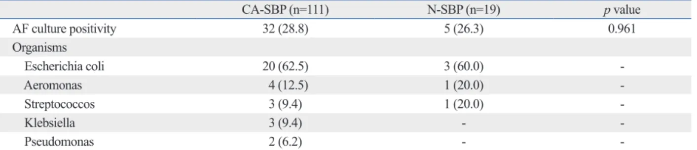
ic fluid samples [32 (28.8%) with CA-SBP vs. 5 (26.3%) with N-SBP]. The most common organism in patients with CA- SBP and N-SBP was Escherichia coli [20 (62.5%) with CA- SBP vs. 3 (60.0%) with N-SBP]. No significant differences in microorganisms were identified between the two groups (p=0.961).
Clinical course during hospitalization
Table 3 shows the clinical courses of patients with CA-SBP and N-SBP during hospitalization. The incidence of liver-re- lated complications such as variceal bleeding, HE, and renal failure did not differ between the two groups (all p>0.05).
Furthermore, blood culture positivity (p=0.578), antibiotic switching (p=0.066), and in-hospital mortality (p=0.163) did not differ.
abdominal distension (n=7), and abdominal pain (n=4), N- SBP patients received a second paracentesis procedure. Af- ter 8.2 days (range, 3.8-12.2) of hospital admission, all 19 patients were diagnosed with N-SBP.
A scheduled follow-up paracentesis at 48 hours after the initial antibiotic administration was performed in 76 pa- tients [67 (60.4%) with CA-SBP vs. 9 (47.4%) with N-SBP, p=0.321]. In another 6 (4.6%) patients, paracentesis was re- peated because they did not show clinical improvement in spite of a decrease in ascitic fluid PMN count [4 (3.6%) with CA-SBP vs. 2 (10.5%) with N-SBP, p=0.092].
Microbiological characteristics of ascitic fluid and blood The organisms cultured from the ascitic fluid are listed in Table 2. Pathogens were isolated in 37 (28.5%) of 130 ascit- Table 2. Ascitic Fluid Culture Positivity and Isolated Organisms
CA-SBP (n=111) N-SBP (n=19) p value
AF culture positivity 32 (28.8) 5 (26.3) 0.961
Organisms
Escherichia coli 20 (62.5) 3 (60.0) -
Aeromonas 4 (12.5) 1 (20.0) -
Streptococcos 3 (9.4) 1 (20.0) -
Klebsiella 3 (9.4) - -
Pseudomonas 2 (6.2) - -
CA-SBP, community acquired spontaneous bacterial peritonitis; N-SBP, nosocomial spontaneous bacterial peritonitis; AF, ascitic fluid.
Variables are expressed as n (%).
Table 1. Baseline Characteristics of Patients with CA-SBP and Patients with N-SBP at the Time of Diagnosis
Total (n=130) CA-SBP (n=111, 85.4%) N-SBP (n=19, 14.6%) p value
Age (yrs) 53.3±8.4 53.8±8.1 50.1±9.4 0.086
Male 88 (67.7) 72 (64.9) 16 (84.2) 0.108
History of previous variceal bleeding 35 (26.9) 31 (27.9) 4 (21.1) 0.534
History of previous hepatic encephalopathy 35 (26.9) 29 (26.1) 6 (31.6) 0.621
Child-Pugh score 10.7±1.9 10.9±1.9 9.9±1.7 0.407
Serum analysis
WBC count (/mm3) 7985±4864 8197±4978 4780±2244 0.006
Albumin (g/dL) 2.5±0.5 2.5±0.5 2.6±0.4 0.476
Total bilirubin (mg/dL) 5.7±4.4 5.6±4.5 6.3±3.7 0.572
Creatinine (mg/dL) 1.3±1.0 1.3±1.1 1.2±0.4 0.725
Sodium (mmol/L) 131.8±5.8 131.6±5.8 135.4±5.8 0.007
Prothrombin time (INR) 2.16±1.15 2.17±1.18 2.10±0.99 0.756
CRP (mg/L) 4.42±3.78 4.02±3.25 5.57±4.19 0.217
Ascitic fluid analysis
WBC count (/mm3) 7006±6231 7457±6510 4372±2393 0.861
PMN count (/mm3) 6390±5611 6327±5886 6756±3615 0.873
Protein (g/dL) 1.21±0.98 1.22±1.05 1.18±0.50 0.833
Albumin (g/dL) 0.55±0.32 0.54±0.31 0.58±0.38 0.689
CA-SBP, community acquired spontaneous bacterial peritonitis; N-SBP, nosocomial spontaneous bacterial peritonitis; WBC, white blood cell; CRP, C-reac- tive protein; PMN, polymorphonuclear leukocytes; INR, international normalized ratio.
Variables are expressed as mean±SD or n (%).
iate analysis identified ascitic fluid culture positivity as the only independent predictor of in-hospital mortality (p=0.036;
HR, 5.392; 95% CI, 1.208-24.061).
Time to recurrence
In total, 104 (93.7%) patients with CA-SBP and 16 (84.2%) with N-SBP survived their first episode of SBP. After dis- charge, SBP recurred in 42 (40.4%) of 104 patients with CA-SBP and 6 (37.5%) of 16 with N-SBP after a median period of 4.7 months (range, 0.8-29.5) and 3.6 months (range, 1.3-12.8), respectively (p=0.925) (Fig. 1).
Overall survival and its predictors
The median survival time of the study population was 6.5 months (range, 0.1-136.1); 117 (90.0%) patients had died by the end of the follow-up period. The median survival time of Antibiotics were switched in 11 (8.5%) patients, due to
cefotaxime resistance, treatment failure, or persistent clini- cal deterioration. Cefotaxime resistance developed in three (8.1%) of 37 patients with positive ascitic fluid culture [2 (6.3%) with CA-SBP vs. 1 (20.0%) with N-SBP, p=0.233].
Cefotaxime was switched to carbapenem in 10 patients and to ciprofloxacin in one. Among these 11 patients, nine pa- tients died during hospitalization, while two recovered.
Predictors of in hospital mortality
Table 4 presents the results of the univariate and multivari- ate analyses to identify the independent predictors of in- hospital mortality. The univariate analysis demonstrated that variceal bleeding during hospitalization and ascitic fluid culture positivity significantly predicted in-hospital mortali- ty (p=0.035 and p=0.031, respectively). However, multivar-
Table 3. Clinical Course during Hospitalization in Patients with CA-SBP and Patients with N-SBP
Variables CA-SBP (n=111) N-SBP (n=19) p value
Variceal bleeding 6 (5.4) 1 (5.3) 0.980
Hepatic encephalopathy 8 (7.2) 3 (15.8) 0.203
Renal failure 5 (4.5) 1 (5.3) 0.146
Blood culture positivity 28 (25.2) 6 (31.6) 0.578
Antibiotic switching 7 (6.3) 4 (21.1) 0.066
In hospital mortality 7 (6.3) 3 (15.8) 0.163
CA-SBP, community acquired spontaneous bacterial peritonitis; N-SBP, nosocomial spontaneous bacterial peritonitis.
Variables are expressed as n (%).
Table 4. Predictors of In-Hospital Mortality
Variables Died
(n=10, 7.7%) Survived
(n=120, 92.3%) Univariate Multivariate
p value p value HR 95% CI
Age (yrs) 52.2±9.2 53.4±8.6 0.679 - - -
Male 5 (50.0) 83 (69.2) 0.223 - - -
Variceal bleeding 3 (30.0) 4 (3.3) 0.035 0.054 18.297 0.526-19.551
Antibiotic switching 2 (20.0) 9 (7.5) 0.201 - - -
Child-Pugh score 11.7±1.9 10.6±1.8 0.070 - - -
Serum
WBC (/mm3) 9792±7106 7798±4620 0.403 - - -
Albumin (g/dL) 2.4±0.30 2.4±0.48 0.907 - - -
Total bilirubin (mg/dL) 6.82±4.78 5.19±4.39 0.116 - - -
Sodium (mmol/L) 131.8±5.9 132.0±5.8 0.596 - - -
Prothrombin time (INR) 2.54±1.06 2.10±2.16 0.138 - - -
CRP (mg/L) 4.98±4.57 4.26±4.33 0.216 - - -
Culture positivity 1 (10.0) 6 (5.0) 0.132 - - -
Ascitic fluid
WBC (/mm3) 8125±4253 7786±4630 0.406 - - -
PMN (/mm3) 7070±4642 5284±5709 0.686 - - -
Albumin (g/dL) 2.5±0.7 2.4±0.6 0.853 - - -
Culture positivity 6 (60.0) 31 (25.8) 0.031 0.036 5.392 1.208-24.061
N-SBP 3 (30.0) 16 (13.3) 0.166 - - -
HR, hazard ratio; CI, confidence interval; WBC, white blood cell; CRP, C-reactive protein; PMN, polymorphonuclear leukocytes; N-SBP, nosocomial sponta- neous bacterial peritonitis; INR, international normalized ratio.
Variables are expressed as mean±SD or n (%).
course during hospitalization, time to recurrence and over- all survival, as well as causes of mortality. These results sug- gest that these two types of SBP are not different disease entities and, furthermore, that the acquisition site of the pathogen (community-acquired vs. nosocomial) does not affect the prognosis of SBP patients.
To date, few studies have compared the characteristics of CA-SBP and N-SBP. Bert, et al.13 found that nosocomial isolates were significantly more resistant to amoxicillin/cla- vulanic acid and cefotaxime than community-acquired iso- lates and, thus, insisted that N-SBP requires a wider spec- trum of antibiotics than CA-SBP. Moreover, Cheong, et al.11 concluded that nosocomial acquisition of SBP patho- gens adversely affected the clinical outcomes of SBP, and that N-SBP mortality was accordingly higher. In contrast, Song, et al.12 reported that acquisition sites of infection did not have prognostic significance in SBP, and Umgelter, et al.17 concluded that the microbiological patterns and out- comes of CA-SBP and N-SBP did not differ. Therefore, dif- ferences in the prognosis of CA-SBP and N-SBP still re- main unresolved. We believe that the discrepancies among these previous studies are a result of heterogeneity in their study populations caused by differences in baseline liver functions between patients with CA-SBP and N-SBP, the inclusion of patients with a history of previous SBP, coex- isting HCC in some patients at baseline, and various etiolo- gies of liver cirrhosis.11-13
Our study differs from previous studies in several ways.
First, the baseline characteristics, including liver function, patients with CA-SBP was 6.6 months (range, 0.1-128.1),
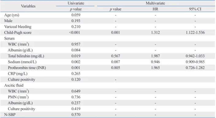
and that of patients with N-SBP was 6.2 months (range, 0.2- 136.1) (p=0.569) (Fig. 2). The 1-, 6-, and 12-month cumula- tive mortality rates of patients with CA-SBP were 7.2%, 48.7% and 64.2%, respectively, and those for patients with N-SBP were 15.8%, 47.6%, and 70.9%, respectively. The Cox-regression hazard model revealed that Child-Pugh score (p=0.001; HR, 1.312; 95% CI, 1.122-1.536) and serum sodium level (p=0.007; HR, 0.946; 95% CI, 0.909-0.985) were independent predictors of overall survival (Table 5).
Causes of mortality
Causes of in-hospital and overall mortality are summarized in Table 6. Septic shock was the most common cause of in- hospital mortality [three (42.9%) with CA-SBP and one (33.3%) with N-SBP, p=0.087]. Concerning overall mortal- ity, gastrointestinal bleeding (21.5%), hepatic failure (21.5%) and septic shock (20.4%) were common in patients with CA-SBP, whereas gastrointestinal bleeding (21.4%), septic shock (21.4%), renal failure (21.4%), and HE (21.4%) were common in those with N-SBP. Causes of in-hospital and overall mortality did not significantly differ between the two groups (p=0.917 and 0.375, respectively).
DISCUSSION
Patients with CA-SBP and N-SBP in this study showed similar clinical and microbiological characteristics, clinical
0.0 0.0
0.2 0.2
0.4 0.4
0.6 0.6
0.8 0.8
1.0 1.0
Cumulative recurrence-free rate Cumulative overall survival rate
0.00 5.00 10.00 15.00 20.00 25.00 30.00 0.0 20.0 40.0 60.0 80.0 100.0 120.0 140.0
Time to recurrence (months) Overall survival time (months)
Fig. 1. Cumulative recurrence-free curves of patients with CA-SBP and pa- tients with N-SBP. Time to recurrence was not different between the groups [median 4.7 months (range, 0.8-29.5) vs. 3.6 months (range, 1.3-12.8), p=0.925]. CA-SBP, community acquired spontaneous bacterial peritonitis;
N-SBP, nosocomial spontaneous bacterial peritonitis;
Fig. 2. Cumulative overall survival curves of patients with CA-SBP and pa- tients with N-SBP. Overall survival was not different between the groups [median 6.6 months (range, 0.1-128.1) vs. 6.2 months (range, 0.2-136.1), p=
0.569]. CA-SBP, community acquired spontaneous bacterial peritonitis;
N-SBP, nosocomial spontaneous bacterial peritonitis;
CA-SBP
N-SBP CA-SBP
N-SBP
preserved, indicating that CA-SBP and N-SBP may be of the same spectrum of SBP and that vulnerability to SBP is determined only by liver function status throughout the course of liver disease. This idea is also supported by the fact that SBP is a problem of increased gut permeability and bacterial translocation resulting from the intrinsic patho- physiological process of each individual patient and is not caused by pathogens from outside of the body.24
Second, our study included only patients who had experi- enced their first episode of SBP. The chance of exposure to more resistant pathogen strains increases as patients experi- ence repeated SBP events and admissions.17 Therefore, it is difficult to purely compare the effect of acquisition site of were similar between enrolled patients with CA-SBP and
N-SBP. Therefore, we could exclude the potential confound- ing effects of different liver function on the prognosis of CA-SBP and N-SBP. Although a previous study revealed that patients with N-SBP showed poorer prognosis and were infected by more virulent organisms than those with CA-SBP,11 baseline liver function was more favorable in patients with CA-SBP. Thus, the poor prognosis for patients with N-SBP might have been caused by poorer baseline liv- er function alone, and not by any inherent characteristics of N-SBP. According to our results, discrimination between CA-SBP and N-SBP may be meaningless, at least for the first episode of SBP, when liver function is relatively well Table 5. Predictors of Overall Survival
Variables Univariate Multivariate
p value p value HR 95% CI
Age (yrs) 0.059 - - -
Male 0.193 - - -
Variceal bleeding 0.210 - - -
Child-Pugh score <0.001 0.001 1.312 1.122-1.536
Serum
WBC (/mm3) 0.957 - - -
Albumin (g/dL) 0.084 - - -
Total bilirubin (mg/dL) 0.019 0.567 1.987 0.942-1.033
Sodium (mmol/L) 0.002 0.007 0.946 0.909-0.985
Prothrombin time (INR) 0.001 0.805 1.965 0.726-1.282
CRP (mg/L) 0.265
Culture positivity 0.120 -
Ascitic fluid
WBC (/mm3) 0.649 - - -
PMN (/mm3) 0.736 - - -
Albumin (g/dL) 0.237 - - -
Culture positivity 0.419 - - -
N-SBP 0.570 - - -
HR, hazard ratio; CI, confidence interval; WBC, white blood cell; CRP, C-reactive protein; PMN, polymorphonuclear leukocytes; N-SBP, nosocomial sponta- neous bacterial peritonitis; INR, international normalized ratio.
Variables are expressed as mean±SD or n (%).
Table 6. Causes of Mortality
In hospital mortality Overall mortality
CA-SBP (n=7) N-SBP (n=3) CA-SBP (n=93) N-SBP (n=14)
Septic shock 3 (42.9) 1 (33.3) 19 (20.4) 3 (21.4)
Gastrointestinal bleeding 1 (14.3) 0 (0.0) 20 (21.5) 3 (21.4)
Hepatic failure 0 (0.0) 1 (33.3) 20 (21.5) 1 (7.2)
Hepatocellular carcinoma 0 (0.0) 0 (0.0) 4 (4.3) 1 (7.2)
Renal failure 0 (0.0) 1 (33.3) 5 (5.4) 3 (21.4)
Hepatic encephalopathy 2 (28.5) 0 (0.0) 13 (14.0) 3 (21.4)
Other 1 (14.3) 0 (0.0) 3 (3.2) 0 (0.0)
Unknown 0 (0.0) 0 (0.0) 9 (9.7) 0 (0.0)
CA-SBP, community acquired spontaneous bacterial peritonitis; N-SBP, nosocomial spontaneous bacterial peritonitis.
Variables are expressed as n (%).
terial infection, and that the third-generation cephalosporin cefotaxime is sufficient as a first-line treatment in patients experiencing their first episode of SBP, regardless of the ac- quisition site of infection.
The acquisition site of SBP pathogen did not affect the clinical course during hospitalization or in-hospital mortali- ty in our study. Ascitic fluid culture positivity was the only significant predictor of in-hospital mortality.20 Furthermore, only Child-Pugh scores and serum sodium levels signifi- cantly predicted overall survival, while the acquisition site of the infection did not. Moreover, we found no statistical differences between patients with CA-SBP and N-SBP re- garding overall survival, time to recurrence, and causes of mortality. All these results suggest that these two types of SBP are similar disease entities.
Despite the unique features of the present study, there are some limitations. First, because this study was retrospective- ly designed, the sample size is relatively small, particularly the group of patients with N-SBP. Moreover, follow-up paracentesis, 48 hours after antibiotics administration, was performed in only 58.5% of patients (60.4% with CA-SBP and 47.4% with N-SBP). Lastly, the microbiology of SBP may have changed over the long study period from 1999 to 2008. We were not able to stratify the organisms of SBP ac- cording to the time period because the number of isolated organisms was too small. Therefore, future prospectively designed studies incorporating a larger number of patients should be performed to overcome these limitations.
In conclusion, the acquisition site of infection (communi- ty-acquired vs. nosocomial) did not affect the clinical out- comes and prognosis of patients with HBV-related liver cir- rhosis who had experienced their first episode of SBP. Third- generation cephalosporins may be effective in empirically treating these patients, regardless of the acquisition site of infection.
ACKNOWLEDGEMENTS
This study was supported by a grant of the Korea Health- care technology R & D project, Ministry of Health and Welfare, Republic of Korea (A102065).
REFERENCES
1. Akriviadis EA, Runyon BA. Utility of an algorithm in differentiat-
infection on prognosis, if a study population includes pa- tients with a history of previous SBP. However, certain en- vironmental changes occurring after the first episode of SBP, such as changes in intestinal bacteria leading to vul- nerability to invasive pathogens, a weakened immune sys- tem by progressive deterioration of liver function, and inter- actions between these factors, might result in the differing prognoses between CA-SBP and N-SBP. Thus, further in- vestigations of microorganism alterations and antibiotic susceptibility in recurring episodes of SBP after surviving the first episode of SBP should be performed.
Third, this study excluded patients with coexisting HCC at the time of SBP diagnosis, which might influence the nat- ural course of SBP, because increased hospitalization peri- ods for HCC management can increase the chance of devel- oping N-SBP.25 Finally, we focused on patients with HBV- related cirrhosis because the natural history of patients with cirrhosis differs according to the etiology of the liver dis- ease,18 and HBV is the most prevalent (57-73%) etiology for liver cirrhosis in Korea.26,27 Hepatitis C virus-related cirrho- sis usually progresses more slowly than HBV-related cirrho- sis, and abstinence from alcohol intake can prolong the sur- vival of patients with alcohol-related cirrhosis.18,28
Although some studies have found that Streptococcus pneumoniae is the most common organisms in patients with CA-SBP and that higher levels of gram-positive patho- gens are present in those with N-SBP,29 the most commonly occurring organisms in SBP are usually enteric gram-nega- tive rods such as Escherichia coli.30 Our study also identi- fied gram-negative pathogens as the dominant pathogens in both patients with CA-SBP and patients with N-SBP. Also, no significant differences in the isolation of microorganisms were found between two groups. This result, which again demonstrates the similarities of CA-SBP and N-SBP, may be explained by the blurring of environmental boundaries between CA-SBP and N-SBP, as patients with cirrhosis ac- companied by ascites receive frequent hospital assistance, including outpatient visits, home nursing care by health- care providers, and repeated hospital admission.31
Since 1985, third-generation cephalosporins have been the most frequently used antibiotics for the management of SBP.32 All patients in the present study initially received ce- fotaxime. Treatment failure and cefotaxime resistance oc- curred in 3.9% and 8.1% of patients respectively, and the antibiotic regimen was switched in only 8.5% of patients.
These results indicate that our study population was not yet susceptible to third-generation cephalosporin-resistant bac-
18. Kim SU, Han KH, Nam CM, Park JY, Kim do Y, Chon CY, et al.
Natural history of hepatitis B virus-related cirrhotic patients hospi- talized to control ascites. J Gastroenterol Hepatol 2008;23:1722-7.
19. Sort P, Navasa M, Arroyo V, Aldeguer X, Planas R, Ruiz-del-Ar- bol L, et al. Effect of intravenous albumin on renal impairment and mortality in patients with cirrhosis and spontaneous bacterial peritonitis. N Engl J Med 1999;341:403-9.
20. Kim SU, Kim do Y, Lee CK, Park JY, Kim SH, Kim HM, et al.
Ascitic fluid infection in patients with hepatitis B virus-related liv- er cirrhosis: culture-negative neutrocytic ascites versus spontane- ous bacterial peritonitis. J Gastroenterol Hepatol 2010;25:122-8.
21. Rimola A, García-Tsao G, Navasa M, Piddock LJ, Planas R, Ber- nard B, et al. Diagnosis, treatment and prophylaxis of spontaneous bacterial peritonitis: a consensus document. International Ascites Club. J Hepatol 2000;32:142-53.
22. al Amri SM, Allam AR, al Mofleh IA. Spontaneous bacterial peri- tonitis and culture negative neutrocytic ascites in patients with non- alcoholic liver cirrhosis. J Gastroenterol Hepatol 1994;9:433-6.
23. Novella M, Solà R, Soriano G, Andreu M, Gana J, Ortiz J, et al.
Continuous versus inpatient prophylaxis of the first episode of spontaneous bacterial peritonitis with norfloxacin. Hepatology 1997;25:532-6.
24. Llovet JM, Bartolí R, March F, Planas R, Viñado B, Cabré E, et al.
Translocated intestinal bacteria cause spontaneous bacterial peri- tonitis in cirrhotic rats: molecular epidemiologic evidence. J Hepa- tol 1998;28:307-13.
25. Park YH, Lee HC, Song HG, Jung S, Ryu SH, Shin JW, et al. Re- cent increase in antibiotic-resistant microorganisms in patients with spontaneous bacterial peritonitis adversely affects the clinical outcome in Korea. J Gastroenterol Hepatol 2003;18:927-33.
26. Kim YS, Um SH, Ryu HS, Lee JB, Lee JW, Park DK, et al. The prognosis of liver cirrhosis in recent years in Korea. J Korean Med Sci 2003;18:833-41.
27. Han YS, Kim BH, Baek IY, Lee DK, Kim KJ, Dong SH, et al.
The change of the etiology, complications and cause of death of the liver cirrhosis in 1990s. Korean J Hepatol 2000;6:328-39.
28. Kaymakoglu S, Eraksoy H, Okten A, Demir K, Calangu S, Cakalo- glu Y, et al. Spontaneous ascitic infection in different cirrhotic groups: prevalence, risk factors and the efficacy of cefotaxime therapy. Eur J Gastroenterol Hepatol 1997;9:71-6.
29. Johnson CC, Baldessarre J, Levison ME. Peritonitis: update on pathophysiology, clinical manifestations, and management. Clin Infect Dis 1997;24:1035-45.
30. Bhuva M, Ganger D, Jensen D. Spontaneous bacterial peritonitis:
an update on evaluation, management, and prevention. Am J Med 1994;97:169-75.
31. Angeloni S, Leboffe C, Parente A, Venditti M, Giordano A, Merli M, et al. Efficacy of current guidelines for the treatment of sponta- neous bacterial peritonitis in the clinical practice. World J Gastro- enterol 2008;14:2757-62.
32. Felisart J, Rimola A, Arroyo V, Perez-Ayuso RM, Quintero E, Gines P, et al. Cefotaxime is more effective than is ampicillin-to- bramycin in cirrhotics with severe infections. Hepatology 1985;
5:457-62.
ing spontaneous from secondary bacterial peritonitis. Gastroenter- ology 1990;98:127-33.
2. Runyon BA. Spontaneous bacterial peritonitis: an explosion of in- formation. Hepatology 1988;8:171-5.
3. Such J, Runyon BA. Spontaneous bacterial peritonitis. Clin Infect Dis 1998;27:669-74.
4. Navasa M, Follo A, Llovet JM, Clemente G, Vargas V, Rimola A, et al. Randomized, comparative study of oral ofloxacin versus in- travenous cefotaxime in spontaneous bacterial peritonitis. Gastro- enterology 1996;111:1011-7.
5. Runyon BA, McHutchison JG, Antillon MR, Akriviadis EA, Montano AA. Short-course versus long-course antibiotic treat- ment of spontaneous bacterial peritonitis. A randomized controlled study of 100 patients. Gastroenterology 1991;100:1737-42.
6. Altman C, Grangé JD, Amiot X, Pelletier G, Lacaine F, Bodin F, et al. Survival after a first episode of spontaneous bacterial perito- nitis. Prognosis of potential candidates for orthotopic liver trans- plantation. J Gastroenterol Hepatol 1995;10:47-50.
7. Sheer TA, Runyon BA. Spontaneous bacterial peritonitis. Dig Dis 2005;23:39-46.
8. Guarner C, Runyon BA, Young S, Heck M, Sheikh MY. Intestinal bacterial overgrowth and bacterial translocation in cirrhotic rats with ascites. J Hepatol 1997;26:1372-8.
9. Fiuza C, Salcedo M, Clemente G, Tellado JM. In vivo neutrophil dysfunction in cirrhotic patients with advanced liver disease. J In- fect Dis 2000;182:526-33.
10. Runyon BA. Patients with deficient ascitic fluid opsonic activity are predisposed to spontaneous bacterial peritonitis. Hepatology 1988;8:632-5.
11. Cheong HS, Kang CI, Lee JA, Moon SY, Joung MK, Chung DR, et al. Clinical significance and outcome of nosocomial acquisition of spontaneous bacterial peritonitis in patients with liver cirrhosis.
Clin Infect Dis 2009;48:1230-6.
12. Song JY, Jung SJ, Park CW, Sohn JW, Kim WJ, Kim MJ, et al.
Prognostic significance of infection acquisition sites in spontane- ous bacterial peritonitis: nosocomial versus community acquired.
J Korean Med Sci 2006;21:666-71.
13. Bert F, Andreu M, Durand F, Degos F, Galdbart JO, Moreau R, et al. Nosocomial and community-acquired spontaneous bacterial peritonitis: comparative microbiology and therapeutic implica- tions. Eur J Clin Microbiol Infect Dis 2003;22:10-5.
14. Terg R, Levi D, Lopez P, Rafaelli C, Rojter S, Abecasis R, et al.
Analysis of clinical course and prognosis of culture-positive spon- taneous bacterial peritonitis and neutrocytic ascites. Evidence of the same disease. Dig Dis Sci 1992;37:1499-504.
15. Hoefs JC, Canawati HN, Sapico FL, Hopkins RR, Weiner J, Montgomerie JZ. Spontaneous bacterial peritonitis. Hepatology 1982;2:399-407.
16. Runyon BA, Hoefs JC. Culture-negative neutrocytic ascites: a variant of spontaneous bacterial peritonitis. Hepatology 1984;4:
1209-11.
17. Umgelter A, Reindl W, Miedaner M, Schmid RM, Huber W. Fail- ure of current antibiotic first-line regimens and mortality in hospi- talized patients with spontaneous bacterial peritonitis. Infection 2009;37:2-8.