© 2018 The Korean Ophthalmological Society
This is an Open Access article distributed under the terms of the Creative Commons Attribution Non-Commercial License (http://creativecommons.org/licenses /by-nc/3.0/) which permits unrestricted non-commercial use, distribution, and reproduction in any medium, provided the original work is properly cited.
Original Article
Lamina Cribrosa Changes after Laser In Situ Keratomileusis in Myopic Eyes
Soomin Lee, Da-Ye Diana Choi, Dong Hui Lim, Tae Young Chung, Jong Chul Han, Changwon Kee
Department of Ophthalmology, Samsung Medical Center, Sungkyunkwan University School of Medicine, Seoul, Korea
Purpose: To determine deep optic nerve head structure changes after transient intraocular pressure elevation during laser in situ keratomileusis (LASIK) for myopia
Methods: Enhanced depth imaging-optical coherence tomography was performed in each myopic eye that under- went LASIK surgery. Enhanced depth imaging-optical coherence tomography images were created at postoper- ative 1 day, 1 week, 2 weeks, and 1 month. Lamina cribrosa (LC) thickness, LC depth and prelaminar thickness at the superior, middle and inferior portions of the optic nerve head were measured by two investigators.
Results: Forty eyes in 40 patients were included in the present study. During follow-up, there were no significant differences in prelaminar thickness or LC depth. The LC demonstrated increased thickness at postoperative 1 day at all three locations (superior, middle, and inferior) (p < 0.001, p < 0.001, p < 0.001, respectively). However, no significant changes were observed at postoperative 1 week, 2 weeks, and 1 month.
Conclusions: The LC thickness could increase at 1 day after LASIK surgery. However, the thickness will gradually return to baseline morphology. Temporary intraocular pressure increase during LASIK does not appear to induce irreversible LC thickness changes.
Key Words: Lamina cribrosa, Laser in situ keratomileusis, Optic disk
Laser in situ keratomileusis (LASIK) is a commonly pre- ferred refractive surgery to correct myopia [1]. However, my- opia is a well-known risk factor of glaucoma. High intraocu- lar pressure (IOP) can occur due to pneumatic suction during LASIK surgery, and additional safety issues of LASIK in myopic patients have been addressed previously [2,3].
The lamina cribrosa (LC), an array of collagen beams with intervening pores, is the primary site of glaucomatous damage [4]. When IOP is increased, pores in the LC might undergo conformational changes due to IOP-related me- chanical strain, eventually causing axonal transport block- age [5]. Therefore, it is important to investigate structural changes in LC to identify whether high IOP during LASIK surgery might induce early glaucomatous changes in myo- pic eyes. However, changes in deep structures after LASIK surgery have not yet been reported.
Therefore, the objective of this study was to determine changes in deep structures of the optic nerve head after
Received: September 8, 2017 Accepted: December 28, 2017
Corresponding Author: Changwon Kee, MD, PhD. Department of Oph- thalmology, Samsung Medical Center, Sungkyunkwan University School of Medicine, #81 Irwon-ro, Gangnam-gu, Seoul 06351, Korea. Tel: 82-2- 3410-3548, Fax: 82-2-3410-0074, E-mail: [email protected]
LASIK surgery using enhanced depth imaging spectral-do- main (SD) optical coherence tomography (OCT). Results of this study could improve understanding of the impact of high IOP during LASIK surgery on deep structures in the optic nerve head (ONH).
Materials and Methods
A total of 52 eyes of 52 participants were initially investi- gated in this prospective observational study. These partici- pants underwent LASIK surgeries performed by a single surgeon (TYC) between July 2013 and September 2013.
This study followed all guidelines for experimental investi- gation in human subjects and was approved by Samsung Medical Center institutional review board (2013-06-061).
This study adhered to the tenets of the Declaration of Helsin- ki and was registered at ClinicalTrials.gov (NCT02676557).
Informed consent was obtained from all participants. Inclu- sion criterion for this study was eyes undergoing LASIK surgery for myopia. Eyes were excluded if images were dif- ficult to investigate, including an unclear LC margin, severe shadowing due to overlying vessels, or poor image quality due to cataracts. Participants underwent a comprehensive ophthalmic assessment, including slit-lamp biomicroscopy, Goldmann applanation tonometry, manifested refraction, dilated fundus photography, and enhanced depth imaging (EDI) SD-OCT (Spectralis OCT; Heidelberg Engineering, Heidelberg, Germany). Eyes were excluded if there had pre- vious ocular disease, including glaucoma. Transient IOP el- evation was induced between 15 and 20 seconds in the eye of each subject by a modified LASIK automated corneal shaper (Mel80; Zeiss Meditec, Dublin, CA, USA) after a lo- cal anesthetic was administered. Because a LASIK suction ring could potentially produce IOP in excess of 90 mmHg, the rigid plastic tubing connecting the suction ring to the main pump was punctured by two 18-gauge needles that were left in situ to reduce potential intraluminal negative pressure and limit possible peak IOP elevation [6].
Measurement of deep structures of the optic nerve head
The Heidelberg Spectralis OCT EDI mode was used for all images and was set to image a 10° × 15° rectangle for horizontal scans centered on the optic disc. This rectangle
was scanned with 65 sections. Each section had 43 average OCT frames based on a previous study [7]. The OCT ma- chine scaling was adjusted to 1 : 1 μm before measurement.
The magnification error was corrected using a formula pro- vided by the manufacturer, based on the auto refraction keratometry results and the focus setting during image ac- quisition. The Heidelberg Spectralis program includes an EDI mode, that automatically places the OCT reference plane toward the bottom of the OCT acquisition screen. The zero delay is placed at the bottom of the OCT screen, which enhances images from the deeper layers of the ONH.
Prelaminar (PL) thickness was defined as the shortest distance between the anterior border of the PL tissue and the anterior border of the LC. LC thickness was defined as the shortest distance between the anterior border and the posterior border of the LC. The anterior and posterior LC margins were defined by a highly reflective structure below the optic cup. Anterior and posterior LC margins were de- fined as the line that connected the peripheral points of the anterior and posterior LC margins. The line that connected the two termination points of Bruch’s membrane was used as a baseline reference. The LC depth was measured by cal- culating the average at each periphery of the anterior LC margin. When the shadow of the vessel trunk restricted ob- servation of the peripheral LC margin in the nasal area, the observed LC margin was used to measure the peripheral point of the LC margin. The distance from the reference line to the level of the anterior border of the LC was mea- sured perpendicular to the reference line. Therefore, the av- erage value of each peripheral LC depth was used as the LC depth (Fig. 1A). The same methods were used to mea- sure other parameters, such as the LC thickness, LC depth and PL thickness in the eyes with curved or irregular LC margins.
PL thickness, LC thickness, and LC depth were measured in central, superior, and inferior mid-periphery ONH. Three B-scans in each portion were measured, and the average LC thickness and depth of nine images were used for analysis (Fig. 1B). All images were analyzed and measured twice by two examiners (SML and DYC) who independently deter- mined LC thickness, LC depth, and PL thickness.
Statistical analysis
Descriptive statistics including the number and percent- age of each categorical variable and mean ± standard devia-
tion of each continuous variable were evaluated to show demographic and ocular parameters. The PL thickness, LC thickness, and depth values were compared to baseline val- ues at each time point: postoperative 1 day, 1 week, 2 weeks, and 1 month. Repeated ANOVA test was performed at each time point to examine differences before and after surgery. Univariate and multivariate logistic regression analyses were performed to identify factors associated with LC thickening after LASIK surgery. Independent variables were age, spherical equivalent (SE), central corneal thick- ness (CCT), IOP and baseline average LC thickness. Pa- rameters with a p-value less than 0.3 in univariate analysis were included in multivariate logistic analysis. The IBM SPSS ver. 23.0 (IBM Corp., Armonk, NY, USA) was used for all statistical analyses of this study. A p-values less than 0.05 was considered statistically significant.
Results
Forty eyes in 40 participants were included in this study.
Thirteen of the participants were male (32.5%), and the av- erage age of all participants was 28 years (range, 19 to 51 years). The average CCT was 556 μm (range, 506 to 627 μm), and the average SE was -6 diopter (range, -10 to -3 di- opter). The average IOP was 17 mmHg (range, 10 to 23 mmHg) (Table 1).
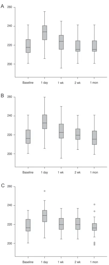
During follow-up, there was no significant difference in PL thickness or LC depth compared to those at preopera- tive exams. LC thickness values at the superior, middle and inferior portions were significantly increased at postopera- tive 1 day (all p < 0.001) (Fig. 2A-2C). However, no signifi- cant changes were observed at postoperative 1 week, 2 weeks, or 1 month compared to baseline LC thickness (Ta- ble 2).
The LC thickness change at postoperative 1 day was sig- nificant in this present study. Therefore, the factors associ- ated with the presence of significant LC thickening at postoperative 1 day after LASIK surgery were examined.
Significant LC thickening was defined as an increase over 10 μm, the standard deviation of baseline LC thickness in this study. In univariate logistic regression analysis, age, Table 1. Preoperative baseline characteristics
Variable Value
Subject 40
Male 13 (32.5)
Age (yr) 28 ± 9 (19–51)
Central corneal thickness (μm) 556 ± 30 (506–627) Spherical equivalent (diopter) -6 (-10 – -3) Intraocular pressure (mmHg) 17 ± 3 (10–23) Values are presented as number, number (%), or mean ± standard deviation (range).
Fig. 1. Delineation of the border of the lamina cribrosa (LC). (A) Prelaminar (PL) thickness was defined as the distance between the anteri- or border of PL tissue and the anterior LC border (green line arrow). LC thickness was defined as the distance between the anterior border and the posterior border of the LC (red line arrow). LC depth was defined as the average distances (two dotted red line arrows) between the peripheral points of the anterior LC margins and the reference line connecting both the Bruch’s membrane openings (white line). When the shadow of the vessel trunk restricts observation of the peripheral LC margin in the nasal area, the observed LC margin was used to measure the peripheral point of the LC margin. (B) LC thickness and depth were measured at the presumed vertical center of three areas (superior, center, and inferior).
A B
Table 2. LC thickness, depth of LC, and PL thickness of myopic eyes after laser in situ keratomileusis surgery Before surgery1 Day after surgery1 Week after surgery2 Weeks after surgery1 Month after surgery p-value† Mean ± SD(reference)Mean ± SDp-value* Mean ± SDp-value* Mean ± SDp-value* Mean ± SDp-value* PL thickness (µm) Inferior 98 ± 11100 ± 110.3699 ± 110.57100 ± 120.41101 ± 130.250.31 Middle103 ± 1499 ± 130.11102 ± 120.67102 ± 140.71101 ± 140.520.25 Superior100 ± 13101 ± 120.24102 ± 120.1598 ± 110.1998 ± 110.180.23 LC thickness (µm) Inferior 218 ± 10234 ± 13<0.001221 ± 80.16220 ± 80.22218 ± 100.89<0.001 Middle218 ± 10230 ± 10<0.001220 ± 90.19219 ± 90.31217 ± 90.32<0.001 Superior219 ± 10233 ± 11<0.001222 ± 80.17221 ± 80.27218 ±100.85<0.001 LC depth (µm) Inferior 426 ± 38427 ± 360.24426 ± 380.46430 ± 380.06428 ± 37 0.140.22 Middle435 ± 41436 ± 400.19433 ± 380.13433 ± 390.15435 ± 380.740.34 Superior427 ± 37428 ± 380.21430 ± 380.09431 ± 390.054428 ± 370.260.18 LC = lamina cribrosa; PL = prelaminar; SD = standard deviation. * Repeated ANOVA test for differences between LC before surgery (reference) and after surgery (time 2, time 3, time 4, and time 5); † Repeated ANOVA testing for different LC values at all time points.
Fig. 2. Lamina cribrosa thickness (µm) changes. Lamina cribrosa thickness at postoperative day 1 was significantly increased com- pared to that at baseline. However, this value was not significantly different compared with other time points. (A) Superior, (B) mid- dle, and (C) inferior.
260
240
220
200
Baseline 1 day 1 wk 2 wk 1 mon
260
240
220
200
Baseline 1 day 1 wk 2 wk 1 mon
260
240
220
200
Baseline 1 day 1 wk 2 wk 1 mon
30 30
22 33 20
6 30
30
A
B
C
SE, CCT, IOP, and baseline average LC thickness were not associated with LC thickening at 1 day after LASIK sur- gery (p = 0.674, p = 0.868, p = 0.244, p = 0.475, p = 0.075, respectively). In multivariate logistic regression analysis, CCT and baseline average LC thickness were not associat- ed with LC thickening at 1 day after LASIK surgery (p = 0.256, p = 0.064) (Table 3).
Discussion
The results of this study demonstrate that LC thickness can increase after baseline at 1 day after LASIK surgery.
However, there were no irreversible changes in deep struc- tures such as PL tissue or LC (Fig. 3). However, just be- cause there are no changes in the LC structure, does not confirm that there is no glaucomatous damage after LASIK. This is because ischemic damages without retinal
Table 3. Logistic regression analysis for variables associated with lamina cribrosa thickening at 1 day after laser in situ keratomile- usis surgery
Univariate analysis Multivariate model
Parameter
OR 95% CI p-value OR 95% CI p-value
Age 0.015 0.945–1.091 0.674 - - -
Spherical equivalent (D) -0.142 0.673–1.119 0.868 - - -
Central corneal thickness (μm) 0.060 0.772–1.352 0.244 0.016 0.989–1.044 0.256
Intraocular pressure (mmHg) -0.086 0.724–1.162 0.475 - - -
Baseline average LC thickness (μm) -0.053 0.886–1.015 0.075 -0.052 0.887–1.017 0.064
Baseline average LC depth (μm) 0.950 0.888–1.016 0.333 - - -
Baseline average PL thickness (μm) 0.961 0.899–1.028 0.349 - - -
OR = odds ratio; CI = confidence interval; LC = lamina cribrosa; PL = prelaminar.
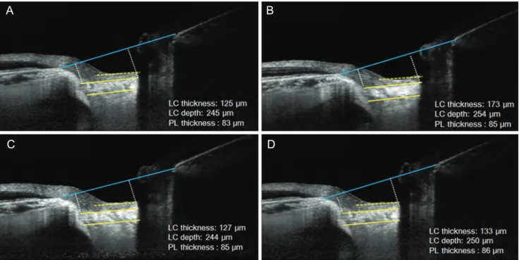
Fig. 3. A representative case. Enhanced depth imaging-optical coherence tomography images of the optic nerve head before and after laser in situ keratomileusis surgery of a 24-year-old female with -4.5 diopters of spherical equivalent. When the images were compared before and after surgery, lamina cribrosa (LC) thickness slightly increased (A) from 125 μm (B) to 173 μm at postoperative 1 day and returned to baseline LC thickness at postoperative (C) 1 week and (D) 1 month. Other parameters such as LC depth or prelaminar (PL) thickness were consistent, based on the results.
A B
C D
ganglion cell apoptosis can also induce damages on retinal nerve fiber layer (RNFL) [8]. However, our results showed no change in LC, supporting the hypothesis that LASIK might not induce irreversible changes in LC in healthy myopic eyes. Although there could be damages to the RNFL due to ischemic and mechanical damages after LASIK surgery, permanent changes in LC structures, such as LC thickness, LC depth, and PL thickness, may not oc- cur after LASIK surgery. To the best of our knowledge, this is the first report that visualizes structural changes in optic nerve tissues after LASIK surgery.
A pneumatic suction ring is usually used for a few sec- onds during LASIK surgery. While applying suction, the IOP should be increased to greater than 65 mmHg to cre- ate a lamellar flap [9]. This type of increase in IOP during suction can cause damages to ocular structures, such as retinal vessels, retinal nerve fibers, and the LC. Retinal complications following LASIK have been previously re- ported [10-12]. Maden et al. [10] have reported non-arteritic ischemic optic neuropathy after LASIK with femtosecond laser flap creation. Smith et al. [11] have shown hemi-reti- nal vein occlusion following LASIK. Lee et al. [12] have reported four cases of optic neuropathy following LASIK.
Glaucomatous changes after LASIK surgery have also been suggested [13-15]. However, most studies showed that there were no changes in RNFL after LASIK surgery [16- 18]. Previously, Kim et al. [19] have investigated eyes with or without refractive surgery and found no differences in glaucoma progression between the two groups. Because LC is related to the pathogenesis of glaucoma, the results of the present study support the results of previous studies.
Changes in LC have been reported in previous studies of eyes with glaucoma. A histologic study by Jonas et al. [20]
suggested that LCs in eyes with glaucoma are thinner than in normal controls. This suggestion was supported by an in vivo study that used EDI-OCT. Park et al. [21] have shown that normal tension glaucoma (NTG) has thinner LC than normal controls. Kwun et al. [22] have reported that NTG eyes have thinner LC than contralateral normal eyes in monocular NTG patients, while LC is thinner at the loca- tion of visual field defect. Kim et al. [19] have shown that open-angle glaucoma has deeper peripheral LC depth than normal controls. In addition, they suggested that this pro- vides evidence of remodeling at the peripheral region of LC. In this study, we did not identify permanent changes in LC after LASIK surgery. This means that LASIK might
not induce irreversible structural changes associated with glaucoma in LC.
The reason why high IOP could cause LC thickening has not previously been explained. In non-human experiments, LC that has been exposed to high IOP for several weeks is thicker than baseline LC [23,24]. The authors of previous studies have suggested that this might be due to LC re- modeling associated with high IOP. However, temporary thicker LC might not be associated with LC remodeling because the exposure time is relatively short (approximately 1 minute). Instead, transient changes in LC thickness might be due to compensatory blood flow after temporary ischemia in deep ONH [25]. Previously, Ozdamar and Oc- akoglu [25] have suggested that LASIK could cause tem- porary increases in blood flow at the LC region in healthy myopic eyes. They proposed that a compensatory mecha- nism for maintaining blood flow after LASIK-induced ischemia in the LC could count for the increase of blood flow following LASIK. If LC temporary thickening is re- lated to an increased blood flow associated compensatory mechanism, it provides a clinical hypothesis for the mech- anism for high IOP to influence deep ONH circulation, in- cluding LC. This supports the hypothesis that high IOP can induce temporary ischemia on deep ONH and offers reasons for ischemic complications after LASIK. However, in our study, only LC thickness increased, but no changes were found in PL thickness. Because vascular affluence in peripapillary choroidal vessels may have greater effects on the prelaminar tissue than LC, it is currently difficult to fully explain this result [26]. Thus, additional studies are needed to determine why only LC thickness changes, but no changes in PL thickness were found in the present study.
There were several limitations to our study. First, cur- rent findings have not yet been corroborated by histologic evaluations. In vivo imaging offers a non-invasive tech- nique to investigate LC changes; however, this modality cannot specify reasons for associated histologic changes in LC. Second, the present study was conducted only in young healthy subjects. Considering that LC stiffness can differ according to age, our study results should be inter- preted with consideration of the specific age demograph- ics. Third, the amount and duration of IOP increase were not measured in the present study. Therefore, different amounts and durations of IOP increase might have affect- ed the results. Fourth, EDI-OCT images obtained at post-
operative 1 day are prone to artifacts due to tearing or sub- tle corneal haze, which can interfere and cause inaccurate evaluation of the optic nerve structures. Images were taken again when they were deemed insufficient for evaluation.
Fifth, evaluation of the LC structure in the myopic tilted optic disc was not accurate. However, because we mea- sured the same position during observation periods in the same eye, we could compare the differences of LC struc- tures even in eyes with disc tilt. In addition, the follow-up period was relatively short in this study, and changes in LC were observed for 1 month. However, LC is a dynamic and changeable structure. Lee et al. [27] have examined the responses of LC and PL thickness to glaucoma surgery.
Their study showed a significant reduction in posterior dis- placement of the LC with increases in PL thickness and LC thickness. Since glaucoma is considered a long-term disease, it is currently unclear whether structural changes in LC would occur after long term follow-up.
In conclusion, LC thickness could increase at 1 day after LASIK surgery. However, the thickness will eventually re- turn to the baseline morphology. Therefore, these changes in LC thickness could be attributable to compensatory blood flow after temporary ischemia on LC.
Conflict of Interest
No potential conflict of interest relevant to this article was reported.
References
1. Yuen LH, Chan WK, Koh J, et al. A 10-year prospective audit of LASIK outcomes for myopia in 37,932 eyes at a single institution in Asia. Ophthalmology 2010;117:1236-44.
2. Chan KC, Poostchi A, Wong T, et al. Visual field changes after transient elevation of intraocular pressure in eyes with and without glaucoma. Ophthalmology 2008;115:667- 72.
3. Lleo-Perez A, Sanchis Gimeno J. Changes in the visual field following laser in situ keratomileusis for myopia.
Ophthalmic Physiol Opt 2007;27:201-9.
4. Quigley HA, Addicks EM, Green WR, Maumenee AE.
Optic nerve damage in human glaucoma. II. The site of in- jury and susceptibility to damage. Arch Ophthalmol
1981;99:635-49.
5. Minckler DS, Bunt AH, Johanson GW. Orthograde and retrograde axoplasmic transport during acute ocular hy- pertension in the monkey. Invest Ophthalmol Vis Sci 1977;16:426-41.
6. Kasetsuwan N, Pangilinan RT, Moreira LL, et al. Real time intraocular pressure and lamellar corneal flap thickness in keratomileusis. Cornea 2001;20:41-4.
7. Han JC, Choi DY, Kwun YK, et al. Evaluation of lamina cribrosa thickness and depth in ocular hypertension. Jpn J Ophthalmol 2016;60:14-9.
8. Bernstein SL, Johnson MA, Miller NR. Nonarteritic ante- rior ischemic optic neuropathy (NAION) and its experi- mental models. Prog Retin Eye Res 2011;30:167-87.
9. Cameron BD, Saffra NA, Strominger MB. Laser in situ keratomileusis-induced optic neuropathy. Ophthalmology 2001;108:660-5.
10. Maden A, Yilmaz S, Yurdakul NS. Nonarteritic ischemic optic neuropathy after LASIK with femtosecond laser flap creation. J Neuroophthalmol 2008;28:242-3.
11. Smith BT, Park CH, Fekrat S. Hemi-retinal vein occlusion following LASIK. Ann Ophthalmol (Skokie) 2006;38:139- 40.
12. Lee AG, Kohnen T, Ebner R, et al. Optic neuropathy asso- ciated with laser in situ keratomileusis. J Cataract Refract Surg 2000;26:1581-4.
13. Shaikh NM, Shaikh S, Singh K, Manche E. Progression to end-stage glaucoma after laser in situ keratomileusis. J Cataract Refract Surg 2002;28:356-9.
14. Bushley DM, Parmley VC, Paglen P. Visual field defect as- sociated with laser in situ keratomileusis. Am J Ophthalmol 2000;129:668-71.
15. Weiss HS, Rubinfeld RS, Anderschat JF. Case reports and small case series: LASIK-associated visual field loss in a glaucoma suspect. Arch Ophthalmol 2001;119:774-5.
16. Whitson JT, McCulley JP, Cavanagh HD, et al. Effect of laser in situ keratomileusis on optic nerve head topography and retinal nerve fiber layer thickness. J Cataract Refract Surg 2003;29:2302-5.
17. Hamada N, Kaiya T, Oshika T, et al. Optic disc and retinal nerve fiber layer analysis with scanning laser tomography after LASIK. J Refract Surg 2006;22:372-5.
18. Zangwill LM, Abunto T, Bowd C, et al. Scanning laser po- larimetry retinal nerve fiber layer thickness measurements after LASIK. Ophthalmology 2005;112:200-7.
19. Kim YJ, Yun SC, Na JH, et al. Glaucoma progression in
eyes with a history of refractive corneal surgery. Invest Ophthalmol Vis Sci 2012;53:4485-9.
20. Jonas JB, Jonas SB, Jonas RA, et al. Histology of the parapap- illary region in high myopia. Am J Ophthalmol 2011;152:1021- 9.
21. Park HY, Jeon SH, Park CK. Enhanced depth imaging de- tects lamina cribrosa thickness differences in normal ten- sion glaucoma and primary open-angle glaucoma. Oph- thalmology 2012;119:10-20.
22. Kwun Y, Han JC, Kee C. Comparison of lamina cribrosa thickness in normal tension glaucoma patients with unilat- eral visual field defect. Am J Ophthalmol 2015;159:512-8.
23. Roberts MD, Grau V, Grimm J, et al. Remodeling of the connective tissue microarchitecture of the lamina cribrosa in early experimental glaucoma. Invest Ophthalmol Vis Sci
2009;50:681-90.
24. Yang H, Thompson H, Roberts MD, et al. Deformation of the early glaucomatous monkey optic nerve head connec- tive tissue after acute IOP elevation in 3-D histomorphomet- ric reconstructions. Invest Ophthalmol Vis Sci 2011;52:345- 63.
25. Ozdamar A, Ocakoglu O. Optic nerve head blood flow us- ing scanning laser Doppler flowmetry after laser in situ keratomileusis. J Refract Surg 2003;19:433-7.
26. Hayreh SS. Blood supply of the optic nerve head and its role in optic atrophy, glaucoma, and oedema of the optic disc. Br J Ophthalmol 1969;53:721-48.
27. Lee EJ, Kim TW, Weinreb RN. Reversal of lamina cribrosa displacement and thickness after trabeculectomy in glau- coma. Ophthalmology 2012;119:1359-66.