J O U R N A L O F
V e te ri n ary S ci e n ce
A BS TRA CT
8 )T h r e e d o g s w e r e e x p e r i m e n t a ll y i n fe c t e d w i t h
D i r o f i l a r i a i m m i t i s .A ll d o g s w e r e e u t h a n i s e d a t 3 0 , 3 6 an d 37 w e e k s afte r in o cu la tion o f D. im m itis fo r th e re c ov e ry o f ad u lt w orm s . Th re e ca se s a cc ou n te d to 42.91 % re co ve ry of in oc u la te d w o rm s. Se ru m s am p le s from do gs e xp e rim e n tally in o cu late d w ith D. im m it is w e re an a lyze d by ELISA an d im m u n o blottin g m e th od s. An tibod y tite rs of d og s d e te cte d by ELIS A pe ak e d be tw e e n 7 a n d 14 w e e k s th e n de cre a se d be tw e e n w e e ks 15 to 24 follow e d by a n oth e r in c re as e du rin g w e e ks 25 to 30 an d pe rsis te d th rou g h ou t th e re m a in de o f th e e x pe rim e n t p e rio d. An a ly sis of ad u lt D. im m it is p rote in s tain e d w ith Coo m as sie brillia n t blu e R-250 in d ica te d se p arate ly m o re th an 10 ba n ds , an d th e m a jo r ban d s w e re 22, 40, 46, 56, 70, 72 an d 89 kD a. An tig e n ic id e n tific atio n of e xtra cts a n tige n s of ad u lts D. im m it is by im m u n oblo ttin g an a ly sis re v e ale d se ve ra l ban d s fro m p oo le d s e ra o f pa te n t in fe ctio n (30 w e e k s afte r in o cu latio n ). Th e d e te c te d ban d s w e re 24, 70, 80 a n d 110 kD a, 22, 72 a n d 84 kDa , an d 58 a n d 72 kDa in do gs 1, 2 an d 3, re s pe c tive ly.
Re su lts of an tibo dy tite rs re ac h e d h ig h le ve ls o n th e 4th m o ltin g sta ge a fte r in o cu la tion of in fe ctiv e la rv a (L3), an d re in force d pre vio u s fin d in gs th at h ig h m ole cu lar w e igh t re gio n s are de te c te d in yo u n g an im a ls .
Ke y w ord s: Dirofilaria im m it
is ,exp e r im en t a l in fe ct ion , E L I S A, im m u n ob lot t in g
*Corresponding author:
Tel: +82-42-821-6788, Fax: +82-42-821-4216 E-mail: [email protected]
In tro d u ctio n
D ir ofi l a ri a i m m it is ,
com m on ly ca lled ca n in e h e a r t w or m , is t h e ca u s e of a s er iou s p a r a s it ic d is ea s e of d ogs e n d e m ic in t em p er a t e , s u b t r op ica l a n d t r op ica l cou n t r ies [4 ]. I n ep izoot iologic s u r ve ys of d ir ofila r ia s is in d ogs , va r iou s m et h od s for d et er m in in g in fe ct ion s t a t u s h a ve b ee n u s e d , in clu d in g m icr os cop ic exa m in a t ion of blood s m e a r s , b lood s a m p le con cen t r a t ion t ech n iq u e s , K n ot t 's t e s t for d et e ct in g cir cu la t in g m icr ofila r ia e, e xa m in a t ion for id e n t ifica t ion of a du lt wor m s a t n ecr op sy, a n d r a diogr a ph ic a n d a n giogr a p h ic eva lu a t ion s [2 ,2 3,2 6]. A m a jor p r ob lem in m a k in g d ia gn os is is th e dir ofila r ia sis wit hou t micr ofila r emia (occu lt d ir ofila r ia s is ) w h ich h a s b ee n s h ow n t o occu r in 1 0 % t o 67 % of d ogs t h a t h a ve be en in fe ct e d n a t u r a lly. T h is h a s b ee n a t t r ib u t ed t o s in gle -s ex in fect ion , p r e s e n ce of im m a t u r e a d u lt s or im m u n e- media t ed clea r a n ce of micr ofila r iae [21]. Th ese occu lt in fe ct ion s a r e ve r y d ifficu lt t o d ia gn os e by m icr os cop ic e xa m in a t ion of blood , s o t h a t , t h e vet e r in a r y p r a ct it ion e r s u s u a lly m a k e t h e d ia gn os is b a s e d on clin ica l s ign s , r a d iogr a p h ic evid e n ce , a n d ot h e r in d ir e ct in d ica t ion of in fe ct ion [2 1].
T h e occu lt D . i m m i t i s in fe ct ion s p os e t h e m os t s er iou s d ia gn os t ic ch a lle n ge. An d , s om e t im es , e ve n in p a t en t in fect ion s , ob s t a cles cou ld b e a void e d in t h e d ia gn os is of d ir ofila r ia s is d u e t o t h e low n u m be r s of m icr ofila r ia e or t h e d ifficu lt y in d is t in gu is h in g m icr ofila r ia e of D . im m i t i s fr om t h os e of D i p en t a l on em a recon d i t u m [5 ]. T o ove r com e t h e s e p r oble m s , va r iou s im m u n ologica l t ech n iq u e s h a ve r ecen t ly be en d eve lop e d [3,5 ,6 ,8 -12 ,1 4,15 ,18 ,1 9,28 ]. Am on g t h es e m et h od s, en zym e-lin k ed im m u n osor ben t a s sa y (E LI S A) a n d im m u n oblot t in g a n a lys is h a ve be en r e cogn ized a s s im p le, s en s it ive a n d p a r t icu la r ly s u it a b le for t h e p a r a s it ic d is ea s es lik e D . i m m it is in fect ion [5 ,6 ,9 ,11 ]. Th e p r es e n t s t u d y w a s p er for m e d t o e lu cid a t e t h e im m u n ologica l r e s p on s es of d ogs exp e r im en t a lly in fe ct e d w it h in fe ct ive la r va e of D . i m m it is u s in g E LI S A a n d im m u n ob lot t in g.
Immunological Responses of Dogs Experimentally Infected with Dirofilaria immitis
Kun-Ho Song, Mineo Hayasaki1, Chusnul Choliq2, Kyu-Woan Cho*, Hong-Ryul Han3, Bung-Hyun Jeong4, Moo-Hyung Jeon5, Bae-Kun Park6 and Duck-Hwan Kim
Departm ent of Veterinary Internal M edicine,
5D epartm ent of Veterinary M icrobiology and
6D epartm ent of Veterinary Parasitology, C ollege of Veterinary M edicine, Chungnam N ational University, D aejeon 305-764, K orea 1Veterinary C linical C enter, School of Veterinary Medicine, Yam aguchi U niversity, Yam aguchi 753-8515, Japan
2Veterinary C linical C enter, Faculty of Veterinary M edicine, Bogor Agricultural U niversity, Bogor 16151, Indonesia
3D epartm ent of Veterinary Internal M edicine, College of Veterinary M edicine, Seoul N ational University, Seoul 151-742, K orea
4D epartm ent of Veterinary Internal M edicine, College of Veterinary M edicine, Kon-kuk U niversity, Seoul 143-701, K orea
Rece ived F eb. 3, 2002 / Accep t ed Ma y 7, 2002M ate ri als an d M e th o d s E x p e r i m e n t a l a n i m a ls
F ive h ea lt h y m on gr el ju ven ile d ogs of a p p r oxim a t ely t h r ee m on t h s of a ge w e r e h ou s ed in m os qu it o-p r oof r u n s , a n d t r e a t ed w it h p ip e r a zin e for in t e s t in a l p a r a s it es p r ior t o exp e r im en t a t ion .
E x p e r i m e n t a l i n fe c t i o n
Th r e e d ogs w er e exp e r im en t a lly in fe ct ed w it h D . i m m i t i s by t h e p r oce d u r e s d es cr ibe d by H a ya s a k i [8]. B r iefly, m os q u it oe s , Aed es t ogoi, colle ct ed d u r in g t h e la r va l s t a ge fr om t h eir n a t u r a l s p a w n in g a r ea s w e r e r e a r e d u n d er la b or a t or y con d it ion s a n d in ocu la t ed b y fe ed in g on a d og w it h a bou t 20 0 cir cu la t in g m icr ofila r ia l (M f) cou n t s p er 20 ㎕ of blood (Table 1). Two dogs were used as the negative control. Infective larvae (L3) of D. im m itis were recovered from the proboscis of the infected mosquitoes 10 to 14 days after blood feeding and suspended in saline during microscopic observation. Three dogs were subcutaneously injected in the inguinal region with each dog receiving 228 (dog No. 1), 278 (dog No. 2) and 248 (dog No. 3) infective larvae (L3), respectively. Experimental dogs were euthanised at 30, 36 and 37 after inoculation for the recovery of adult worms.
Se ru m sa m ple s
The blood was collected from the cephalic vein once a week, and serum separated and stored at -80 until analysis.
Circ u la tin g Mf co u n ts
Dogs were screened for Mf by concentration method, and microfilarial density in 20㎕of blood was measured by counting Mf on methylene blue stained smears [20].
P re pa ratio n o f D. im m it is an tig e n
The crude extracts of D. im m itis were prepared as previously described [7]. Briefly, the antigens used in this study were extracted from adult worms of D. im m itis by phosphate buffered saline (PBS, pH7.2 , 0 .1 M ). T h e s e w or m s w e r e h om oge n ize d a n d s on ica t e d b y t is s u e h om ogen ize r
(1 5m in , 4 ) a n d u lt r a s on ica t or (5 0 w a t t , 15 m in , 4 ), r e s p e ct ively, a n d t h en a llow ed t o in cu ba t e over n igh t a t 4 . Aft e r cen t r ifu ga t ion a t 1 8,0 00 g, t h e s u p er n a t a n t a s a n t igen w a s colle ct e d a n d k ep t a t -8 0 . Th e p r ot ein con ce n t r a t ion of t h e a n t igen w a s d et er m in ed u s in g t h e m et h od s of L ow r y et a l.[1 7].
E n z y m e -L i n k e d I m m u n o s o r b e n t A s s a y (E L I S A ) E LI S A w a s p e r for m ed by m et h od s of G r ie ve et a l.[6]. D .
im m i t i sa n t igen w a s d ilu t e d t o 1 0 ㎍/㎖ in PBS. 100㎕ of antigen was dispensed into each well and incubated for 30 minutes at room temperature. 200㎕ of 1% bovine serum albumin(BSA) solution in PBS was added and the plates were incubated at 4 overnight. The wells were washed three times with 200㎕ of 0.1% Tween 20 in PBS (TPBS) in 3 minutes for each wash. 100㎕ of the serum from the test subject was diluted to 1:100 in PBS and added to each well.
The microplates were incubated at 37 for 1 hour, washed with TPBS, and 100㎕ of peroxidase-conjugated goat anti-dog IgG (ICN Pharmaceuticals, Inc., USA) diluted to 1:200 in PBS, was added to each well and incubated at 37 for 1 hour, then washed with TPBS three times (3 minutes for each wash). A fresh preparation of substrate working solution was made from 1% o-phenylenediamine in methanol, 0.1M citrate buffer (pH4) and 3% hydrogen peroxide, and 100㎕ of the solution was added to each well and left at room temperature for 30 minutes. The enzyme reaction was stopped by adding 50㎕ 4 N sulfuric acid and values of optical density (OD) were read at 498㎚ (Sanko J unyaku co. J apan). The specificity of the assay was evaluated by cross-checking with the DiroCHEK kit (Synbiotics co., San Diego, USA) using five of the known positive and ten known negative serum samples.
S od iu m do de c yl su lfa te -p olya cry la m ide g e l e le ctro- p h ore sis (SD S-P AGE)
A minislab gel consisting of 12.5% acrylamide and 0.1%
SDS was used as described by Laemmli [16]. The serum samples were diluted to 500㎍ protein/㎖ in the sample buffer (0.0625 M Tris-HCl, pH 6.8, 2% SDS, 10% glycerol, 5% 2-mercaptoethanol, 0.00125% bromophenol blue) and heated for 5 minutes in boiling water. 10㎎ of protein was
Ta ble 1 . W or m b u r d en of d ogs exp e r im en t a lly in ocu la t ed w it h D . i m m it is
D o g N o . N o . o f L 3 i n je c t e d D u r a t i o n o f i n fe c t i o n (w e e k s )
N o . o f a d u l t s w o r m r e c o v e r e d
(F /M ) R e c o v e r y r a t e
1 2 3
24 8 27 8 22 8
37 31 38
15 1(95 /9 6) 3 1(16 /1 5) 12 7(66 /6 1)
6 0.8 8 1 1.1 5 5 6.7 0
M ea n (%) 4 2.9 1
L 3: I n fe ct ive la r va e
F /M : F em a le /M a le
t h e n loa d ed in e a ch w e ll, follow e d b y ele ct r op h or e s is on 20 ㎃ currents at 4 for 120 minutes. The gels were subsequently stained with Coomassie brilliant blue R-250 (Katayama chemical, J apan). Approximate molecular weight of s e p a r a t e d ba n d s w er e e s t im a t e d u s in g m ole cu la r w e igh t m a r k e r s (B I O -R AD , U S A).
Im m u n o b l o t t i n g
F ollow in g S D S -P AG E , t h e p r ot ein b a n d s in t h e p olya cr yla m id e ge l w er e t r a n s fe r r e d ele ct r op h or et ica lly t o a n it r ocellu los e m em br a n e s h e et s (p or e s ize 0.4 5 ㎛, BIO-RAD, USA) at 80 volts at 4 for 120 minutes, with a Tris-glycine electro-transfer buffer (0.025 M Tris, 0.192 M glycine, 20%
methanol and 0.1% SDS). The nitrocellulose sheets were blocked for overnight in Tris-buffered saline (TBS) (0.02M Tris-HCl, pH 7.5, 0.5M NaCl) containing 3% gelatin (BIO-RAD, USA). The nitrocellulose sheets were cut into strips and placed into p la s t ic t r a ys . S t r ip s w e r e s e p a r a t e ly r ea ct ed t o t e s t s a m p le s t h a t w er e d ilu t ed 1:50 0 in TB S con t a in in g 1 % gela t in , a t r oom t e m p e r a t u r e for 2 h r s , per oxida se-conjugat ed goat a n ti dog IgG (ICN P h a r m a ceu t ica ls I n c, U S A) s e r u m d ilu t e d t o 1 :5 00 in T B S con t a in in g 1 % gela t in . S t r ip s w e r e t h e n w a s h ed w it h T B S . S u b s t r a t e s olu t ion con s is t in g of 15 ㎎ of 4-chloro-1-naphtol dissolved in 5㎖ methanol, 15㎕ hydrogen peroxide, was added to each well and allowed to develop at room temperature for 20 minutes. The reactions were stopped by replacing the substrate with TBS.
Results
Re c ov e ry o f w o rm s at n e c rop sy
Dogs were euthanised on week 30, 36 and 37 post inoculation for the recovery of adult worms. At necropsy, 151 adult worms (95 females and 56 males) in Dog No.1, 31 adult worms (16 females and 15 males) in Dog No.2 and 127 adult worms (66 females and 61 males) in Dog No.3. were recovered from the right ventricle and pulmonary arteries. These represent approximately 42.91% recovery of inoculated worms.
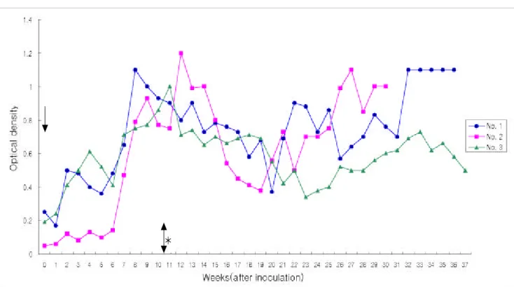
ELIS A of se ra from e xp e rim e n ta lly in fe c te d d og s In the Dog No.1, No.2 and No.3, antibody titers in the sera of inoculated dogs were significantly detected 7 weeks post inoculation and persisted until 14 weeks. Antibody titers of dogs detected by ELISA peaked between 7 and 14 weeks then decreased between weeks 15 to 24 followed by another increase during weeks 25 to 30 and persisted throughout the remainde of the experiment period.
An alys is o f a du lt D. im m it is p rote in by Coo m as sie brillian t blu e R-250 sta in in g
D. im m itis proteins were separated into more than 10 bands stained with Coomassie brilliant blue R-250 (CBB) staining, the major bands were 22, 40, 46, 56, 70, 72 and 89 kDa.
Fig 1. Antibody responses of dogs to experimental D. im m itis infection by ELISA using D. im m itis antigens.
*The range of optical density of control group. The arrow indicates the day of inoculation.
*
112 Kun-Ho Song, Mineo Hayasaki, Chusnul Choliq, Kyu-Woan Cho, Hong-Ryul Han, Bung-Hyun Jeong, Moo-Hyung Jeon, Bae-Kun Park and Duck-Hwan Kim
An tig e n ic ide n tific atio n o f cru de e xtrac t a n tige n s of a du lt D. im m it is by im m u n oblo ttin g
Pooled sera of patent infection (30 weeks after inoculation) from Dog No.1 revealed four antigenic bands with 24, 70, 80 and 110 kDa. Dog No.2 showed three antigenic bands of 22, 72 and 84 kDa, while two antigenic bands were detected in Dog No. 3 with 58 and 72 kDa.
Discussion
Experimental D. im m itis infections in dogs have been performed by various investigators [6, 8, 27, 29]. The recovery rate of adult worms from dogs experimentally inoculated with the infective larvae of D. im m itis were 45%, Hayasaki [8], 66%, Thrall et al.[27] and 50%, Wong et al.[29], respectively. In this study, three cases accounted to 42.91 % recovery of inoculated worms. Although this study did not confirm the correlation between antibody titer and the number of adult worm, Grieve et al.[6] reported that there was no relationship between them.
The ELISA used in conjunction with a Knott's test, exposure history, clinical signs, laboratory results, and radiographic changes are useful for studying seroepizootiologic pattern and risk factors of heartworm infection. ELISA testing has been shown to be capable of identifying prepatent
infections [6]. Diagnosis of prepatent infection or asymptomatic occult infection is important to the practitioner who may be preparing to initiate a heartworm preventive program based on negative results of a Knott`s test. In addition, experimental studies of beagles have shown abnormalities of pulmonary arteriograms 6 months after inoculation with infective D. im m itis larvae, at which time the dogs were still amicrofilaremic and results of indirect fluorescent antibody testing for antimicrofilarial antibodies were negative [22].
ELISA testing for the detection of D. im m itis provide a useful and improved assay for serological characterization and detection of infection. Grieve et al.[6] reported that ELISA titers were not significantly increased until 11 or 16 weeks, and remained at maximum levels for the duration of the observation period. However, the present study revealed that antibody levels were detected as early as 7 weeks post inoculation, and developed continuously to 14 weeks, and then diminished to 15 to 24 weeks, then climbed again to 25 to 30 weeks, and high titer levels persisted throughout later experiment time. One explanation of these different results may be the different responses of individual dogs to the variable extent and duration of parasitic exposure [13].
Another explanation may be that different antibodies are being detected by different protocols used [11].
Konno et al.[15] reported that protein bands in extracted
fig . 2. Protein fractions of adult D. im m itis were separated by SDS- PAG E s t a in e d w it h C oom a s s ie b r illia n t blu e
R -25 0(A), a n d D . im m i t i s a n t ibod y r e s p on s es w it h D . i m m i t i s a n t igen by im m u n ob lot t in g a n a lys is (B ). La n e
1:s t a n d a r d m a r k e r , la n e 2-4: n ega t ive con t r ol, la n e 5-7 : p a t e n t in fe ct ion (la n e 2 ,5: N o.1 , la n e 3,6: N o.2 , la n e
4,7: N o.3).
a n t ige n of D . im m i t i s w a s d et e ct ed by C oom a s s ie b r illia n t blu e R -2 50 s t a in in g w e r e 44 ba n d s (1 4 t o 2 30 k D a ). T h e pr esen t s t u dy r evea led t h a t D . im m it is pr ot ein wa s s e p a r a t e d in t o m or e t h a n 1 0 ba n d s (22 t o 8 9 k D a ) by C oom a s s ie br illia n t blu e R -2 50 s t a in in g, t h e m a jor ba n d s d e t e ct e d w e r e 22 , 4 0, 4 6, 5 6, 70 , 72 a n d 89 k D a . Alt h ou gh t h e n u m b er of p r ot ein ba n d s w a s les s t h a n t h o s e of Konno et al.[15], similar protein bands were detected by Coomassie brilliant blue R-250 staining. Immunoblotting in th e D . im m i t i s infection helps to clarify the relation of antigen- antibody in immune response, a more effective application of immunologic diagnosis [9,12,24]. Tamashiro et al.[24] and Boto et al.[1]
reported that low molecular weight reactivity as more prevalent in older and /or more heavily infected dogs in experimental D. i
m m itisinfect io n by means of immunoblotting.
I n n a t u r a l D . im m i tis infection, Tanaka et al.[25] reported the trends in the distribution of immunoblotting patterns with either young animals and/or mild infection tended to reveal antibodies with reactivity to antigen in the higher molecular weight regions of immunoblotting (more than 80 kDa), he also reported that the majority of antibodies with reactivity to antigens in the low molecular weight regions (less than 37 kDa), occurred more frequently in older dogs and/or heavier infections. The results of Tamashiro et al.[24], Tanaka et al.[25] and Boto et al.[1] suggested that immunoblotting will produce similar results in both experimental and natural infections.
In the present s t u d y, a n t ige n ic id e n t ifica t ion of cr u d e ext r a ct a n t ige n s of a d u lt s D . i m m it is b y im m u n ob lot t in g a n a lys is r e ve a le d s eve r a l b a n d s fr om t h e p ooled s e r a of p a t e n t in fe ct ion (30 w ee k s a ft er in ocu la t ion ). Th e d et ect ed ba n d s w er e 2 4, 7 0, 8 0 a n d 11 0 k D a in D og N o.1 , 22 , 72 a n d 84 k D a in D og N o.2 , a n d 5 8 a n d 7 2 k D a in D og N o.3 . T h e r es u lt s p r e s en t ed h e r e a r e s im ila r t o t h os e of T a m a s h ir o e t a l.[24 ] a n d B ot o et a l.[1 ], in w h ich t h e h igh e r m olecu la r w e igh t r e gion s w e r e d e t e ct e d by im m u n oblot t in g in you n g a n im a ls (m or e t h a n 70 k D a ).
I n s u m m a r y, a n t ibod y t it e r s by E LI S A r e a ch h igh le vels a t t h e 4 t h m olt in g s t a ge a ft e r in ocu la t ion of L3 , a n d t h e h igh er m olecu la r w e igh t r e gion s a r e m a in ly d et e ct ed by im m u n ob lot t in g in you n g a n im a ls . F u r t h e r s t u d ie s by ot h er t ech n iqu es a r e n e ed ed t o e lu cid a t e im m u n ologica l r es p on s es in D . i m m i t i s -in fe ct e d d ogs .
A ck n o w le d g e m e n t
Th is s t u d y w a s fin a n cia lly s u p p or t ed b y 20 01 of Aca d em ic R e s e a r ch & S ch ola r s h ip F ou n d a t ion , C h u n gn a m N a t ion a l U n iver s it y.
Re fe re n ce s
1 . B o t o , W. M ., P o w e r , K . G . a n d L e v y , D . A. An t igen s of D i rofi l a r ia i m m it is w h ich a r e im m u n oge n ic in t h e canine host: detection by immunostaining of protein
blots with the antibodies of occult d ogs . J . I m m u n ol.
1 98 4, 1 3 3 , 9 75 -9 80 .
2. C a r l i s le , C . H . C a n in e d ir ofila r ia s is . I t s r a d iogr a p h ic a p p ea r a n ce . Vet . R a d iol.1 98 0, 2 1 , 1 23 -13 0.
3. D z i m i a n s k i , M . T ., M c T e r , T . L . a n d M c C a ll, J . W.
1 98 9. E va lu a t ion of t w o a d u lt h e a r t w or m a n t ige n d ia gn os t ic t e s t k it s u s in g w ell-d e fin ed d og a n d ca t s e r a . I n : P r oc 3 4t h An n u M e et Am As s oc Vet P a r a s it ol.
O r le n d o: Am . As s oc. Vet . P a r a s it ol. 3 3.
4. E t t i n g e r , S . J . a n d F e ld m a n , E . S . T ext book of vet e r in a r y in t e r n a l m ed icin e: D i sease of the dog and cat.
pp. 937-939. 5th ed. WB Saunders, Philadelphia, 2000.
5. Glick m an , L.T., Grie ve , R. B. a n d B re its ch w e rdt, E.
B . Serological pattern of canine heartworm (D
i rof il a r iaim m itis). Am. J . Vet. Res. 1984, 45, 1178-1183.
6. Grie ve , R. B ., J o h n so n , M. M. an d J ac obs on , R. H.
Enzyme-linked immunosorbent assay for measurement of a n tibody responses to Dirofilaria im m itis in experi- mentally infected dogs. Am. J . Vet. Res. 1981, 42, 66-69.
7. Hay as ak i, M. Indirect hemagglutination test for diagnosis of canine filariasis. J pn. Vet. Sci. 1981, 43, 21-26.
8. Hay as ak i, M. Reaginic and hemmaglutinating antibody production in dogs infected with Dirofilaria im m itis.
J pn. J . Vet. Sci. 1982, 44, 63-70.
9. Hay as ak i, M., Na ka m u ra, F . an d Kats u h iko , K.
Immunoblotting analysis of somatic components of Dirofilaria im m itis. J . Vet. Med. Sci. 1994, 56, 1181-1183.
10. Ho ov e r, J . P ., Fo x, J . C. an d Clay po ol, P . L.
Comparison of visual interpretation and optical density measurements of two antigen tests for heartworm infections in dogs. Canine Pract. 1996, 21, 12-2 0 . 11. Ka ga n , I. G. Advances in immunodiagnosis of
parasistic infections. Z. Parasitenkd. 1974, 45, 163-175.
12. Ka n e ko , H., Ha ya sa ki, M. an d Oh ish i, I. Antigenic identification of excretory-secretory products of adult Dirofilaria im m itis. J pn. J . Vet. Sci. 1990, 52, 995-1000.
13. Ke n n e d y, M. W., Mcin to sh , A. E. a n d Blair, A. J . M H C (R T 1) r es t r ict ion of a n t ibod y r e p er t oir e t o in fe ct ion w it h t h e n em a t od e N ip p os t r on g yl u s b ra s i li en s is in the rat. Immunology 1990, 71, 317-320.
14. Ko h le r, G. an d Milste in , C. Continuous culture of fused cells secreting antibody of predefined specificity.
Nature 1975, 256, 495-497.
15. Ko n n o, K. a n d Ha ya sa ki, M. Antigenic cross reactivity among Dirofilaria im m iti
sa n d t h e fou r in t e s t in a l p a r a s it e-s p e cie s in t h e d og. J p n . J . P a r a s it ol.
1 99 5, 4 4 , 1 61 -1 64 .
16 . L a e m m l i , U . K. C lea va ge of s t r u ct u r a l p r ot e in s d u r in g t h e a s s e m bly of t h e h e a d b a ct er iop h a ge T 4. N a t u r e 1 97 0, 2 2 7 , 6 80 -6 85 .
17 . L o w r y , O . H ., R o s e n b r o u g h , N . J ., F a r r , A. L . a n d
R a n d a l l, R . J . P r ot e in m ea s u r e m e n t w it h t h e folin
p h en ol r ea ge n t . J . B iol. C h em . 19 51 , 1 9 3 , 26 5-2 75 .
18 . M a r t i n i , M . C a p e lli , G . a n d P o g la y e n , G . T h e va lid it y of s om e h a em a t ologica l a n d E L I S A m et h od s for t h e d ia gn os is of ca n in e h e a r t w or m d is ea s e . Ve t . R e s . C om m u n . 1 99 6, 2 0 , 3 31 -33 9.
19 . M a t h e r n e , C . M ., G r e e n , S . P . a n d C o r w i n , R . M . D et e ct ion of cir cu la t in g D ir of i la ri a i m m it is a n t igen s in r a n d om s ou r ce la b or a t or y d ogs : eva lu a t ion of t w o com m e r cia l s er od ia gn os t ic t e s t s . L a b . An im . S ci. 19 88 , 3 8 , 5 84 -5 87 .
20 . O h i s h i , I., K o b a y a s h i , S . a n d Ku m e , S . S t u d ies on t h e d ia gn os is of ca n in e fila r ia s is . I I I . C on cen t r a t ion of m icr ofila r ia in t h e t e s t b lood . J p n . J . Vet . M e d . As s oc.
1 95 9, 12 , 1 49 -1 53 .
21 . O t t o , G . F . 1 97 8. T h e s ign ifica n ce of m icr ofila r ia in t h e d ia gn os is of h e a r t w or m in fe ct ion , in H . C . M or ga n , G . F . O t t o, R . F . J a ck s on (e d s ): P r ocee d in gs of t h e h e a r t w or m sym posiu m `77. Bon ner Spr ings, Ka n, Vet er in a r y M ed icin e P u b lis h in g. p p . 22 -3 0.
22 . R o w l i n g s , C . A., D a w e , D . L . a n d M c C a l l, J . W. F ou r t yp e s of occu lt D ir of i la ri a im m i t i s in fe ct ion in d ogs . J . A. V. M . A. 19 82 , 1 8 0 , 13 23 -1 32 6.
23 . R a w l i n g s , C . A., M c C a l l, J . W. a n d L e w i s , R . E . Th e r e s p on s e of t h e ca n in e h ea r t a n d lu n g t o D i r of il a r i a
i m m i t i s .