- 79 -
서 론
타액선 종양은 두경부 악성 종양의 약 5%를 차지하며,1,2) 점 액표피양 암종은 이 중 가장 흔한 형태의 악성 종양으로 타액 선 종양의 약 12~29%를 차지한다.3) 주 타액선에서는 이하선 에 가장 호발하며,1,2) 소타액선에선 약 46%에서 구강 내에서 발생한다. 구강 내 발생 빈도는 구개가 가장 흔하며, 협부, 하 악, 입술, 그리고 혀 등의 순이다.1) 구후 삼각부, 구인두, 그리 고 이소성 타액선에 발생하는 경우는 매우 드물다. 국내에서 는 현재까지 구후 삼각부 점액표피양 암종은 보고된 예가 없다.
이에 저자들은 구강 내 이물감과 전경부 종물을 주소로 내원 한 65세 여자 환자에서 구후 삼각부의 점액표피양 암종으로 진단된 증례를 치험하여 문헌고찰과 함께 보고하고자 한다.
증 례
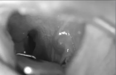
66세 여자환자가 내원 약 3개월 전부터 시작된 좌측 구강 내 이물감과 전경부의 종물로 타병원 진료 후 본원 이비인후과 를 방문하였다. 신체검사상 좌측 구후 삼각부에 약 1.5×1 cm 크기의 푸른빛을 띄는 딱딱한 종물(Fig. 1)과 전경부에 약 3
×3 cm 크기의 무통성 고정성 종물이 관찰되었다. 두경부에 비정상적인 림프절은 촉지되지 않았고, 내시경 검사상 인후두 에 특이소견 없었다. 경부 전산화 단층촬영에서 좌측 구후삼 각부에 약 1.2×1 cm 크기의 종물(Fig. 2)과 우측 갑상선에 석 회화를 동반한 약 3×3 cm 크기의 결절과 좌측 갑상선에는 2.5×2 cm 크기의 종물이 관찰되었다. 구후삼각부 종물은 혈 관종보다 딱딱하였으나, 색깔이 푸른빛이어서 혈관성 종양을 의심하여 세침흡인검사는 시행하지 않았다. 우측 갑상선에서 세침흡인검사를 시행하여 Bethesda 분류상 category III로 보고되어, 구후삼각부 종괴에 대한 절제생검과 우측 갑상선 절제술을 계획하였다. 전신 마취 하에 구강 내로 접근하였다.
구강 종물은 비교적 주변과 잘 박리되었으나, 입천장혀근(pal- atoglossus)과 상인두수축근(superior pharyngeal constrictor) 에 부분적으로 유착이 있는 양상이었다. 구강 종물은 동결절 대한 두경부 종양 학회지
제 30 권 제 2 호 2014
구후 삼각부 종물 양상의 점액표피암종 1예
중앙보훈병원 이비인후과,1 병리과2
곽슬기1·김춘동1·김은주2·김승우1
=Abstract =
A Case of Mucoepidermoid Carcinoma Presenting as a Retromolar Trigonal Mass
Seul Gi Kwak, MD1, Choon Dong Kim, MD1, Eun Ju Kim, MD2, Seung Woo Kim, MD1 Departments of Otolaryngology-Head and Neck Surgery1 and Pathology,2 Veterans Health Service Medical Center,
Seoul, Korea
Salivary gland tumors take possession of almost 5% in head and neck malignancies. Among these, mucoepider- moid carcinoma(MEC) is most common malignany in major salivary glands(12-29%) and the parotid gland is most predilection site. Intra-oral MEC has a tendency to various locations, and the predilection sites are palate, cheek, mandible, lip and tongue in order of frequency. A few cases of MEC are occurred in with retromolar trigone, oro- pharynx, and ectopic salivary gland. Recently, we experienced a-65-year old woman with retromolar trigonal mass, and she was finally diagnosed as MEC. We report it with review of literature.
KEY WORDS:Mucoepidermoid carcinomaㆍOral cavity.
Received : September 15, 2014 Accepted : September 22, 2014
교신저자 : 김승우, 134-791 서울 강동구 진황도로 61길 53 중앙보훈병원 이비인후과
전화 : (02) 2225-1384 ・ 전송 : (02) 2225-1385 E-mail : [email protected]
online©MLComm
- 80 - 편 검사를 시행하지 않았으며, 우측 갑상선에선 저명한 악성 소견은 보이지 않아 좌측 갑상선 절제술은 시행하지 않고 수 술을 종료하였다. 최종 조직검사에서 구후삼각부 종괴는 저 등급 점액표피양 암종으로(Fig. 3), 우측 갑상선은 광범위 침
습 여포암종으로 확진되었다. 점액표피양 암종은 변연부 침 범은 보이지 않았으나, 무종양의 안전 경계가 2 mm로 보고되 어 술 후 일주일 째 좌측 구후 삼각부에 대해 입천장혀근과 상인두수축근을 일부 포함하여 광범위 절제 및 좌측 완성 갑상선 절제술을 시행하였다. 두번째 수술의 조직검사결과 절제부위에서 악성소견은 보이지 않았으며, 좌측 갑상선은 결 절성 과증식으로 보고되었다. 수술 후 양전자단층촬영상 특이 소견 보이지 않았고, 술 후 10개월이 지난 현재 재발 소견 없이 외래 추적관찰 중이다.
고 찰
구강에서 종양이 가장 호발하는 곳은 구강설과 구강저이고, 구후삼각을 침범하는 경우는 드물다.4) 구강을 침범하는 악성 종양 중 약 7%에서 구후삼각을 침범하며, 그 중 대부분은 편 평세포 암종이고, 소타액선 종양이 발견되는 경우도 있다.5)
점액표피양 암종은 가장 흔한 타액선 종양으로 이중 약 46%
는 구강내의 다양한 위치에 분포하는 소타액선에서 발생한 다.1) 구개는 소타액선에 발생한 점액표피양 암종이 가장 호발 하며 약 55%를 차지하며,1) 그 외에도 구후삼각부, 구강저, 협 점막, 입술과 혀 등에서 발생할 수 있다.6) Lutcavage 등은 소타 액선 종양환자 265명 중 6.4%에서 구후삼각부에서 발생하였 다는 보고하였다.7)
점액표피양 암종은 일반적으로 수년에 걸쳐 천천히 자라 는 무통성 종물로 나타나게 되며 그 외에도 통증, 궤양, 변색, 안면마비 등의 증상이 나타날 수 있다.8) 저악성도의 종양에서 는 그 크기가 5 cm을 넘는 경우는 드물고, 고악성도의 종양에 서는 빠르게 자라는 무통성의 종물이 주변조직으로 침윤되는 양상으로 나타나며, 원격전이와 궤양을 동반하는 경우도 있 다.2) 구강내의 종양은 표층에 위치하기 때문에 점액 낭종이나 혈관성 종양과 비슷하게 푸르거나 붉은 빛깔을 띤다.9,10) 임상 증상은 비특이적이며 대부분 천천히 자라는 무통성 종괴로 나타나게 되는데, 통증의 동반, 빠르게 자라는 경우는 고등 급 점액표피양암종 을 의심해 볼 수 있다. 감별질환으로는 편
Fig. 1. The external photography shows 1.5×1 cm sized bluish mass in retromolar trigone.
Fig. 2. Preoperative contrast-enhanced axial CT scan shows low density mass, and it is located medial to the mandibualr angle (arrow).
Fig. 3. Pathologic findings. A : Cut surface of surgical specimen. B : It shows that low grade mucoepidermoic carcinoma composed of mucous cells lining cystic space(H & E, ×200). C : The photo shows epidermal cells with very few mucous cells and minimal cystic changes suggestive of mucoepidermoid carcinoma(H & E, ×400).
A B C
- 81 - 평세포암, 선편평세포암, 타액관 암종, 전이성 신세포암 등이 있다.1)
컴퓨터 전산화단층촬영으로 골 침윤을 확인할 수 있고, 자 기공명영상은 연부조직 침윤에 효과적이다. 크기가 작고 쉽 게 접근할 수 있는 종양은 구개나 구후삼각에 위치하는 경우 를 제외하고는 영상학적 평가가 필수적이지 않으나, 크기가 크거나 재발한 종양 또는 뇌신경의 침범이 의심되는 경우는 영상학적 평가가 필수적이다.11) 세침흡인검사가 진단에 도움 을 주며, 악성의 정도가 세침흡인검사의 결과 평가 영향을 미친 다고 알려져 있다.12) Klijanienko 등에 의하면 저악성도(68%) 에 비해 고악성도(87%)의 종양이 진단율이 더 높다고 한다.12)
조직학적으로 점액표피양 암종은 3단계로 구분되며 저악성 도(48%), 중등 악성도(13.3%), 고악성도(38.7%)로 분류된다.2) 다양한 성장양식과 세포형태가 특징적이며, 이 두 가지 요소가 점액표피양 암종의 진단과 등급을 결정하게 된다. 종양에는 낭종과 고형 형태로 배열된 점액 세포, 표피양 세포, 중간 세 포, 원주 세포 및 투명세포가 분포하며, 이 세포들의 함량이 종 양의 등급을 분류하는데 중요하다.1) 저악성도의 종양은 점액 세포의 비율이 높고 덜 침습적인 반면 고악성도의 종양은 점 액세포의 비율이 낮다.2)
해부학적으로 접근 가능한 점액표피양 암종의 치료방법은 완전한 수술적 절제이다.13) 저악성도 종양은 수술적 치료만으 로도 충분한 것으로 알려져 있다.14) 소타액선에 발생한 작은 크기의 저악성도 점액표피양 암종은 국소 광범위 절제술만으 로 충분한 경우가 많지만, 범위가 넓은 경우 하악골 절제술, 상 악골 절제술, 유양돌기
절제술, 측두하와 접근법, 또는 전방 두개 안면부 절제술 등 이 필요한 경우도 있다.15) 절제 연의 침범이 있는 경우 높은 재 발율을 보이므로 적절한 절제범위 선택이 필요하다.6) 경부 림 프절의 전이는 나쁜 예후인자이며,16) Chen 등의 보고에 의하 면 고악성도 종양에서는 34%, 중등-악성도에서는 8.1%, 저악 성도에서는 3.3%에서 경부림프절의 전이를 보였다.17) 방사선 치료는 고악성도 종양의 환자와 수술 절제연이 불분명한 경우 또는 경부 림프절 전이가 있는 경우에 수술적 치료에 추가적으 로 사용된다.18,19)
예후는 5년 생존율이 중등도(97.4%)와 저악성도(98.8%) 종 양은 차이가 없으며 고악성도 종양은 67%로 의미있게 낮다.17) 나이가 젊은 환자, 여자환자에서 예후가 좋은 것으로 알려져 있으며, Ki-67 항원은 증식성 표지인자로 높게 발현될수록 조 직학적 등급이 증가된다.20,21)
본 증례에서의 교훈은 소타액선 종양이 구후삼각부에서 발 생할 수 있으며, 종양의 성상이 혈관종이 의심되는 경우에도, 비교적 딱딱하고, 수술 시 주변과 유착이 있는 경우는 소타액 선 종양을 의심해야 하겠다.
중심 단어 : 점액표피양 암종 ・구강.
References
1) Ranganath MK, Matmari V, Narayanaswamy UD, Bavle RM.
Mucoepidermoid carcinoma presenting as a retromolar muco- cele. Ann Maxillofac Surg. 2011;1(1):66-69.
2) Devaraju R, Gantala R, Aitha H, Gotoor SG. Mucoepidermoid carcinoma. BMJ Case Rep. 2014 Aug 1;2014:bcr-2013-202776. doi:
10.1136/bcr-2013-202776.
3) Spiro RH, Huvos AG, Berk R, Strong EW. Muoepidermoid car- cinoma of salivary gland origin. A clinicopathologic study of 367 cases. Am J Surg. 1978;136(4):461-468.
4) Genden EM, Ferlito A, Shaha AR, Rinaldo A. Management of cancer of the retromola trigone. Oral Oncol. 2003;39(7):633-637.
5) Crecco M, Vidiri A, Angelone ML, Palma O, Morello R. Retro- molar trigone tumors: Evaluation by magnetic resonance imag- ing and correlation with pathological data. Eur J Radiol. 1999;
32(3):182-188.
6) Triantafillidou K, Dimitrakopoulos J, Iordanidis F, Koufogiannis D. Mucoepidermoid carcinoma of minor salivary glands: A clin- ical study of 16 cases and review of the literature. Oral Dis. 2006;
12(4):364-370.
7) Lutcavage GJ, Schaberg SJ, Fulbright DK, Nelson CL. Retromo- lar trigone mass. J Oral Maxillofac Surg. 1993;51(9):1024-1029.
8) Plambeck K, Friedrich RE, Schmelzle R. Mucoepidermoid car- cinoma of salivary gland origin: Classification, clinical-patho- logical correlation, treatment results and long-term follow-up in 55 patients. J Cranioaxillofac Surg. 1996;24(3)133-139.
9) Eversole LR. Mucoepidermoid carcinoma: Review of 815 re- ported cases. J Oral Surg. 1970;28(7):490-494.
10) Melrose RJ, Abrams AM, Howell FV. Mucoepidermoid tumors of the intraoral minor salivary glands: A clinicopathologic study of 54 cases. J Oral Pathol. 1973;2(6):314-325.
11) Sharma P, Jain TK, Singh H, Suman SK, Faizi NA, Kumar R, et al. Utility of (18)F-FDG PET-CT in staging and restaging of pa- tients with malignant salivary gland tumours: A single-institu- tional experience. Nucl Med Commun. 2013;34(3):211-219.
12) Klijanienko J, Vielh P. Fine-needle sampling of salivary gland lesions. IV. Review of 50 cases of mucoepidermoid carcinoma with histologic correlation. Diagn Cytopathol. 1997;17(2)92-98.
13) Vander Poorten V, Hunt J, Bradley PF, Haigentz M Jr, Menden- hall WM, Suarez C, et al. Recent trends in the management of minor salivary gland carcinoma. Head Neck. 2014;36(3):444- 455.
14) Chen AM, Lau VH, Farwell DG, Luu Q, Donald PJ. Mucoepi- dermoid carcinoma of the parotid gland treated by surgery and postoperative radiation therapy: Clinicopathologic correlates of outcome. Laryngoscope. 2013;123(12):3049-3055.
15) Byrd SA, Spector ME, Carey TE, Bradford CR, McHugh JB.
Predictors of recurrence and survival for head and neck muco- epidermoid carcinoma. Otolaryngol Head Neck Surg. 2013;149 (3):402-408.
- 82 -
16) Armstrong JG, Harrison LB, Thaler HT, Friedlander-Klar H, Fass DE, Zelefsky MJ, et al. The indications for elective treatment of the neck in cancer of the major salivary glands. Cancer. 1992;
69(3):615-619.
17) Chen MM, Roman SA, Sosa JA, Judson BL. Histologic grade as prognostic indicator for mucoepidermoid carcinoma: A popu- lation-level analysis of 2400 patients. Head Neck. 2014;36(2):
158-163.
18) Hosokawa Y, Shirato H, Kagei K, Nishioka T, Tei K, Ono M, et al. Role of radiotherapy for mucoepidermoid carcinoma of sal- ivary gland. Oral Oncol. 1999;35(1):105-111.
19) Ghosh-Laskar S, Murthy V, Wadasadawala T, Agarwal J, Budruk- kar A, Patil N, et al. Mucoepidermoid carcinoma of the parot- id gland: Factors affecting outcome. Head Neck. 2011;33(4):497- 503.
20) O’Brien CJ, Soong SJ, Herrera GA, Urist MM, Maddox WA.
Malignant salivary tumors-analysis of prognostic factors and survival. Head Neck Surg. 1986;9(2):82-92.
21) Tran L, Sadeghi A, Hanson D, Juillard G, Mackintosh R, Calca- terra TC, et al. Major salivary gland tumors: Treatment results and prognostic factors. Laryngoscope. 1986;96(10):1139-1144.